Peripheral Blood Morphology Flashcards
the area of the slide that you would exam on a peripheral smear
from the monolayer to the feathered edge
RBC should be the size of a _______ nucleus
lymphocyte
the ares of the central pallor in a RBC should be ___ of the total RBC diameter
1/3
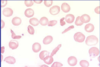
anisocytosis
refers to the red cells which vary widely in size
RDW
red cell distribution width; measures the range of red cell sizes ; measures anisocytosis
microcytosis
refers to RBC that are small
MCV
mean cell volume; measures the individuals volume of red blood cells; measures microcytosis and macrocytosis
differential diagnosis for microcytosis (6)
- Iron deficiency 2. Anemia of chronic disease 3. Thalassemia 4. Hemoglobin C disease 5. Lead Poisoning 6. Sideroblastic anemia
macrocytosis
RBC are too large; use MCV
differential diagnosis of macrocytosis (8)
- B12/FOLATE DEFICIENCY 2. Liver disease 3. Thyroid disease 4. MDS myelodysplastic syndrome 5. Anti-retrovirals 6. Aplastic anemia 7. Chemotherapy 8. Elevated reticulocyte count
hypochromasia
RBC with too little hemoglobin
hypochromasia RBC have a central pallor that is less/more than 1/3 the total red cell diameter
more
hypochromasia is measured by
MCH mean cell hemoglobin
polychromasia
RBC that have more of a blueish tinge; probably reticulocytes
poikilocytosis
RBC that vary in shape
target cells
look like bulls-eyes
differential diagnosis of target cell (4)
- liver disease (most common) 2. thalassemias 3. hemoglobin C 4. After a splenectomy
spherocytes
have loss of central pallor
Spherocytes can be seen in
- autoimmune hemolysis 2. hereditary spherocytosis
schistocytes
red cell fragments with sharp edges
schistocytes are hallmark of
microangiopathic hemolytic anemia (MAHA)
sickle cell
seen in sickle cell anemia
echinocytes
- aka burr cell - small, regular projections - seen in renal disease
acanthocytes
- aka spur cells - large, irregular projections - seen in liver disease
teardrop cells
myelophthisic processes, which are diseases of marrow infiltration
teardrop cells can be seen in (5)
- splenomegaly 2. Leukemia and lymphoma 3. Granulomatous disease 4. tumor metastatic to marrow 5. myelofibrosis
Howell Jolly Bodies
peripheral, round, small purple inclusions within red cells that represent nuclear remnants
when are Howell Jolly Bodies most likely seen
after splenectomy
rouleaux
linear arrangements of RBC
rouleaux cells are typically seen in disorders with
- increased levels of immunoglobulin (Multiple Myeloma or Waldenstroms macroglobulinemia) 2. severe hypo-albuminemia
agglutination occurs when red cells are coated with
IgM
iron deficiency anemia (3)
- hypochromic 2. microcytic 3. increased number of platelets
megaloblastic anemia
- red cells are macrocytic - hyper-segmented neutrophils
autoimmune hemolytic anemia (AIHA)
- polychromasia - microspherocytes

iron deficiency anemia

a. target cell
b. howell-jolly body
c. nucleated rbc
d. schostpcyte
e. basophilic stippling

megaloblastic anemia

autoimmune hemolytic anemia (AIHA)
black arrows- polychromasia
green arrows- microspherocytes

microangiopathic hemolytic anemia
sickle cell anemia
