Pancreas WB images Flashcards
the aorta and IVC are the posterior landmarks of the pancreas
sagittal plane of the pancreas

the portal venous system is the posterior border of the pancreas


to which vascular structure are the arrows pointing in this image?
SMV is seen along the posterior border of the neck of the pancreas
identify if the image is transvere or longitudinal and what the arrows are pointing to

sagittal
arrows= IVC
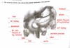
identify the anatomic structure that the arrows are pointing to in this image

the splenic artery, rises from the celiac axis and is seen as the posterior border of the neck of the pancreas
what is the arrow pointing to?

Hepatic Artery
the common bile duct is between the calipers
this is the mickey mouse sign (porta hepatis)
what is the arrow head pointing to?

pancreatic duct
a 45 year old male presents w/ midepigastric pain, elevated amylase and lipase levels and tenderness. ID the sonographic finidings

sonographic findings consist of an enlarged, edematous pancreas consistant with pancreatits
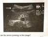
a patient w/ known acute pancreatitis presents w/ continued pain. describe the sonographic findings

sonographic findings consist of pancreatitis w/ a pancreatic pseudocyst
a 56 year old male w/ a 1 week history of jaundice and pain has reported a 3 month history of N&V, weight loss & diarrhea. given this info. what are the sonographic findings?

sonographic findings consist of adenocarcinoma of the pancreas w/ a dilated pancreatic duct
