Muscle Tissue Flashcards
What’s the overall function of muscle tissue?
Movement
What makes movement possible in muscle tissues?
Contractility
How is movement generated in muscle tissues?
Movement is generated by the interaction between actin and myosin proteins
There are three specialized contractile cells. What are they?
- Myoepithelial cells
- Pericytes
- Myofibroblasts
____________________ are specialized contractile cells with secretory functions
Myoepithelial cells
__________________ are smooth-muscle-like cells that surround blood vessels
Pericytes
__________________ are cells that secrete collagen and have a true contractile role
Myofibroblasts
A _____________ is a collection of multicellular contractile units
Muscle
Muscle tissue is composed of cells differentiated for __________________ with microfilaments and associated proteins that generate the force necessary for such a function
Contractility
Nearly all cells of muscle tissue are of __________________ origin
Mesodermal/mesenchymal
Muscle cells differentiate from mesenchymal stem cells by increasing cell ________________ and _____________ protein synthesis
Length
Myofibrillar
How many types of muscle tissues are there?
Three: cardiac, skeletal, smooth
_______________ muscle tissue has cross-striations and is composed of elongated, branched individual cells
Cardiac
At sites of end-to-end contact of cardiac muscle cells, there are _______________________
Intercalated discs
Contraction of cardiac muscle is _______________, vigorous, and _______________
Involuntary
Rhythmic
___________________ tissue consists of collections of __________________ cells that do not show straitions
Smooth muscle cells
Fusiform
In what type of muscle tissue are contraction processes slow and not subject to voluntary control?
Smooth muscle tissue
____________________ muscle tissue is composed of bundles of very long, cylindrical, __________________ cells that show ________________
Multinucleated
Cross-striations
The contraction of skeletal muscle cells is quick, forceful, and usually under _________________ control
Voluntary
The interaction between thin _______________ filaments and thick ___________ filaments results in skeletal muscle contraction
Actin
Myosin
What is the fine transparent tubular sheath which envelops the fibers of skeletal muscles called?
Sarcolemna
Mature cardiac muscle cells exhibit a cross-striated banding pattern comparable to that of skeletal muscle.Unlike multinucleated skeletal muscle, however, each cardiac muscle cell possesses _____________ centrally located pale-staining nuclei
Only one or two
Surrounding muscle cells is a delicate sheat of _____________________ containing a rich capillary network
Endomysium
What’s the difference between the sacrolemna and endomysium?
The sarcolemma is the cell (plasma) membrane of the muscle fiber itself. The endomysium is a thin sleeve of fibrous connective tissue over the muscle fiber
What is a unique and distinguishing characteristic of cardiac muscle?
The presence of dark-staining transverse lines that cross the chains of cardiac cells are irregular intervals
What type of muscle tissue is shown?
What does “I” point to? “N”? “D”?
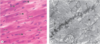
Cardiac muscle fibers
I = intercalcated disc
N = nuclei
D = desmosome (in EM)
____________________ is the interface between adjacent cardiac muscle cells where many junctional complexes are present
Intercalated discus
Transverse regions of intercalated discus have many _______________ and ______________________, which resemble the zonula adherentes between epithelial cells
Desmosomes
Fascia adherentes
What’s the purpose of desmosomes and fascia adherentes in cardiac muscle cells?
To bind cardiac cells firmly together and to prevent their ripping under constant contractile activity
Longitudinal portions of each intercalated disc have multiple ______________________, which provide ionic continuity between adjacent cells and act as electrical synapses, of sorts
Gap junctions
Gap junctions in cardiac muscle cells allow for such cells to act as a ______________________ with contraction signals passing in waves from cell to cell
Multinucleated syncytium
_____________________ consists of muscle fibers, long, cylindrical multinucleated cells
Skeletal muscle
Why are skeletal muscle cells multinucleated?
Because they result from fusion of embryonic mesenchymal cells (myoblasts)
The nuclei of skeletal muscle cells are ______________ in shape and found at the ______________ of the cell
Oval
Periphery
What’s the cytoplasm of muscle cells called?
Sarcoplasm
What’s the smooth ER of muscle cells called?
Sarcoplasmic reticulum
What’s the plasma membrane of muscle cells called?
Sacrolemma
Skeletal muscle cells develop from mesenchymal cells called _________________ that align and fuse together, making long, multinucleate tubes called ______________, which then synthesize proteins to create myofilaments and continue differentiation into functional microfilaments
Myoblasts
Myotubes
_________________ is the external sheath of dense connective tissue that surroundings the entire muscle
Epimysium
____________________ is the thin septa of connective tissue that extends inward from the epimysium, surrounds the __________________ or fiber bindles within a muscle, and contains a rich capillary network
Endomysium
Fascicles
____________________ is the connective tissue that surrounds each fascicle
Perimysium
What’s the purpose of the connective tissues surrounding muscle and its fibers and cells?
To transmit the mechanical forces generated by contracting muscle cells and fiber
Most muscles taper off at their ________________
Extremities
At the ________________ junction, the connective tissue components of the epimysium associate with tendons
Myotendinous junction
At the myotendinous junction, collage fibers of the ______________ insert themselves among the muscle fibers and associate with the _______________
Tendon
Sarcolemma
Myotendinous junctions join muscles to the __________________ of bones
Periosteum
_________________ muscle fibers are elongated, tapered, and non-striated cells
Smooth
Each smooth muscle cell is enclosed by a thin _________________ and a fine network of _______________, which serve to combine the forces generated by each smooth muscle fiber into a concerted action (e.g., peristalsis in the intestine)
Basal lamina
Reticular fibers
Where would you find the nucleus of a smooth muscle cell?
At the center of the cell’s broadest part
Muscle cells are pseudo-_______________, where the narrow part of one cell lies adjacent to the broad part of another cell (i.e., like pringles)
Staggered
The borders of smooth muscle cells become __________________ when they contract and the nucleus becomes _______________
Scalloped
Distored
In skeletal muslce, there are alternating light and dark bands. What are the darker bands called?
A bands for anisotropic
What are the lighter bands called?
I bands for isotropic
What’s a way to remember that the darker bands are A bands, the lighter bands are I bands?
dArker = A bands
lighter = I bands
Each band of a muscle fiver is bisected by a dark transverse line called the ___________ line
Z line
The functional unit of the muscle fiber is the ______________ and extends from ____ line to ____ line
Sarcomere
Z line to Z line
Each muscle fiber contains several parallel bundles of ___________________
Myofibrils
Myofibrils are long series of ________________ with thick and thin filaments separated by ____________
Sarcomeres
Z discs
What causes the A and I banding pattern in sarcomeres?
The regular arrangement of two types of myofilaments, thick and thin
________________ make up myofibrils
Myofilaments
There are two types of myofilaments: thin filaments, which are composed of _____________, and thick filaments, which are composed of ____________
Actin
Myosin
Thick filaments are composed primarily of _____________________
Myosin
_____ bands are mainly thick filaments but also have overlapping portions of thin filaments. These are the darker bands
A bands
The ______ is where there are no thin filaments present
H zone
The ________ is in the middle of the H zone and is a region of connection between adjacent thick filaments
M line
What is the major protein of the M line?
Myomesin
What binds to myosin and holds thick filaments in place?
Myomesin
_________________ has a molecular mass of around 500 kDa and can be dissociated into two identical heavy chains and two light chains. The heavy chains are thin, rod-like molecules, which twist together to form _______________ tails
Myosin
Myosin
Thin filaments are composed of _____________, the long filamentous polymers of its globular monomer
F-actin
What shape does F-actin have?
Double helix shape
Polymerization of F-actin produces a filament with ______________
Polarity
F-actin is associated with ________________ and ________________
Tropomyosin
Troponin
______________________ is a long and thin protein composed of two polypeptide chains that assemble into longer polymers within the groove between two twisted actin strands
Tropomyosin
____________________ is a protein complex of three subunits that attaches to tropomyosin at regular intervals
Troponin
In muscle cells, the sarcoplasmic reticulum is specialized for _____________ sequestration
Caclium ion
Where does the depolarization of the sarcoplasmic reticulum occur?
The myoneuronal junction on the surface of the muscle cell
To provide for uniform contraction, skeletal muscle fivers have a system of ___________________________
Transverse (T) tubules
What a transverse (T) tubules?
Fingerlike invaginations of the sarcolemma that form a complex network of tubules that encircle every myofibril near the A and I band boundaries of each sarcomere
Which of the muscle tissue types have the greatest regeneration potential and why?
Smooth muscle tissue because it’s composed of simpler, mononucleated cells that are capable of more active regenerative response. After injury, viable cells undergo mitosis and replace damaged tissue, and pericytes can help repair vascular smooth muscle
__________________ are cells that can help repair vascular smooth muscle
Pericytes
Of the three muscle tissue types, whcih is the least capable of regeneration? Why?
Cardiac muscle tissue because it lacks satellite cells so there is no regenerative capacity beyond early childhood
Why do skeletal muscle tissues have limited regeneration?
Because their nuclei do not undergo mitosis as they develop from fusion of early muscle cells
In skeletal muscle, there is a small population of ___________________________, an inactive researve of myoblasts remaining after muscle differentiation, that can be activated after injury or other stimuli
Mesenchymal satellite cells
