Molavi's Chapter 21 - Lymph Node and Spleen Flashcards
A patient on rituximab may. . .
. . . lose expression of CD20 in a B cell lymphoma
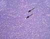
Components of a germinal center
- Tingible body macrophages (circle)
- Centroblasts with prominent nucleoli (arrow)
- Surrounding mantle of mature B cells (arrowhead)
- Proliferating B cells and follicular T cells and dendritic cells (CD10+)

Lymphomas and associated leukemias (Table)

Solid tumor disease in association with myeloid leukemias
Uncommon, but may occur
Examples include the chloroma and the granulocytic sarcoma
Categories of lymphoma
- Hodgkin’s
- Non-Hodgkin’s
- High-grade lymphomas (B and T)
- Low-grade B cell lymphomas
- Lymphoblastic lymphomas
- “Other” T cell lymphomas
- Non-B, non-T cell lymphomas
Clues to an extra-nodal lymphoma
- Discohesive cells
- Homogeneity
- Sheet-like growth
- Nuclei that are highly irregular in shape
- Accentuation of cell density around vessels (DLBCL in the brain is shown as an example)

In Italy, all roads lead to Rome.
In lymphoma, all roads lead to __.
In Italy, all roads lead to Rome.
In lymphoma, all roads lead to DLBCL.
It is the “terminal” lymphoma, the “anaplastic sarcoma” of lymphomas. As such, it has no real characteristic translocation – rather it is just any sufficiently mutated and aggressive B cell lymphoma.

Classifying DLBCLs
- of Germinal Center origin
- of Activated B Cell origin (non-GC, worse prognosis)
- “High-grade B cell lymphoma”
- Double-hit (2 driver rearrangements)
- Triple-hit (3 driver rearrangements)
Burkitt’s lymphoma
Distinct high-grade B cell lymphoma often identified by its mitotic rate (Ki67 index of nearly 100%) and population of medium-sized, densely packed lymphocytes with intermixed tingible body macrophages and apoptotic bodies. Often described as a “starry night” background.
Genetics: Most cases involve t(8;14), myc under the IgH promoter. EBV-associated.
Tingible body macrophage
Type of macrophage found in germinal centers or lymphomas that contains many phagocytosed, apoptotic cells in various stages of degradation.
“Tingible” means “stainable”
“Liscensed” or “empowered” to phagocytose by FDCs. FDCs provide germinal center macrophages with Milk fat globule-EGF factor 8 (Mfge8, aka lactadherin). Opsonizes phosphatidylserine on apoptotic cells (eat me signal) and binds to integrins on phagocytes to facilitate efferocytosis.


Follicular lymphoma
Second most common non-Hodgkin’s lymphoma (to DLBCL).
Appears as a nodular pattern of back-to-back follicles that fill a lymph node. On high-power, these follicles are full of neoplastic centrocytes (smaller, arrowhead) and centroblasts (larger, arrow). The relative proportion determines lymphoma grade. These lymphomas can also have areas of diffuse growth.
When circulating, the centrocyte nucleus has a folded, “baby’s butt” appearance.
Genetic: Defined by t(14;18), bcl2 under the IgH promoter. Bcl2 normally turns off in germinal centers, making these cells susceptible to apoptosis and priming them for germinal center selection and affinity maturation.

How can you distinguish benign follicular hyperplasia from follicular lymphoma?
Features seen in benign follicular hyperplasia but not lymphoma:
- Germinal centers of variable sizes cuffed by mantle cell zones (not back to back)
- Polarity of germinal centers (centroblasts and centrocytes opposite one another)
- Presence of tingible body macrophages
- “Open” sinuses of histiocytes
- Abundant mitoses and apoptoses (normal in a GC, but in FL there is much less apoptosis!)
- Bcl2 negativity within the germinal center (relative to mantle cell zone)

Small lymphocytic lymphoma
The lymphoma form of CLL/SLL, which are essentially equivalent diagnoses and usually simulatneously present.
On low-power, appears as a very homogenous, blue lymph node. The normal architecture is replaced by a sheet of normal appearing lymphocytes, sometimes with a vague suggestion of nodularity containing proliferative cells (pseudofollicles).
On high-power, SLL cells have chromatin that may remind you of a plasma cell, with small, round, regular clockface nuclei.
Genetics: Variable. No single defining translocation, however the t(11;14) – cyclin D1 under the IgH promoter – which is often characteristic of mantle cell lymphoma may be seen in CLL/SLL. Deletions, including del q11, q13, and q17 are sometimes seen. Trisomy 12 is sometimes seen. t(2;14) – Bcl-11A under the IgH promoter – is sometimes seen.
Markers: These cells abnormally express CD23 and CD5.

Marginal zone lymphoma
Named after the more prominent and identifiable marginal zone of the spleen.
Cells have a pale look on low-power due to a prominent ring of clear cytoplasm, giving them a “fried egg” appearance on high power. This morphology is called “monocytoid.” Lymphomas of this morphology are divided into three categories: Splenic MZL, nodal MZL, and extranodal MZL of MALT.
Genetics: See image. Complicated.
Markers: MZL-MALTmay sometimes be distinguished on the basis ofabnormal CD43 expression.

MZL-MALT genetics
Some subsets are associated with:
- t(11;18) – API2/MALT1 fusion protein (this one is SPECIFIC to MZL-MALT)
- t(1;14) – Bcl-10 under the IgH promoter
- t(14;18) – MALT1 under the IgH promoter
- t(3;14) – FOXP1 under the IgH promoter

Mantle Cell Lymphoma
Although it is often classified among the “low grade” B cell lymphoma histologic type, is much more aggressive than other low grades.
On low power it is reminiscent of SLL - sheets of lymphocytes effacing the node. In a node that’s not entirely replaced, you may be able to see the expansion from the mantle zone. Other features: hyalinized vessels, crinkled/angular nuclear membranes (unlike SLL), nuclear size variation.
Genetics: Characterized by t(11;14) – cyclin D1 under the IgH promoter. This is classic for MCL, but can also be seen in MZL, so watch out. Rarely, CCND2 or CCND3 may be seen as the translocation target instead of CCND1. Note: CCND1 = cyclin D1 = bcl-1
Markers:
Standard lymphoma IHC stain panel
- CD3 (pan-T)
- CD20 (pan-B)
- CD5 (additional T)
- CD10 (follicular origin)
- CD43 (additional T)

Acute lymphoblastic lymphoma
Not usually diagnosed as lymphomas – the leukemia forms tend to present first or alone (usually as B-ALL, or T-ALL if in the thymus).
Nuclei are larger than a normal lymphocyte and chromatin is immature (widely dispersed throughout nucleus). Unlike DLBCL, there are no prominent nucleoli or thick nuclear membranes.
Markers: Blast markers (TdT, CD34). These tumors are known to occasionally lose expression of CD45. So, CD45 negativity – especially in a kid – does not rule out lymphoma.
Lymphoblastic leukemia + lymph node mass:
Lymphoblastic leukemia + thymic mass:
Lymphoblastic leukemia + lymph node mass: B-ALL
Lymphoblastic leukemia + thymic mass: T-ALL

Hodgkin’s lymphoma
Reed-Sternberg cells are present.
Diagrams of the subtype are attached

What’s going on with this lymph node biopsy?

Nodular sclerosing-type Hodgkin’s lymphoma
An almost “cirrhotic”-looking lymph node.
This diagnosis should be suspected on low power, then zoom in and hunt for Reed-Sternberg cells.

Peripheral T cell lymphoma NOS
Most common T cell lymphoma. A neoplasm of mature T cells.
Paracortical or diffuse infiltrate with effacement of the normal architecture by medium to large sized cells with pleomorphic nuclei, vesicular chromatin, prominent nucleoli and frequent mitoses.Clear cell morphologyandReed-Sternberg-like cells may sometimes be seen.
Genetics/variants: TBX21 -subtype is associated with polymorphous inflammatory background. GATA3-subtype is associated with minimal inflammatory background. Lymphoepithelial variant is associated with diffuse small cells with subtyle nuclear atypia and CD8-features with epithelioid hstocytes.

Angioimmunoblastic T cell lymphoma (AITL)
Neoplasm of mature T cells. Notable for associated proliferation of capillaries and DCs within the node. Arise from follicular helper T cells.

Anaplastic large cell lymphoma (ALCL)
The T cell equivalent of DLBCL.
Large, pleomorphous, activated-type cells with horse-shoe or wreath nuclei and a peripheral eosinophilic region (Golgi apparatus). Overall there is a cohesive growth pattern. Several subtypes exist.
Markers: T cell specific markers and CD30 are characteristic, however may lose markers due to poor differentiation.
“Diffuse sheets of small mature lymphocytes” in a lymph node
- SLL/CLL
- MCL
- MZL
Prominent follicular/nodular pattern
- Follicular lymphoma
- Reactive lymph node
Diffuse sheets of large atypical cells
- DLBCL
- Anaplastic large cell lymphoma (ALCL)
Mitotically active, primitive cells resembling small cell carcinoma
- Lymphoblastic lymphoma
- Burkitt’s lymphoma
A pink and/or ganulomatous mixed infiltrate
Mixed cellularity Hodgkin’s lymphoma
Fibrous bands dividing a lymph node
Nodular sclerosing Hodgkin’s lymphoma

Circulating Follicular Lymphoma cell
Sometimes described as “baby’s butt” nucleus
LPL typically involves. . .
. . .the bone marrow
In fact, it involves the marrow even moreso than lymph nodes in most cases. Bone marrow involvement is characterized by a nodular, dffuse, and/or interstitial infiltrate composed of predominantly small lymphocytes admixed with variable numbers of plasma cells/plamacytoid lymphocytes. Increased mast cells are often also present.
What’s going on in this bone marrow aspirate from a patient with pancytopenia?

This is an excellent example of LPL. There are a couple of mature plasma cells, but the mixture is mostly composed of more lymphoid cells with variable plasma cell-like/plasmacytoid features:
- Slightly eccentric nuclei
- Clockface nuclei with scant cytoplasm
- Small perinuclear hof
- Dutcher/Russell bodies in otherwise lymphoid-appearing cells (attached)
- Increased mast cells
In LPL, you are expecting to see replacement of normal elements by a range from lymphocytic to full plasmacytic differentiation

What characterizes a normal splenic marginal zone?
The marginal zone is a light zone surrounding splenic follicles which contains post follicular memory B cells (MUM1+) derived after stimulation of recirculating cells from T cell dependent antigen

“Regernerative” lymphoid nodules in bone marrow
Proposed as a feature of regenerative change following chemotherapy or other bone marrow insult – especially following anti-CD20 therapy
An emerging concept, still debated
Characteristics include a well-circuscribed, reactive lymphoid nodule composed primarily of small T cells with a few B cells, and with surrounding eosinophils
Epithelioid granulomas in bone marrow
Can be seen as a reaction to BM stress, as in chemotherapy for lymphoma/leukemia
However, if you see one of these you are obligated to rule out mycobacterial disease, even if the diagnosis of lymphoma is known. After all, lymphoma-related immunosuppression increases the risk of TB or systemic MAC.
