Blood and BM Path Chapter 18 - Acute Myeloid Leukemia Flashcards

1
Q

Adults tend to get ___ leukemias.

Kids tend to get ___ leukemias.

A

Adults tend to get myeloid leukemias.

Kids tend to get lymphoid leukemias.

2
Q

WHO categories of AML

A
  • AML with recurrent cytogenetic abnormalities
  • AML with myelodysplasia-related changes
  • AML with therapy-related changes (history of methylating agent or topoisomerase II inhibitor chemotherapy exposure)
  • AML not otherwise specified
3
Q

Why was the required BM blast percentage decreased from 30% to 20% in the 2001 edition of the WHO guidelines?

A

Because it has been demonstrated that “MDS-EBII” patients with greater than 20% blasts had a prognosis identical to those with AML.

4
Q

Why is diagnosing APML such an emergency?

A

Because if it is not differentiated from another form of AML and the patient starts conventional chemotherapy, there is a high risk of DIC.

5
Q

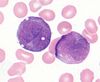
The only definitive morphologic finding that differentiates myeloid blasts from lymphoid blasts

A

Auer rods

6
Q

Immature vs mature myeloid surface antigens

A

Immature: CD13, CD33, CD117

Mature: CD15

7
Q

Megakaryocytic surface markers

A

CD41a and CD61

These are also surface markers for platelets!

8
Q

“AML without maturation” vs “AML with minimal differentiation”

A

“without maturation”: <3% MPO, SBB-negative blasts, no Auer rods (which contain MPO and SBB!)

“with minimal differentiation”: >3% MPO, SBB-positive, Auer rods present

9
Q

Subcategories of “acute myeloid leukemia with recurrent genetic abnormalities”

A
  • Balanced translocations
  • Inversions
  • Mutations of NPM1 or CEPBA genes
10
Q

Prognosis of various AML with recurrent genetic abnormalities

A
11
Q

3 AML diagnoses that do not require 20% blasts in the marrow

A
  1. t(15:17) PML-RARa
  2. inv(16) OR t(16:16) CBFB-MYH11
  3. t(8:21) RUNX1-RUNX1T1
12
Q

Recurrent genetic abnormalities that strattle the line between MDS and AML

A

inv(3), t(3:3), or t(6:9)

These are all associated with multilineage dysplasia and may be a cause of primary MDS, may present as MDS-EBSI or -EBSII, or may present as frank AML (>20% blasts) with multilineage dysplasia

13
Q
A
  • AML with RUNX1-RUNX1T1, Associated with t(8:21)
  • RUNX1 encodes the core binding factor alpha protein (CBFA), one component of a heterodimeric hematopoietic TF. Abnormal fusion to RUNX1T1 (aka ETO for eight twenty one) impairs or misdirects gene regulatory activity.
  • Morphologically appear with blasts displaying ample cytoplasm and abundant granules, sometimes with Auer rods. “Sunset cell” morphology is characteristic.
  • Dyspoietic morphology of granulocytes is common (Pelger-Huetoid)
  • Tell-tale features:
    • Corkscrew Auer rods
    • Prominent perinuclear hofs (green arrows)
    • Large salmon pink granules (black arrow) intermixed with typical dark granules
    • Together, the above make a “sunset cell”
  • Expression profile:
    • Frequent aberrant expression of B cell markers (CD19, Pax5, cytoplasmic CD79a)
    • CD34 overexpression
    • Myeloid profile: CD13, CD33, MPO
  • Prognostics:
    • High presenting WBC count (>20) - poor
    • KIT mutation - intermediate
14
Q
A
  • AML with CBFB-MYH11, associated with inv(16) or t(16:16)
  • Involve the gene for the second half of the core binding factor TF, CBFB. It is fused to a smooth muscle myosin heavy chain (MYH11).
  • Tend to occur in younger patients
  • Most (but not all) show granulocytic or monocytic blasts accompanied by abnormal eosinophil precursors within the marrow
    • These eosinophil precursors show occasional to numerous large purple or basophilic granules of variable shape intermixed with eosinophilic granules
  • Expression profile:
    • Often abnormally express CD2
15
Q
A
  • Acute promyelocytic leukemia with PML-RARa. Associated with t(15:17)
  • Known for life-threatening bleeding or clotting events in association with DIC
    • If the diagnosis is even suspected, all trans retinoic acid is initiated even before confirmation
  • Most common in middle-age. 2/3 of cases present with low WBC counts.
  • Morpholoci features:
    • “Hypergranular” morphology
    • Abnormal promyelocytes with bilobed (“sliding discs”) nuclei
    • Frequent Auer rods, often multiple per cell
  • Expression characteristics:
    • Promyelocytes often with CD34-HLA-DR- immunophenotype
    • Bright CD33, MPO, and SBB
    • Variable CD13
    • May abberantly express CD64 or CD2
16
Q

Less common genetic abberations of the retinoic acid receptor

A
  • ATRA-resistant
    • t(11:17) ZBTB16-RARa
    • t(17:17) STAT5B-RARa
  • ATRA-susceptible
    • t(11:17) NUMA1-RARa
    • t(5:17) NPM1-RARa

These are reported as “AML with a variant RARa translocation.” They also may have very different morphologies.

17
Q
A
  • AML with MLLT3-MLL, associated with t(9:11)
  • Recently separated from the group of 11q23 MLL gene translocations to reflect its better prognosis (others have poor prognosis)
  • MLL has histone methyltransferase activity. Likely it plays a role in gene regulation via epigenetic mechanisms.
  • Usually seen in pediatric patients
  • Morphologic features
    • Blasts with monocytic or myelomonocytic morphology
    • Somewhat bland, blasty appearance
  • Surface expression
    • No special patterns
    • CD34- and CD117-
    • MPO positive
18
Q
A
  • AML with DEK-NUP214, associated with t(6:9)
  • DEK is a chromatin-associated protein and NUP214 a nuclear pore protein. The mechanism of the fusion’s contribution to oncogenesis has not been well described, but is thought to involve abnormal nuclear transport.
  • Uncommon translocation that may affect a wide age range of patients. Prognosis is poor.
  • Usually in the context of pancytopenia with myelodysplasia in all lineages
  • Morphologic features:
    • Distinguished by presence of BM or blood basophilia (>2% basophils), seen in 50% of cases
    • Blasts may be myeloid or monocytic
  • Surface expression:
    • Unremarkable myeloid or monocytic immunophenotype
19
Q
A
  • AML with RPN1-EVI1, associated with inv(3) or t(3:3)
  • EVI1 (ectopic virus integration-1) is a proto-oncogene with zing finger protein homology, proposed to have transcriptional repression activity via recruitment of histone deacetylases. Fusion to RPN1, a proteasome component, may serve to drive EVI1 overexpression.
  • Relatively rare form of AML. Primarily occurs in adults. Prognosis is poor.
  • Associated with myelodysplastic findings in granulocytes and platelets, usually with relative sparing of erythroids
  • Patients may present before the blast count exceeds 20%
  • Morphologic features:
    • Presence of monolobated or bilobated megakaryocytes, which may be increased in number
    • Blasts are myeloid, monocytic, or megakaryocytic
  • Surface expression:
    • Unremarkable myeloid blast immunopthenotype
    • Megakaryoblastic cases express CD41 and CD61
20
Q
A
  • AML with RBM15-MKL1, associated with t(1:22). Often called “megakaryoblastic AML”
  • RBM15 encodes a protein with RNA-binding motifs and MKL1 encodes a transcriptional coactivator of serum response factor (SRF). The mechanism of this translocation’s oncogenicity is unknown.
  • Very rare form of infant leukemia. Presents as HSM or other organomegaly.
  • Morphologic features:
    • Megakaryocytic differentiation with medium-sized or larger cells that ahve round nuclei and fine reticular chromatin, visble nuclei, agranular basophilic cytoplasm, and occasional cytoplasmic blebbing (arrows)
    • Marrow fibrosis is common (as with other megakaryoblastic leukemias)
  • Surface expression:
    • Blasts lacking CD45, CD34, and HLA-DR
    • Express myeloid markers CD13 and CD33
    • Express megakaryocyte markers CD41, CD61, and sometimes CD42
21
Q

RTK mutations in AML

A

KIT and FLT3 are commonly mutated in AML, particularly in cases with normal karyotypes.

These take the form of internal tandem duplications and point mutations of the tyrosine kinase domain. They correlate with a poor prognosis.

KIT mutations are especially common in t(8:21) RUNX1-RUNX1T1 and inv(16), which herald a poor prognosis.

22
Q
A
  • AML with mutated NPM1 (with or without associated FLT3 mutation)
  • NPM1 encodes a nuclear shuttling protein reported to have roles in robosome and centrosome biology as well as regulation of the ARF-TP53 pathway.
  • More common in adult AML (30% of adult AML, 5% pediatric AML)
  • Typically have a normal karyotype.
  • Morphologic features:
    • Myelomonocytic or monocytic
    • No myelodysplastic findings
    • May take the form of AML without maturation or AML with maturation and erythroleukopenia
    • Blasts with cup-shaped nuclear invaginations are associated with this mutation, especially when it co-occurs with FLT3 mutations.
  • Surface expression:
    • Blast immunophenotype with loss of CD34
    • Staining for NPM1 reveals overexpression on IHC
  • Diagnosis
    • Typically detected by PCR-based methods
    • Exception is tetranuceotide insertion in exon 12 creating frameshift, which interferes with nuclear localization signaling and interrupts export
23
Q

AML with no chromosomal anomalies, CD34+MHCII+, and aberrant expression of CD7. Erythropoiesis is preserved but patient has severe thrombocytopenia with purpura.

A
  • AML with mutated CEBPA
  • CEBPA is a TSG and transcription factor implicated in the differentiation of many disparate cell lineages, including granulocytes
  • Detected in 10% of all AML cases, ~25% of cases co-occur with FLT3-ITD mutations
  • No apparent association with age or sex.
  • Relative preservation of erythropoiesis, but worse thrombocytopenia and higher circulating blast count. Extramedullary disease is less frequent.
  • Morphologic features:
    • FAB M1 (no maturation) and M2 (with maturation) blast morphology
    • Rarer cases have myelomonocytic or monocytic morphology
  • Surface expression:
    • CD34+HLA-DR+
    • Often aberrantly express CD7
  • Diagnosis:
    • Requires gene sequencing, as over 100 distinct mutations have been described
24
Q
A
  • AML with myelodysplasia-related changes
  • Characterized by: Myeloid blast count >20% in the blood OR bone marrow of a patient WITHOUT history of cytotoxic chemotherapy/radiation therapy and WITH at least one of the following:
    1. Prior Hx of MDS or MDS/MPN
    2. Characteristic myelodysplasia-associated cytogenetic abnormalities
    3. Multilineage dysplasia in at least 50% of cells in at least 2 lineages
    • Must not be explained by a recurrent genetic abnormality (this takes diagnostic precedence)
  • Clinical features:
    • Dominated by severe cytopenias
  • Morphologic features:
    • Neutrophils with hypogranularity, hyposegmented nuclei, or bizarre nuclear segmentation patterns
    • Erythroid precursors with megaloblastoid changes (delayed nuclear maturation relative to cytoplasmic hemoglobinization), multiple nuclei, or irregular nuclear contours, ringed sideroblasts, cytoplasmic inclusions, PAS+ vacuoles
    • Megakaryocytes with micromegakaryocyte morphology, hyposegmented nuclei, or widely separated nuclear lobes
    • Blast morphology is variable, but AML with maturation and acute myelomonocytic leukemia morphologies are most common
  • Surface markers:
    • Limited utility
    • Non-specific aberrancies such as expression of CD7 or TdT
25
Q

“Hypocellular AML”

A

May occur in multiple contexts

Basically means <30% BM cellularity (or <20% in patients over 60)

Often represents progression of hypocellular MDS and occurs in the setting of MDS with myelodysplasia-related changes.

26
Q

Window for developing therapy-related AML following alkylating agent chemotherapy

A

5-7 years post-treatment and preceded by a period of MDS

27
Q

Window for developing therapy-related AML following topoisomerase II inhibitor chemotherapy

A

1-3 years following treatment

28
Q

Characteristics of therapy-related AML

A
  • 15% of all AMLs
  • Show multilineage dysplasia and myelodysplasia-associated cytogenetic changes
  • Prognosis is generally poor, but depends upon which initial malignancy the patient had
29
Q

AML not otherwise specified

A

Now represents a diagnosis of exclusion

Blood OR bone marrow must demonstrate >20% blasts to make this diagnosis

30
Q

AML with minimal differentiation

A
  • Most common at the extremes of age
  • Morphologic features:
    • Medium-sized blasts with dispersed or partially condensed chromatin, inconspicuous nucleoli, and minimal agranular basophilic cytoplasm w/o Auer rods
    • DDx includes lymphoblastic leukemia, megakaryoblastic AML, and blastic lymphomas
  • Surface markers:
    • By definition, MPO is negative or <3% of blasts
    • Myeloid blastic markers: CD13, CD117, CD33
    • TdT, CD34, CD38, and HLA-DR are often positive
    • Without mature myeloid markers: CD11b, CD15, CD65
    • Without monocytic markers: CD14, CD64
    • May express TdT, but without other lymphoid markers
31
Q

AML without maturation

A
  • 90% of blasts are nonerythroid cells and there is minimal differentiation to more granulocyte forms
  • 5-10% of all AML cases
  • Morphologic features
    • Similar to AML with minimal differentiation, but basophilic cytoplasmic granules or Auer rods may be present
  • Surface markers:
    • By definition, at least 3% of cells are MPO+ or SSB+
    • Myeloid markers: CD13, CD33, CD117, sometimes CD11b
    • CD34 and HLA-DR are usually positive
    • Mature granulocyte markers are often negative: CD15 and CD65
    • Mature monocyte markers are negative: CD14, CD64
    • Aberrant surface or cytoplasmic CD7, cCD3, cCD79a, and cCD22 are common,
32
Q

AML with maturation

A
  • Distinguished from AML without maturation by the presence of maturing granulocytes making up more than 10% of nonerythroid BM nucleated cells
    • ​<20% monocytic lineage
    • myeloblasts comprising 20-89% of nonerythroid cells
  • Morphologic features:
    • Blasts commonly show cytoplasmic granules and/or Auer rods
  • Surface markers:
    • Show typical myeloid phenotype including mature myeloid markers: CD13, CD33, CD117, CD11b, CD15, CD65
    • Monocytic markers are usually absent: CD14, CD64
33
Q

Mature myeloid markers vs mature monocytic markers

A

Mature myeloid: CD15, CD65

Mature monocytic: CD14, CD64

34
Q
A
  • Acute myelomonocytic leukemia
  • Distinguished from AML with maturation by the presence of >20% monocyte-lineage cells and >20% maturing granulocytic lineage cells among non-erythroid BM nucleated cells.
  • Myeloblasts, monoblasts, or promonocytes must comprise >20% of total nucleated cells in blood OR in bone marrow.
  • It can sometimes be difficult to distinguish CMML from AMML, as it requires the distinction of promonocytes from monocytes.
  • Morphology:
    • Promonocytes tend to have a more lobular (often kidney bean-shaped or cerebriform) nucleus and bluer cytoplasm compared to monocytes.
35
Q
A
  • Acute erythroid leukemia
  • In the more common erythroid/myeloid variant, erythroid precursors represent at least 50% of total BM nucleated cells and myeloblasts are at least 20% of the remaining non-erythroid cells. Presents with pancytopenia and circulating nucleated red cells.
  • The pure erythroid form is just that. . . pure erythroid.
  • Surface markers:
    • CD34- HLA-DR-
    • Negative for myeloid antigens
    • CD71 dim, glycophorin +, HbA +
    • May aberrantly express megakaryoblastic markers CD41 and CD61
36
Q
A
  • Acute monoblastic and monocytic leukemia
  • Monoblasts, monocytes, or pro-monocytes comprise 80% of non-erythroid nucleated BM cells, while maturing neutrophil lineage is <20%.
  • Clinically there is a tendency for extramedullary disease, often involving the skin, gums, or CNS
  • Must be differentiated from MLL3 translocations, most often t(9:11) MLLT3:MLL, which can also have a monoblastic/promonocytic morphology.
  • If HLH is present, this may represent t(8:16). This does not constitute AML with recurrent genetic abnormalities, but is often a translocation associated with this phenotype.
  • Surface markers:
    • CD34 negative (usually)
    • HLA-DR+
    • Myeloid markers: CD13, CD15, CD65, CD33
    • Monocytic markers: CD14, CD64, CD11b, CD11c, CD4, CD36, CD64, CD163, lysozyme
37
Q
A
  • Acute megakaryoblastic leukemia
  • At least 50% of blasts must show evidence of megakaryoblastic differentiation.
  • Most cases with megakaryoblasts meet criteria for AML with t(1:22), AML with myelodysplasia-related changes, or Down-syndrome associated AML
    • Once these have been ruled out, you are left with true primary megakaryoblastic leukemias
  • Megakaryoblastic leukemias will feature marrow fibrosis, and so aspirate will often be a dry tap
  • Morphologic features:
    • Promegakaryocytes have smooth, dense chromatin, variable nucleoli, and scant to moderate cytoplasm. Cytoplasmic membrane may show irregularities, such as blebs. Ultrastructure shows demarcation membranes or bull’s eye granules
    • Megakaryoblasts are smaller than promegekaryocytes and have more abundant cytoplasm
38
Q

Myeloid sarcoma

A

Involvement of a tissue mass by myeloid blasts. Equivalent to a diagnosis of AML irrespective of blast counts in blood or marrow.

May accompany MDS, MPN, or MDS/MPN.

Pediatric cases are less likely to present without a blood or marrow component and more likely to have t(8:21).

CD68 is the most commonly positive marker overall for myeloid sarcomas, but is not particularly specific.

39
Q

Transient abnormal myelopoiesis

A
  • Roughly 1/10 of patients with Down syndrome are born with clinical findings of acute megakaryoblastic leukemia
    • But. . . 75% of cases spontaneously resolve within a few months
    • For the remaining 25% (1/40 of Down patients), this disease is life threatening
  • Clinical manifestations of severe cases:
    • Cardiac or respiratory compromise
    • Hyperviscosity
    • Hepatic or renal dysfunction
    • Splenic necrosis
    • DIC
  • Hematopathologic features are very similar to the FAB M7 megakaryoblastic category with typical myeloid and megakaryoblastic markers along with aberrant CD7 and CD56.
  • In addition to trisomy 21, these cells are often found to have GATA1 mutated. This mutation may be germline or somatic.
40
Q

Megakaryocytic markers

A
  • CD41
  • CD42
  • CD61
  • CD71
41
Q

Myeloid leukemia associated with Down syndrome

A
  • In 20-30% of Down patients who develop TAM, the disease is followed within 1-3 years by a recurrent and non-remitting AML
    • Prognosis is very good compared to conventional childhood AML
  • This is often preceded by peripheral blood cytopenias and BM myelodysplastic features.
  • BM shows extensive fibrosis and extramedullar disease is common (liver, spleen)
  • Similar to TAM, these leukemias show trisomy 21 with GATA1. They also have relatively high rates of partial trisomy 8 and trisomy 1.
    • If it doesn’t have GATA1 mutated, it is just a regular old AML – NOT Down-associated.
42
Q

Blastic plasmacytoid DC neoplasm

A
  • An aggressive neoplasm presenting within the skin with frequent BM and systemic leukemic involvement. In 65% of cases, at least some ciruclating tumor cells are seen.
  • Derived from plasmacytoid DCs in the T-cell zones of lymphoid tissues (that are the predominant source of type 1 IFN during viral infections)
  • Morphologic features:
    • Medium sized blasts with irregular nuclear contours, one to several visible nucleoli, scant agranular blue-gray cytoplasm. Sometimes contain microvacuoles.
  • Surface markers:
    • CD34-CD117-
    • Express CD4, CD56, TCL1, CD43, CD45RA
    • Plasmacytoid DC marker CD123+
    • Express CD68 as small cytoplasmic dots
43
Q

Etiology of the “cup-like” morphology in AML with NPM1 mutation

A

Can occur with NPM1 mutation alone, but more often occurs when NPM1 and FLT3-ITD are both mutated

On ultrastructural studies, it seems to be due to nuclear invagination with vesiculation.

44
Q

MPO and SBB in categorizing AMLs

A

“M0” by the old classification system was MPO and SBB negative – the most immature type of AML

Nowadays, we define AML without maturation as AML with >3% MPO and SBB staining (to demonstrate lineage) without evidence of more mature precursors

A picture of an M1 cell / AML without maturation blast is shown

45
Q

Almost all AMLs are HLA-DR+, except ___, which is charcteristically HLA-DR-

A

Almost all AMLs are HLA-DR+, except APML, which is charcteristically HLA-DR-

46
Q

Most blasts (both myeloid and lymphoid) are positive for ___, not to be confused with plasma cells

A

Most blasts (both myeloid and lymphoid) are positive for CD38, not to be confused with plasma cells

47
Q

Post-chemotherapy bone marrow changes

A

Increased plasma cells, usually more along the vessels (benign distribution)

Pink, amorphous material from cell death

Relative red cell hyperplasia

48
Q

IRF8

A

Emerging as a specific marker for monocyte/macrophage lineage, useful in the diagnosis of monoblastic leukemias or CMML

49
Q

What’s going on in this patient with pancytopenia? Below is an image from bone marrow aspirate.

A

Pure erythroid leukemia

May be primary or secondary from an MPN. Cells are characterized by royal blue cytoplasm and many round to sausage-shaped cytoplasmic vacuoles. These cells almost never circulate – they are present in the BM.

p53 mutations are common, and biallelic p53 mutations portend a poor prognosis.

50
Q

Blast chromatin tends to have the appearance of. . .

A

. . . a fine sieve

51
Q

“Granulated blast”

A

Somewhere in-between a blast and a promyelocyte

Generally has the real appearance of a blast, but with scant visible granules.

Represents a cell that is undergoing the transition, but is best classified as a blast.

52
Q

Patient presents with flu-like symptoms and posterior cervical lymphadenopathy. Peripheral smear is shown below. On labs, the patient is anemic and thrombocytopenic. What is the most likely diagnosis?

A

Acute monocytic leukemia with differentiation.

Don’t be fooled into thinking this is mononucleosis! The smear shows a spectrum from blast-like to monocytic. Also, patients with mono can have very high WBC counts, but really should not be anemic or thrombocytopenic. This suggests a marrow-infiltrating process, as in acute leukemia.

Helpful diagnostics: Elevated serum lysozyme, bone marrow biopsy and smear, cytogenetics, testing for causes of infectious mono (EBV, CMV, HIV, toxoplasma)