Lecture 16 - Respiratory Flashcards
(33 cards)
Functions of the respiratory system
- Gas conditioning: Warm, moisten and filter air
- Sound productions
- Olfaction
- Defense
Divisions of the respiratory system
Anatomically
- > upper resperatory tract (above larynx)
- > lower respiratory tract (below and including larynx)
Functionally
- > conducting portion
- > respiratory portion
Structures that make up the upper respiratory tract
- nose and nasal cavities
- paranasal sinuses
- pharynx
- > these are all part of the conducting portion of the RS
Nose
- > main conducting airway for inhaled air
- > supported by paired nasal bones superiorly that form the bridge of the nose
- > hyalin keeps nostrils open and for support
- > supported anteroinferorly from bridge by septal and alar cartilages
- > wings of the nose are made from dense CT

Conducting vs Respiratory portions
Conducting - > moves air in and out of the lungs; like veins and arteries, too big to diffuse
Respiratory - > includes the respiratory bronchioles and alveoli an moves respiratory gases in and out of blood; like capillaries
Nasal Cavities
- > the superior, middle and inferior nasal conchae (turbinates) form the lateral wall for each cavity
- > they’re lumpy irregular surfaces so air turbinates in the nasal cavity which slows it down, moistening, filtering and warming the air
- > superior and middle conchanae are part of the ethmoid bone
- > riht and left cavities are divided by the vomer and septal cartilage (part of ethmoid)

Paranasal sinuses
• These spaces make the bones lighter in weight and are named after the bones in which they reside
- > Frontal
- > Ethmoidal
- > Sphenoidal
- > Maxillary

Pharynx
- shared by two organ systems (digestive and respiratory)
- Divided into three regions
- > Nasopharynx
- > Oropharynx
- > Laryngopharynx

Nasopharynx
- > continuous with the nasal cavity and superior to the soft palate
- > opening of the eustachian auditory tubes
- > posterior nasopharynx wall house a single pharyngeal tonsil (adenoid); swollen pharygeal tonsils can cause snoring

Oropharynx
- > begins at the end of the soft palate and ends at the end level of the hyoid
- > the opening of the oral cavity into oropharynx is the fauses, defined by two arches in the back of throat by uvula
- > palatine tonsiles are embedded in lateral wall between areches
- > lingual tonsils located at the base of the tongue
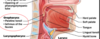
Laryngopharynx
- > starts inferior to hyoid and is continuous with the larynx and esophagus

Lower respiratory tract (conducting portion)
• comprised of (biggest to smallest branches)
- > larynx
- > trachea
- > bronchi
- > Bronchiole

Lower respiratory tract (respiratory portion)
• comprised of the following
- > respiratory bronchioles
- > alveolar ducts
- > alveolar sacs
- > alveoli

Larynx
- connects pharynx to trachea
- also called voice box
- supported by framework of cartilage, ligaments and muscles
- the three major cartilages are:
- Thyroid cartilage
- Cricoid cartilage
- Epiglottis

Thyroid Cartilage
- largest cartilage, but it’s incomplete
- has anterior and lateral wall
- no posterior wall
- v-shaped anterior projection in called the laryngeal prominance (adams apple)
- usually larger in males because of testosterone’s influence on growth of cartilage

Cricoid Cartilage
- only complete ring around trachea
- inferior to thyroid cartilage
- acts as an attachement point for ligaments and muscles for vocal sound production (phonation)
- the fact that you can move your vocal cords in completely dependant on muscle attachements from vocal cords to cricoid cartilage

Epiglottis
- spoon-shaped cartilage that project superiorly into pharynx
- swallowing causes the epiglottis to close the opening to the larynx thus preventing materials from entering the lower respiratory tract
- elastic cartilage allows it to snap back into place

Sound Production
• 2 sets of vocal cords
true vocal cords (vocal folds) - > make sound and move, found in larynx and comprised of vocal ligaments covered by a mucous membrane
false vocal cords (vestibular folds) - > sit superior to and protect true cords
- the opening between vocal folds is the rima glottidis
- vocal fold + rima glottidis = glottis
- when air is forced through the rima glottidis, it causes vibration of the vocal cords whch result in the production of sound

Trachea
- inferior to the larynx, superior to the primary bronchi, and anterior to the esophagus
- supported by c-shaped treacheal cartilage; incomplete hyaline rings which make sure it doesn’t colapse

Bronchial Tree
- the bronchial tree is a highly branched system of air-conducting passages that begin with the primary bronchi and end with the terminal bronchi (conducting portion of RS)
- trachea branches into left/right primary bronchi at carina
- the right primary bronchus is larger and more vertical (more likely to get objects stuck)
- right primary bronchus divided into 3 secondary (lobar) bronchi
- left primary bronchus divides into 2 secondary (lobar) bronchi
- secondary bronchi divide into 8-10 tertiary bronchi (segmental bronchi)

Bronchioles
- walls are composed of a relatively thick layer of smooth muscle
- contraction of the smooth muscle resulting in the contriction of the bronchile is called bronchoconstriction
- relaxation of the smooth muscle resulting in a widening of the bronchioles is called brochidilation
- bronchioles branche into terminal bronchioles, which are the last portion of the conduction portion of the respiratory system
What makes up the respiratory portion of the respiratory system
- respiratory bronchioles
- alveolar ducts
- pulmonary alveoli

Explain the repiratory portion of the respiratory system
- termial bronchioles branch into resperatory bronchioles
- respiratory bronchioles branch into alveolar ducts
- alveolar ducts lead into groups of alveolar sacs (round, hollow respiratory unit)
- the thin wall of the valeolus is the structure where respiratory gases (02 CO2) diffuse between the blood and the air in the lungs

The alveolar wall is formed from which types of cells
- Alveolar type I cells
• simple squamous epithelial cells that promote rapid diffusion of gases
- Alveolar type II cells (Clara cells)
- almost cuboidal in shpae and produce pulmonary surfactant, which decreases surface tension within the alveolus and prevents the colapse of the alveoli
- highly vascularized
- wandering macrophages are present to phagocytize pathogens/debris
- alveolar pores alow pressure equalization so that all alveoli expand/diflate the same amount and don’t burst


