Lecture 15 - Integumentary Flashcards
List all layers of you integument
Two layers
- > Epidermis
- > Dermis
*hypodermis - > not technically part of the skin but can’t talk about the first two without this one*
Size and weight characteristics of integument
- > largest and heaviest organ
- > makes up 7-8% of body weight
- > 1.5-2m^2
Karatenized
skin becomes keratenized, helps become waterproof
Integument functions
- > protection
- > prevention of H20 loss
- > temperature regulation
- > metabolic regulation
- > immune defense
- > sensory reception
- > Excretion/ Secretion
Epidermis
- > most superficial layer
- > avascular
- > keratinized stratified squamous epithelium
- keratin is a water-insoluble protein
- 4-5 layers (strata) of distinct cell types
List the epidermal strata from deep to superficial
- > stratum basale
- > stratum spinosum
- > stratum granulosum
- > stratum lucidum (found only in thick skin)
- > stratum corneum
Stratum Basale; what makes up this layer?
- > only layer that undergoes mitosis
- > sits on basement membrane
- > one layer of cells adjacent to dermis:
- keratinocytes: most abundant; produce keratin to waterproof skin
- Melanocytes: description on another slide
- Tactile cells: sense touch
- Dendritic cells: immunity

Melanocytes
- > found in stratum basale
- > cells with long branched cytoplasmic processes
- > produce a pigment (brown, black or yellow) that absorbs UV light to prevent DNA damage, reducing some forms of skin cancer
- > melanin surrounds nucleus to protect it from radiation

Stratum Spinosum
- > several layers thick
- > daughter cells from the stratum basale
- > differentiate into nondividing (may see a rare mitotic cell), highly specialized keratinocytes
- > develop a spiny/prickly shape

Stratum Granulosum
- > three to five layers of keratinocytes
- > cytoplasm fills with keratin filaments - grainy
- > organelles begin to degrade as cell is fully saturated with keratin
- > fully keratinized cells are dead but strong and water-insoluble

Thin vs Thick skin
Thin skin - > pretty much everywhere, less layers of corneum
Thick skin - > only found in palms and soles of feet, has leucidum
Stratum Lucidum
- > thin, translucent region, two to three layers thick
- > present ONLY in thick skin
- > cells lack organelles, filled with eleidin, a transparent, intermediate product of keratin maturation

Stratum Corneum
- > most superficial layer of epidermis
- > thickness varies from a few to 50 layers thick depending on location on the body
- > comprised solely of dead keratinocytes (corneocytes)
- > slought off by abrasion = dust
- > desquamation: squamous cells, shedding skin
How is skin colour determined?
*NOT DEPENDANT OF # OF MELANOCYTES; BASED ON HOW ACTIVE THEY ARE*
- > melanin: brown-black pigment produced by melaocytes; genetic inheritance, increases with UV light exposure (protects nuclear DNA)
- > hemoglobin: blood pigment; causes complexions to look pink (or blue)
- > carotene: yellow-orange pigment from food that builds up in skin
Types of Epidermal Variations
*ALSO KNOW AS SKIN MARKINGS*
Nevus - > (mole) localized overgrowth of melanocytes
Hemangioma - > (birthmark) proliferation of blood vesses may dissapear (strawberry) in childhood or may persist (port-wine though adulthood)
Friction Ridges - > folds of epidermis/dermis on fingers, palms, toes and soles used for grasping (cause us to leave finger print)
Dermis
- > lies deep to the epidermis
- > two layers of connective tissues
- Papillary: superficial
- Reticular: deeper
*mainly collagen fibres
* contains blood vesses, glands, hair . follicle, nail roots, sensory nerve endings, and smooth muscles

Papillary layer of the dermis
- > directly below statum basale cells of epidermis
- > dermal papillae and epidermal ridges(epidermal heads) interlock (epidermal-dermal junction), increasing surface area between the epidermis and dermis
- > dermal papillae contain capillaries that supply nutrients to the avascular epidermal cells

Reticular layer of the dermis
- > forms the majority of the dermis
- > comprised mainly of dense irregular connective tissue with large collagen bundles, blood vessels, glands, hair follicles and nerves
- > collagen bundles help connect dermis to underlying hypodermis
- > irregular collagen fibres help resist stress from all angels

Lines of cleavage
- > lines in the deep dermis formed by the predictable orientation of collagen bundles
- > incisions parallel to LoC heal faster as perpendictular incisions gape open as the skin in under tension

Innervation of the skin
- > nerve fibres are present in dermis
Functions
- > tactile (touch) receptors
- > control blood flow
- > control glandular secretion
Blood supply/body temp regulation of the integumentary system
Epidermis = AVASCULAR
Dermis = VASCULAR
- important for controlling body temp
- vasocontriction: narrowing of blood vessels to preserve core body temp
- vasodilation: widening blood vessels releases body heat, lowering body temp
Hypodermis
A.K.A Subcutaneous layer
- > deep to, not really part of, integument
- > areolar and adipose(fat) tissue connective tissue
Functions
- > protects underlying structures
- > stores energy
- > thermal insulation

Epidermal Derivatives
• Structures that grow from the epidermis
- > nails
- > hairs
- > glands
Nails
- > Derived from the stratum corneum
- > cells densely packed together filled with parallel fibres of hard keratin
- > the nail body covers a layer of epidermis called the nail bed
- > nail bed appears pink because of underlying capillaires

Nail Anatomy
• Protective structures on digits
Nail body - > flat keratinized cells protecting digets
Nail Bed - > live epidermal cells under nail body
Nail root - > region hidden by cuticle
Nail Matrix - > thickened newly growing part of the nail bed
Lunula - > white semilunar proximal area of nail body caused by thickened underlying stratum basale obscuring capillaries in dermis

parts of a Hair
• Columns of keratinocytes growing from follicles deep in dermis or hypodermis
hair shaft - > exposed, completely keratinized
Hair follicles - > epidermal fold surrounding the hair
Sebaceous gland - > secretes sebum (natural moisturizer) into hair follicle; moisturizes hair and skin

Hair anatomy
Hair bulb: a swelling at the bottom of the hair follicle filled with dividing keratinocytes; increase in hair matrix during growth
Hair Papilla: conntective tissue, nerves and blood vessels below follicle that support the keratinocytes
Arrector pili: involuntary smooth muscle attached to hair shaft; responds to emotional states (fear or rage) and cold temps by contracting; standing hair up and producing goosebumps

What is a blister
when the epidermal-dermal junctions separates and the gap fills with interstitial fluid
Functions of hair
- > protection
- > heat retention
- > facial expression
- > sensory reception
- > visual identification
- > chemical signal dispersal
Skin exocrine glands
• Sweat glands (2 types; produce watery substance)
- > Eccrine sweat glands
- > Apocrine sweat glands
• Specialized gland types
- > sebaceous glands: produce oily secretion (holocrine)
- > ceruminous glands: produce earwax
- > mammary glands: produce milk
Eccrine Sweat glands
• also known as merocrine sweat glands
- > simple coiled tubular glands that secrete into a duct with a pore on skins surface
- > numerous on forehead, palms and soles
- > secretion is 99% water, clear, controlled by NS; NO STANK
• Functions
- > thermoregulation
- > secretion
- > protection

Apocrine sweat glands
• simple coiled tubular glands that secrete into hair follicles around nipples, armpits, groin, and anus
- > secretion is thick clowdy and contains proteins and lipids
- > leads to bacterial growth, causes BO in above regions

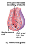
Sebaceous glands
- holocrine glands
- secretes oily sebum into hair follicles
- > cell produces so much sebum that cell dies and flows up with sebum, leads to acne
- > lubricates hair and skin
- > relatively inactive during child hood, sex hormones at puberty cause secretions to increase significantly