Lab Midterm Flashcards
(21 cards)

Vein with valve (arrow)

Small intestine
Plicae circulares
P - plicae
SM - submucosa
V - villi
M - muscularis
S - serosa

Small intestine
DG - duodenal glands
MM - muscularis mucosae
D - excretory ducts

Colon
ME - Muscularis externa with tenia coli at the bottom
S - submucosa
M - mucosa filled with intestinal glands

Colon glands
L - inner tubular lumen
LP - lamina propria
Arrow - lymphocyte penetrating epithelium

Colon intestinal gland
G - goblet cell with mucus
Columnar cells near goblet cells
Mucus in the lumen of the gland

Esophagus
M (red) - mucosa
SM - submucosa
M (black) - muscularis
A - adventitia

Esophagus
E - stratified squamous epithelium of the mucosa
LP - lamina propria with scattered lymphocytes
MM - smooth muscle strands of the muscularis mucosae
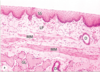
Esophagus
SS - stratified squamous epithelium of the mucosa
LP - lamina propria with scattered lymphocytes
MM - smooth muscle strands of the muscularis mucosae
GL - esophogeal mucous gland in the submucosa
D - duct

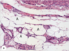
Esophagus muscularis
St - skeletal or striated muscle fibers
Sm - smooth muscle fibers
Both are seen so this is the middle 1/3 of the esophagus

Loose CT and dense irregular CT
L - loose CT
D - dense irregular CT

Loose CT and dense irregular CT
L - loose CT
D - dense irregular CT

Dense irregular CT

Dense irregular CT
Capsule formed by the tissue is seen and covered by a simple epithelium of serous mesothelial cells (S), which produce a hyaluronate-rich lubricant around such organs as the testes
White adipose adipocytes in CT with small bv
A - adipocytes

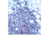
White adipose tissue

White adipose tissue with triglycerides
Stained with osmium tetroxide to preserve triglycerides
L - lipid

White adipose tissue with nuclei and undifferentiated adipocytes
Arrows - nuclei
* - undifferentiated adipocytes
White adipose (top) and brown adipose with many lipid droplets (bottom) surrounding a blood vessel
BV - small bv

Areolar (loose) CT

Reticular CT
