January 25, 2016 - Pulmonary Vasculature Flashcards
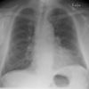
Kerley B Lines
Short parallel lines at the lung periphery. These lines represent interlobular septa which are usually less than 1 cm in length and parallel to one another at right angles of the pleura.
Causes may include pulmonary edema, malignant lymphoma, pneumonia, pulmonary fibrosis, or sarcoidosis.

Pulmonary Circulation
Pumping the blood from the right ventricle through the lungs and to the left atrium.
Pressures are very different between the two pumps.
Pulmonary Vascular Resistance
PVR = (PAmean - LAmean) / CO
PA = pulmonary artery
LA = left atrium
CO = cardiac output

Starling Resistor
Flow is dicated by the pressure difference between P1 and P2, unless Pext is greater than the pressure at P1 or P2.
Think of this like the pressure coming before the lungs, the pressure after the lungs, and the alveolar pressure.

Acute Hypoxemia
Decreased PO2 in the alveoli leads to vasoconstriction of pulmonary arterioles. This reduces perfusion to poorly ventilated regions which is usually good. It will cause a redistribution of cardiac output so other areas get dilated and the resistance drops.
However if this is chronic, there will be generalized hypoxemia which will cause generalized vasoconstriction and increase pulmonary vascular resistance.
Cor Pulmonale
“Pulmonary heart disease”
Is the enlargement and eventual failure of the right ventricle as a response to increased vascular resistance.

Tricuspid Regurgitation
Everyone has a little bit of regurgitation, but in diseased states with higher pressures, more blood will regurgitate through the tricuspid valve.
Through echocardiography we can get a doppler measurement of TR jet peak velocity, which can be used to estimate the pressure in the right ventricle.
Can pick up pulmonary hypertension using non-invasive tests.
Hereditary Hemorrhagic Telangiectasia
Osler-Weber-Rendu Syndrome. Autosomal dominant which affects 1/5000 people.
Often presents with epistaxis, skin telangioectasia, GI blood loss.
There are no capillaries. This can be detected by injecting bubbles into the peripheral vein. By using bubbles, if there was a cardiac shunt you would immediately see them in the RV. If you see them 5 or 6 seconds later, it indicates a arterio-venous malformation as the bubbles do not get filtered out by the capillaries.
Treatment for Arterio-Venous Malformations
Radiologically-guided embolization.


