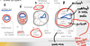
Ear Development Flashcards
1
Q
- During what week do the ears begin developing?
- What is the critical period for ear development?
- Ears develop thru WEEK _
A
- 4
- 4-8
- 20
2
Q
- Parts of the external ear
A
- Auricle (pinna)
- External acoustic meatus
- External layer of tympanic membrane
3
Q
- What are the parts of the middle ear?
A
- Ossicles (malleus, incus, stapes)
- Internal layer of tympanic membrane
- Middle ear cavity
4
Q
- What are the parts of the internal ear?
A
Vestibulocochlear organ
5
Q
- The auricle of the external ear is derived from _
A
- 1st and 2nd pharyngeal arches
6
Q
- Auricular hillocks are _ swellings covered with _
- The neural crest components of the auricular hillocks migrates and pulls surface ectoderm to form parts of the external ear
A
- Mesenchymal, ectoderm
7
Q
- Innervation of the external ear
A
- CN V3: Auriculotemporal
- CN X: Auricular branches
- CN VII: Facial N
- Great Auricular N (sensation)
8
Q
- How do these form?

A
- Abnormal migration of surface ectoderm
- If cartilage is also present in skin tags, there was also abnormal migration of NCCs
9
Q
- What condition is shown in the following image?
- What embryological mechanism explains this?

A
- Anotia
- Auricular cells and NCCs did not migrate or form
10
Q
- What is shown in the following image?
- What embryological mechanism can account for this?

A
- Microtia
- NCCs from 1st and 2nd pharyngeal arch migrated abnormally
11
Q
- The _ (made from ectoderm) invaginates and forms the external auditory meatus
- It fills with cells and around 6 months of age undergoes apoptosis
A
- 1st pharyngeal cleft
12
Q
- What is shown in the following image?

A
- Meatal plug
- Surface ectodermal cells that helped form the external auditory meatus did not undergo apoptosis @ 6 mo
13
Q
- _ is the first part of the ear to develop
- What germ layer is it derived from?
- At what week does it begin to form?
A
- Inner ear
- Ectoderm
- 4th week
14
Q
- During the 4th week the _ invaginates to form the otic pit
- The otic pit then forms the _, which gives rise to the primordium of the membranous labyrinth
A
- Otic placode
- Otic vesicle

15
Q
- What are the two parts of the membranous labyrinth?
A
- Utricle (dorsal)
- Saccule (ventral)
Ball sacks are in the front (ventral)
16
Q
- Development of the utricle is dependent on what genes?
- What are the components of the utricle?
A
- DLX 5 and DLX 6
- Endolymphatic duct and sac
- Semicircular ducts and ampullae (DLX 5 and DLX 6)
- Lateral canal (OTX1)
- Macula of utricle
17
Q
- Development of the saccule is dependent on what genes?
- What are its components?
A
- PAX 6 (Cochlear duct)
- Ductus reunions-connects vestibular cochlea to vestibular apparatus
- Macula of saccule, organ of Corti
18
Q
- Innervation of the inner ear
A
- Hair cells (from surface ectoderm of otic vesicle)
- Ampullae-acceleration
- Macula-gravity
- Organ of Corti-Sound vibration
- Cranial nerve VIII (vestibulocochlear n.)
- Vestibulocochlear ganglion (Surface ectoderm adn NCC component)
- Spiral (cochlear) ganglion
19
Q
- Perilymph
- _ is usually patent and enables free passage from the subarachnoid space into the inner ear
- Similar in ion concentration to _
A
- Perilymphatic duct
- CSF (High Na+, Low K+ and Protein)
20
Q
- Endolymph
- _ produces endolymph
- Similar in ion concentration to _
- _ stores it
A
- Stria vascularis
- Intracellular fluid (High K+ and protein, low Na+)
- Endolymphatic sac
21
Q
- _ duct connects with the subarachnoid space
- Why is this bad?
A
- Perilymphatic
- Inner ear infections can invade the subarachnoid space and lead to meningitis

22
Q
- Function of the bony labyrinth
- Vacuoles form
A
- Protects membranous labyrinth
- Perilymphatic space (semicircular ducts)
- Scala vestibuli
- Scala tympani
23
Q
- The first pharyngeal cleft comes from what embryological germ layer?
- What structure does it form?
A
- Ectoderm
- External auditory meatus
24
Q
- The 1st pharyngeal pouch comes from what embryological germ layer?
- What structure does it form?
A
- Endoderm
- Tubotympanic recess (and eventually tympanic cavity)
25
Q
- The tympanic membrane is derived from what embryological germ layer
A
- Ectoderm (External surface-V3 Trigeminal, Parts of Vagus N.)
- Mesoder
- Endoderm (Internal surface-glossopharyngeal n.)
26
Q
- The malleus and incus form from what embryological germ layer
A
- 1st arch (Neural Crest Cells)
27
Q
- The stapes forms from what embryological germ layer?
A
- 2nd arch (Neural Crest Cells)
28
Q
- The ossicles are covered with _ epithelium
A
- Endodermal (that came from the first pharyngeal pouch)
29
Q
- Tensor tympani
- Comes from which pharyngeal arch
- What innervates it
A
- Action prevents damage from loud sounds
- 1st pharyngeal arch
- Innervated by CN V (Anything from CN V comes from 1st pharyngeal arch)
30
Q
- Stapedius m
- Function
- What pharyngeal arch is it derived from
- Innervation
A
- Pulls stapes posteriorly and tilts its base in the oval window and prevents damage from loud sounds
- 2nd pharyngeal arch
- Innervated by CN VII
31
Q
- Congenital deafness
A
- Inner ear forms independently from middle and external ears
- Causes
- Genetic
- Maldevelopment of sound-conduction apparatus of middle and external ears
- First arch syndrome
- Abnormalities of malleus and incus (1st pharyngeal arch)
- Congenital fixation of the stapes (can’t move or transmit sounds)
- Neurosensory structures of inner ear
- Rubella infection during 7-8 week causes defects of spiral organ and deafness
