Case studies and Quiz Flashcards
How do you make a haematological diagnosis?
Clinical history and exam
Imaging
Cytogenetics
Molecular genetics
Immunophenotyping
Chromosomal Analysis
A 5-year-old boy of Indian ethnic origin presented with lymphadenopathy and a mediastinal mass on chest radiology
WBC 180 × 109/l, Hb 93 g/l and platelet count 43 × 109/l. Blood film shows blast cells.
Think about
What is the most likely diagnosis?
What is the mediastinal mass?
ALL
Mediastinal Mass = Thymoma
Why does a mediastinal mass occur?
The very high WBC (180 × 109/l) in a child means a diagnosis of leukaemia is almost certain
The low Hb (93 g/l) and platelet count (43 × 109/l) are the result of bone marrow infiltration
The mediastinal mass is the thymus, which is infiltrated by T lymphoblasts
What would be the best technique to confirm the diagnosis of ALL?
1 Immunophenotyping
2 Cytochemistry
Immunophenotyping
48-year-old male – railway engineer
2-week history bleeding gums
Attended dentist - severe bleeding
1 episode of haematuria
Minor bruising
Attended Accident and Emergency department
Left subconjunctival haemorrhage
Small bruises over abdomen
No enlarged lymph nodes
No hepatosplenomegaly
What test is most likely to reveal the cause of the problem?
Liver function tests
Creatinine
Coagulation screen
Blood count, film and coagulation screen
Normal Renal function
Minor liver derangement (longstanding)
Alanine transaminase 97 iu/l (0‒37)
Alkaline phosphatase 72 iu/l (30‒130)
Bilirubin 24 μmol/l (0‒17)
FBC
WBC 7.5 × 10⁹/l (4.0‒11)
Hb 109 g/l (130‒170)
MCV 83 fl (83‒01)
Platelets 21 × 109/l (120‒400)
Coagulation screen
PT 13.4 s (9.6‒1.6)
APTT 21.5 s (24‒32)
Fibrinogen 0.97 g/l (1.8‒3.6)
How could you explain a SHORT APTT and a low fibrinogen?
What other test do you need?
Blood Film
Do you think these are myeloid cells or lymphoid?
How would you prove it?

- Granules strongly suggest myeloid
- Proof
–Cytochemistry
–Immunophenotyping
–But note that neither test is actually necessary
Which diagnosis do you suspect?
- Chronic myeloid leukaemia
- Acute promyelocytic leukaemia
- Some other type of acute myeloid leukaemia
- Acute lymphoblastic leukaemia
Acute promyelocytic leukaemia
Which of the following tests would be most useful to confirm the diagnosis of APML?
1Cytochemistry
2Immunophenotyping
3Cytogenetic analysis/FISH/molecular genetic analysis
Cytogenetic analysis etc
- A 68-year-old retired secretary
- Gradual onset of fatigue, lethargy and exertional dyspnoea
- Non-smoker, not much alcohol, good diet
- On examination
–Pallor (conjunctival and nail bed)
–Mild ankle oedema
•What one test would you do next?
FBC
- WBC 4.7 × 10⁹/l (3.7–9.5)
- Hb 76 g/l (115–150)
- MCV 110 fl 82–98 fl
- Neutrophil count 1.4 × 10⁹/l (NR 1.7–6.1)
- Platelet count 182 × 109/l (NR 145–350)
- Which test should be done next?
1Blood film
2Bone marrow aspirate
3Liver function tests
4Thyroid function tests
5Serum vitamin B12 and red cell folate
Blood Film
•Her blood film looked like this, what is it?

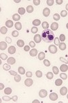
•There were macrocytes but no oval macrocytes or hypersegmentation of neutrophils
- Serum vitamin B12 — normal
- Red cell folate — normal
- Liver function tests — normal
- Thyroid function tests — normal
- Ferritin — 875 μg/l (normal range 20–200)
- What do you suspect and what would you do next?
- Bone marrow aspirate
- 12% blast cells (normal < 5%)
- 45% of erythroblasts were ring sideroblasts
DIAGNOSIS: myelodysplastic syndrome (MDS) (MDS with excess of blasts)
- Initially the patient tolerated the anaemia and required no treatment
- Later she needed red cell transfusion
- Later still she needed platelet transfusions
- Predicted survival was 1.1 years
- However, she was still alive 7 years later
The ferritin was 875 μg/l (NR 20–200) Why?
Does it matter?
1Yes
2No
3Probably not
Probably not
- A 72-year-old Indian woman
- Vegetarian, teetotal, non-smoker
- Shortness of breath on exertion
- Fatigue
- Painful gums and tongue
- Unable to eat spicy food
- On examination: pallor only
- WBC normal
- Hb 52 g/l
- MCV 122 fl
Platelet count normal
What is the most important test
1 Vitamin B12 and folate assays
2Liver function tests
3Thyroid function tests
4Bone marrow aspirate
Blood alcohol level

Vitamin B12 and folate assays
- Vitamin B12 180 ng/l (NR 125‒600)
- Red cell folate 227 pg/l (NR 215‒650)
- Thyroid function normal
- Liver function tests mildly impaired (bilirubin 20 μm/l, AST 110 iu/l (NR 40‒135)
- Lactate dehydrogenase (LDH) 3870 iu/l (NR 50‒450)
- What would you do next?
Bone Marrow Aspirate and look for giant metamyelocyte and megaloblasts

Do you think the patient has a myelodysplastic syndrome?
1 Yes
2 No
No
- Parietal cell antibodies: positive
- Intrinsic factor antibodies: positive
- Schilling test: 0% excretion
- The patient turned out to be a vegan
- You should now know the correct diagnosis and be able to explain how you would treat the patient
•The patient turned out to be a vegan
- A 70-year-old woman was referred to a vascular surgeon because of gangrenous toes
- They looked a bit like this
- History
–Not diabetic
–Had smoked 20‒30 cigarettes a day since age of 18 years
–Breathless on exertion and morning cough
•Examination
–Reduced femoral and distal pulses on side of affected toes
–Not breathless at rest, no cyanosis
–Plethora, conjunctival suffusion
–Spleen not felt
•What simple tests would you do first?

- Blood gases were normal
- Blood count was not
–WBC 18.6 × 10⁹/l
–Hb 180 g/l
–Platelet count 1648 × 109/l
•Ultrasound examination of the abdomen showed normal kidneys and increased splenic size
The most likely diagnosis is
1Chronic myeloid leukaemia
2Essential thrombocythaemia
3Polycythaemia vera
4Chronic obstructive pulmonary disease
Polycythaemia vera
What test would you do next to try to confirm the diagnosis?
1Molecular analysis for JAK2 mutation
2Measure total volume of red cells in circulation
3Bone marrow aspirate and trephine biopsy
?
You now know the diagnosis. How would you treat the patient?
1Venesection alone
2Imatinib
3Venesection plus hydroxycarbamide
432P
Venesection plus hydroxycarbamide
•Why is phlebotomy unsuitable as the only treatment for this patient?
?
What is abnormal?

- Percentages of white cells are meaningless unless you use them to produce an absolute count
- What are the two possible explanations of
Neutrophils 1%
Lymphocytes 99%?
FBC of an 83-year-old man with no abnormal physical findings
This was an incidental discovery, which has persisted. What is the most likely diagnosis?
- Acute lymphoblastic leukaemia
- Chronic lymphocytic leukaemia
- HIV infection
- Infectious mononucleosis
- Whooping cough

Chronic lymphocytic leukaemia

- The RBC, Hb and Hct are high
- The patient appears to be polycythaemic
–Is it a true or a pseudo-polycythaemia?
–Is he hypoxic from chronic lung disease or cyanotic heart disease?
–Does he have inappropriate erythropoietin secretion from a cyst or tumour?
–Does he have an intrinsic bone marrow disease?
•In this blood count there are no clues as to the cause of the polycythaemia
FBC of a 67-year-old woman with facial plethora—most likely diagnosis?

Polycythaemia vera
FBC of a 67-year-old woman with facial plethora
She is a smoker and has coronary artery disease
List all the abnormalities present and state the most likely diagnosis
How would you investigate her further?
- The RBC, Hb and PCV are high
- The WBC, neutrophil count and basophil count are high
- The platelet count is high
- The most likely diagnosis is polycythaemia vera
- Tests that might help to confirm this diagnosis might include
–Analysis for JAK2 V617F mutation
–Bone marrow aspiration and trephine biopsy
–Serum erythropoietin
- You might also want to do other tests to exclude a secondary polycythaemia (are they necessary in this patient?)
- She is a smoker and has coronary artery disease (does this matter?)
- He has extensive atheromatous disease and diabetes mellitus
- He has had an abdominal aortic aneurysm replaced by a graft and has had a stormy post-operative course with
–Sepsis
–Acute renal failure
–Respiratory impairment
His FBC shows:
–WBC 37.5 × 10⁹/l
–Neutrophil count 35 × 109/l
–Lymphocyte count 0.8 × 10⁹/l
–Monocyte count 1.2 × 10⁹/l
–Myelocytes and metamyelocytes 0.5 × 10⁹/l
–Hb 110 g/l
–MCV 89 fl
–Platelet count 514 × 109/l
Which bits are abnormal?
Film comments:
–Toxic granulation and vacuolation
–Left shift
–Rouleaux
- What is the likely cause of the high white cell count?
- Could this be leukaemia?
Reactive neutrophilia

CML
Elderly mak came to haematology OPD with bruising.
On this day his platelet count was 13 × 109/l
A week earlier it had been 3 × 10⁹/l with a normal Hb and WBC
–Abnormalities of the vessels
–Abnormality of the circulating blood

- It is gangrene
- It could be due to
–Abnormalities of the vessels
–Abnormality of the circulating blood
- Think of a vascular abnormality that could cause it
- Think of three abnormalities of the blood that could be responsible
What could be responsible for the bruises in a child?
- Non-accidental injury
- Coagulation abnormality
- Thrombocytopenia
- If this is a coagulation abnormality, what is most likely?
- If it is thrombocytopenia think of two possible explanations in this age group
What does it show?
What questions would you ask?

She has a hypochromic microcytic anaemia
- What causes of microcytosis do you know?
- Does the blood count in this patient help to suggest which is likely?
- Does anything in the history help to suggest or eliminate specific causes?
- Think of four questions you would ask
- Think of one test that you would do first
FBC of a North African woman with an 18-month old baby—most likely diagnosis?
IDA
•History was difficult as the patient spoke only Arabic and French. You might want to have asked about
–Diet
–Menstrual history
–History of pregnancies
–Blood loss
•History was difficult as the patient spoke only Arabic and French. You might want to have asked about
–Diet
–Menstrual history
–History of pregnancies
–Blood loss

- Rheumatoid arthritis
- Anaemia of chronic disease
- Iron deficiency anaemia resulting from use of aspirin or non-steroidal anti-inflammatory drugs (NSAID)
- Neutropenia or thrombocytopenia from drug toxicity
- Felty syndromes
- Increased erythrocyte sedimentation rate (ESR)
- A 10-year-old girl presented with a painful right knee that had started when she knocked her knee in a swimming pool
- The next day she had become unwell with malaise, anorexia and fever
- Her GP prescribed amoxicillin for ‘otitis media’
- Next day her mother took her to an Accident and Emergency Department
- She was afebrile
- Her right knee was painful and swollen
- X-ray of the knee showed patchy changes in density in the right medial tibial plateau
- Blood tests showed
–WBC 6.6 × 10⁹/l
–ESR 60 mm in 1 h (NR 0‒10)
–C-reactive protein (CRP) 27 mg/l (NR 0‒10)
•What is the most likely diagnosis?
osteomyelitis
- The diagnosis was osteomyelitis resulting from Staphylococcus aureus infection
- Other diagnoses you might have considered are septic arthritis, haemorrhage into the joint following minor trauma, non-accidental injury
- The blood tests and radiology were very useful in reaching the correct diagnosis
- Fever is often present and the WBC is often increased in osteomyelitis but not invariably
- There is a history of trauma in 35% so this should not deflect you from this diagnosis
- The CRP is normal in only 2% of cases
- A 1-year-old boy, an only child, presented to an Accident and Emergency department with a swollen right elbow following minor trauma
- On clinical examination and radiology there was no evidence of bony injury
- He was sent home
- Work out a differential diagnosis and as we work through the case history note the errors in management
- What analgesic would you not use?
?
- Three days later he was brought back with increased pain and swelling
- Joint aspiration yielded haemorrhagic fluid
- He was started on antibiotics and sent home
- Four weeks later he was brought back as the effusion had not resolved
- The joint was surgically explored (dark blood) and a biopsy was taken (‘synovitis’)
- Post operatively the wound bled persistently
- What test would you do?
•A coagulation screen showed a normal prothrombin time (PT) and a prolonged activated partial thromboplastin time (aPTT)
Draw out the coagulation cascade

- The aPTT was 96 s (NR 24‒35s)
- The prolonged aPTT was corrected by mixing the infant’s plasma with normal plasma
- What is the most likely diagnosis?
- What is the significance of the mixing test
- A family history was taken—the infant’s maternal grandfather had bled abnormally after minor surgical procedures
- The child’s factor VIII was 2 iu/l (NR 50‒150 iu/l)
- A 21-year-old woman presented with abdominal pain, bruising and altered level of consciousness
- She had a low grade fever
- Her platelet count was 15 × 109/l
- Her bilirubin was increased and LDH was greatly increased
- Her creatinine was marginally increased
- Her blood film looked something like this
What is the nature of the anaemia?
What is the most likely diagnosis?

•Microangiopathic haemolytic anaemia
Thrombotic thrombocytopenic purpura
What is the differential diagnosis?
- Haemolytic uraemic syndrome
- Meningococcal septicaemia
- Thrombotic thrombocytopenic purpura
What is thrombotic thrombocytopenic purpura?
Thrombotic thrombocytopenic purpura is diagnosed from a classic pentad of clinical features
- Microangiopathic haemolytic anaemia
- Thrombocytopenia
- Fever
- Neurological abnormalities
- Renal impairment
What is The underlying defect in thrombotic thrombocytopenic purpura?
- The underlying defect in thrombotic thrombocytopenic purpura is a deficiency of a plasma protein called von Willebrand factor cleaving protease (or ADAMTS13)
- The deficiency is autoimmune in origin
- The low levels of the protease lead to high levels of very large multimers of vWF, which cause platelet thrombi to form
- The widespread platelet thrombi are responsible for the clinicopathological features
- How should the patient be treated?
- Does she need a platelet transfusion?
- Does she need corticosteroids?
- Treatment is by plasma exchange
- Platelet transfusions should NOT be given
- Although the disorder is immune in nature, corticosteroids are not the primary treatment
- A previously healthy 20-year-old man presented with fever, sore throat, malaise, dyspnoea and abdominal pain
- Ten days before admission he had fallen on his left side and had attended Accident and Emergency with pain in the left chest wall
- Chest X-ray had been normal but he was observed overnight
- On this presentation, temperature 39.70C, BP 115/95, pulse rate 96 beats/minute, generalized lymphadenopathy, pharynx inflamed, mild hepatomegaly, spleen palpable 2 cm below left costal margin, abdomen tender
- WBC 11.2 × 10⁹/l, lymphocyte count 7.8 × 10⁹/l, Hb 109 g/l, numerous atypical lymphocytes
- What test would you do?
•A screening test for infectious mononucleosis was positive
•Does he need any further tests to confirm a diagnosis of infectious mononucleosis?
- Not really, but tests were done anyway
- He had IgM antibodies to EBV viral capsid antigen
- What does this tell us?
•He has had a recent EBV infection
•How should he be managed? Is there any cause for concern?
- Yes—he is anaemic, he has tachycardia and he has abdominal pain and tenderness
- Splenic damage and intraperitoneal haemorrhage should be suspected and urgent imaging should be done
- An urgent abdominal ultrasound showed splenomegaly and detachment of the inferolateral part of the spleen, 9 cm from the tip with subcapsular haematoma formation
- There was no intraperitoneal bleeding
- An urgent abdominal ultrasound showed splenomegaly and detachment of the inferolateral part of the spleen, 9 cm from the tip with subcapsular haematoma formation
- There was no intraperitoneal bleeding
- What should be done now?
- Emergency splenectomy was performed
- The spleen weighed 588 g (normal 200 g)
Why is EBV linked to splenomegaly?
- Splenomegaly occurs in c. 50% of patients with infectious mononucleosis
- Splenic rupture occurs in 0.1‒0.5% of patients
- Mortality has been estimated at 30%, mainly because of delayed diagnosis
- There is often a history of recent trauma, which may be very trivial (e.g. coughing, turning over in bed)
•What are the risks of hyposplenism?
–Overwhelming bacterial sepsis (particularly pneumococcal or Haemophilus influenzae)
–Fatal malaria
–Fatal Capnocytophaga canimorsus infection
What is advice post splenectomy?
- Vaccinate for pneumococcus, meningococcus and Haemophilus influenzae
- Vaccinate against influenza
- Prescribe life-long penicillin
- Advise the patient on
–Dog bites
–Travel to malaria zones
–Prompt treatment of infection
•Issue a splenectomy card and information sheet
- 63-year-old retired professional man
- Referred to haematology outpatients from gastroenterology clinic
- Microcytic anaemia ? cause
- Oesophageal reflux, confirmed by barium swallow
- Normal gastroscopy otherwise
- Symptoms resolved on omeprazole
- Also complained of rectal bleeding
- Proctoscopy: prolapsed haemorrhoids
- Colonoscopy: normal
- Presented again a year later
- 3 kg weight loss in one month
- Progressive anaemia
- Cervical lymph node felt and later disappeared
- Serum ferritin 308 μg/l
- Serum vitamin B12 and red cell folate normal
- Haemoglobin electrophoresis: normal Hb A2
- C-reactive protein 87 mg/l
- Repeat gastroscopy and duodenal biopsy: normal
- Chest X-ray and abdominal ultrasound normal
- Referred to haematology
On examination:
Thin
BP 120/100
Chest clear
HS normal
? Palpable spleen
DDx?
- Alpha thalassaemia (beta thalassaemia already excluded)
- Sideroblastic anaemia
- Anaemia of chronic disease
How do we Search for underlying cause in ACD?
–ESR 110 mm in 1 h
–C-reactive protein 149 mg/l
–Normal renal and liver function and calcium
–Protein electrophoresis: increased a1 & a2 globulins
–Auto-antibodies negative
–Normal PSA, CEA and AFP
–CT abdomen: “hepatic haemangiomas”, spleen 16 cm, no lymphadenopathy
- This is what the lymph node looked like
- What can you see?

?
- This is what the lymph node looked like under the microscope
- What is the giant bi-nucleated cell?

Reed Sternberg cells
Reed Sternberg cells, diagnosis?
•Nodular sclerosing Hodgkin lymphoma
What is the pathophysiology of ACD?
- Reduction in red cell lifespan
- Cytokine release
–IFNg, IL1 and TNF
- Reduced proliferation of erythroid precursors
- Suppression of endogenous erythropoietin production
- Impaired iron utilisation
How do you treat ACD?
- Treat underlying disease
- Recombinant erythropoietin
- Transfusion
What is Hodgkin Lymphoma?
- 1% cancer registrations per annum
- 3 per 100 000 in UK
- Peak in adolescence and > 50 years
- Commonly presents with painless supra-diaphragmatic lymphadenopathy
- One third present with B symptoms
–Fever, drenching night sweats or > 10% loss of body weight in last 6 months
What is the aetiology of hodgkins lymphoma?
- Increased risk in families of affected patients
- Association with HLA DPB1
- Epstein‒Barr virus found in >79% of over 50s
How do you decide treatment?
Staging involved anatomical stage (I, II, III, IV)
Absence or presence of B symptoms (A or B)
What is the treatment of 2B disease?
- Stage IIB disease (PET scan showed no activity in spleen)
- Unfavourable disease characteristics
–night sweats
–large mediastinal mass of nodes
–Age > 50 years
–ESR > 50 mm in 1 h
- Combination chemotherapy
- Radiotherapy to residual mediastinal disease

Yes