Autoimmune and Autoinflammatory Diseases 3 Flashcards
What are antinuclear antibodies?
Group of antibodies that bind to nuclear proteins
Test by staining of Hep-2 cells (human epidermoid cancer line)
Very common
Low titre antibodies (<1:80) often found in normal individuals (esp older women)
What may a person with SLE present with?
history of fatigue
Generalised arthralgia, particularly of small joints of hands
Hair fall
Mouth ulcers
Butterfly rash
What is the genetic predisposition of SLE?
Abnormalities in clearance of apoptotic cells
Polymyorphisms in genes encoding complement, MBL, CRP
Abnormalities in cellular activation
Polymorphisms in genes encoding/controlling expression of cytokines, chemokines, co-stimulatory molecules, intracellular signalling molecules
B cell hyperactivity and loss of tolerance
Antibodies directed particularly at intracellular proteins
? Debris from apoptotic cells that have not been cleared
Nuclear antigens - DNA, histones, snRNP
Cytoplasmic antigens - Ribosome, scRNP
What is the pathophysiology of SLE?
Antibodies bind to antigen to form immune complexes
Immune complexes deposit in tissues
Skin, joints, kidney
Immune complexes activate complement (classical pathway)
Immune complexes stimulate cells expressing Fc and complement receptors
How can you compare the disease pathology in Type II and Type III?
Immune complexes deposit in basement membrane in a type III response
Note the contrast in staining pattern compared with a type II response where antibody specific for the basement membrane (rather than immune complexes) despoit.
How do you measure antibody levels?
Measured by titre (the minimal dilution at which the antibody can be detected) or by concentration in standardised units
What are the targets of ANA?
A positive ANA result will trigger the laboratory to investigate for dsDNA and ENA antibodies
dsDNA
Ro, La, Sm, U1RNP Ribonucleoproteins
SCL70
Topoisomerase
Centromere
What is the pattern for ANA in SLE?
Homogeneous staining associated with specificity for dsDNA
Specificity investigated with ELISA based assay
What are anti dsDNA antibodies used for?
Measures antibodies against double stranded DNA
Are highly specific for SLE (95%)
Occur in ~60-70% of SLE patients at some time in their disease
Very high titres are often associated with more severe disease, including renal or central nervous system involvement.
Useful in disease monitoring
an increase in antibody titre is associated with disease activity and may precede disease relapse.
False positive results unusual (<3%)
What is speckled antibody?
Associated with antibodies to extractable nuclear antigens
Specificity is for some ribonucleoproteins (Ro, La, Sm, U1RNP) – confirm with ELISA
What are anti ENA antibodies?
Ro, La, Sm, RNP (all are ribonucleoproteins)
Antibodies may occur in SLE
Anti-Ro and La are also characteristically found in Sjogren’s syndrome
Titres not helpful in monitoring disease activity
What is classical pathway of complement?
Formation of antibody-antigen immune complexes
activate complement cascade via classical pathway
complement components become depleted if constantly consumed
Quantitation of C3 and C4 acts as a surrogate marker of disease activity
[NB we measure UNACTIVATED complement proteins, not activated forms]
What is the complement profile in SLE?

What is APLS?
•Anti-phospholipid syndrome
–Recurrent venous or arterial thrombosis
–Recurrent miscarriage
–May be associated with livedo reticularis, cardiac valve disease
–May occur alone (primary) or in conjunction with autoimmune disease (secondary)
What are the antibody tests for APLS?
•Three antibody tests
–Lupus anti-coagulant
- Prolongation of phospholipid-dependent coagulation tests
- cannot be assessed if the patient is on anticoagulant therapy
–Anti-cardiolipin antibody
•Antibody specific for negatively charged phospholipids
–Anti-B2 glycoprotein 1 antibody
•Antibody specific for glycoprotein found associated with negatively charged phospholipids
Check all three antibodies in individuals presenting with unexplained thrombosis or recurrent pregnancy loss
What is CREST?
•Limited Cutaneous Systemic Sclerosis (CREST)
Skin involvement does not progress beyond forearms
(although it may involve peri-oral skin)
–Calcinosis
–Raynauds
–Oesophageal dysmotility
–Sclerodactyly
–Telangectasia
–
–Primary pulmonary hypertension
What is Diffuse Cutaneous Systemic Sclerosis?
•Diffuse Cutaneous Systemic Sclerosis
Skin involvement does progress beyond forearms
- CREST features
- More extensive gastrointestinal disease
- Interstitial pulmonary disease
- Scleroderma kidney / renal crisis
What is the difference between limited (CREST) and diffuse systemic sclerosis?
Limited:
Anti-centromere antibodies
Diffuse:
Nucleolar pattern
Anti-topoisomerase antibodies (Scl70)
RNA polymerase
Fibrillarin
What is the difference between dermatomyositis and polymositis?
Dermatomyositis
- Muscle Bx: perivascular CD4 T cells and B cells
- Immune complex mediated vasculitis
Polymositis
- Muscle Bx: CD8 T cells surround HLA Class I expressing myofibres
- CD8 T cells kill myofibres via perforin / granzymes
What are the Ix for myositises?
•Positive ANA (in some patients)
– extended myositis panel - Anti Jo-1 positive, Anti-Mi2, Anti-signal recognition peptide antibody
(Extra info)
Anti-aminoacyl transfer RNA synthetase antibody eg Jo-1 (cytoplasmic)
Anti-signal recognition peptide antibody (nuclear and cytoplasmic) (PM)
Anti-Mi2 (nuclear) (DM>PM)
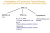
What are investigations for CTDs?
What are the autoantibodies for rheumatological diseases?

What are the systemic vasculitides?

What are the 3 types of ANCA associated SV vasculitis?
Microscopic polyangiitis / Microscopic polyarteritis / MPA
Granulomatosis with polyangiitis / Wegener’s granulomatosis / GPA
Eosinophilic granulomatosis with polyangiitis / Churg-Strauss syndrome / eGPA
What are ANCA?
Antibodies specific for antigens located in primary granules within cytoplasm of neutrophils
Inflammation may lead to expression of these antigens on cell surface of neutrophils
Antibody engagement with cell surface antigens may lead to neutrophil activation (type II hypersensitivity)
Activated neutrophils interact with endothelial cells causing damage to vessels - vasculitis
What is cANCA?
–Cytoplasmic fluorescence
–Associated with antibodies to enzyme proteinase 3
–Occurs in > 90% of patients with granulomatous polyangiitis with renal involvement
What is pANCA?
Perinuclear staining pattern
Associated with antibodies to myeloperoxidase
Less sensitive and specific than cANCA
Associated with microscopic polyangiitis and eosinophilic granulomatous polyangiitis
Investigations show:
Positive antinuclear antibodies (ANA)
Anti-dsDNA+ve
Low C3 and C4
High ESR
Negative results for:
Ro, La, Sm, RNP
SCL70
Centromere
Jo-1
Anti-neutrophil cytoplasmic antibodies (ANCA)
What is the diagnosis?
Systemic sclerosis
Systemic lupus erythematosus
Dermatomyositis
Sjogrens syndrome
ANCA associated vasculitis
SLE
Investigations show:
Positive anti-neutrophil cytoplasmic antibodies
(ANCA)
Negative results for:
Anti-nuclear antibody (ANA)
Normal complement
Raised ESR and CRP
What is the diagnosis?
Systemic sclerosis
Systemic lupus erythematosus
Dermatomyositis
Sjogrens syndrome
ANCA associated vasculitis
ANCA associated vasculitis
