Alimentary Canals Flashcards
What are the predominant type of diseases vary among different species?
-Dogs and cats develop alimentary neoplasia more often than
farm animals.
- Ruminants and pigs develop a wide range of infectious diseases often poorly controlled by vaccination.
- Horses are prone to intestinal displacementscolic
Portals of Entry of pathogenic agents
Ingestion (most common)
Coughed up by the lungs and swallowed Systemic hematogenous route
Migration through the body (parasites)
Defense Mechanisms
Saliva
Resident flora and fauna
Gastric pH
Secreted immunoglobulins
Vomiting
Intestinal proteolytic enzymes
Phagocytes and other effector cells within the mucosa/ submucosa High rate of epithelial turnover
Increased peristalsis resulting in diarrhea
Name and what the complication is

In palatoschisis there is a central
defect in the midline fusion of the palatine shelves resulting in communication between the oral and nasal cavity.
Caused by Veratrum californicum
Complication- aspiration pneumonia

Cheiloschisis (“harelip”) - calf
What is a consequence of this and what is it called?

Prehension, prone to infection
Malocclussions
Failure to the upper and lower incisors to interdigitate properly
May result in difficulties in the prehension and mastication of food.

Short lower jaw (brachygnathia)

Foal, prognathia is protrusion of the lower jaw.

Dental attrition (loss of tooth structure caused by mastication).
The degree of tooth wear depends on the tooth, the animal species and the types of food. Abnormal wearing is most common in herbivores results in “step mouth”.

Periodontal disease
Resident bacterial films and the acid and enzymes they produce lead to enamel, gingival and periodontal ligament damage.
Dental plaque
Dental calculus (tartar mineralized dental plaque)
primary diseases of the oral cavity
Primary diseases are rare; the exception is Actinobacillosis (Actinobacillus lignieresii) or pyogranulatmous glossitis chronic stomatitis – the tongue is often involved:

Actinobacillosis (Actinobacillus lignieresii)
Pyogranulatous glossitis

Radiating clubs of amorphous eosinophilic material: Splendore-Hoeppli phenomenum
A. Lignieresii is a Gram-negative rod

Thrush (Candidiasis)
Candidaspp.(eg.C.albicans)
Often is observed young animals treated with antibiotics for long periods of time, or animals with underlying debilitating diseases
AFFECTS THE KERATIN(SILVER STAIN)- RESULTS IN HYPERKERTAIN

Lingual lesions are often a manifestation of systemic disease like renal disease (uremic glossitis), BVD or other viral infections like FMD (discussed later).

Lymphoplasmacytic gingivitis, stomatitis –cat Many cats are FeLV or FIV positive.
IMMUNE MEDIATED

Feline chronic gingivo-stomatitis (FCGS). Cinical signs: oral pain, dysphagia, ptyalism and weight loss.
Etiology: unclear. Dental plaque, FCV, and immune-mediated mechanisms appear to be involved. FCGS is also common in FIV positive cats.

Chronic ulcerative (lympho-plasmacytic) paradental stomatitis – Most common in older dogs
Thickening of the gingivia
Vesicular stomatitides
Vesicle: a raised lesion (up to 1 cm in the largest dimension) filled with clear (serous) fluid located within the epithelium or between the epithelium and lamina propria). A larger lesion is referred as bulla.
If observed in the oral cavity of dogs & cats:
Rule out immune-mediated diseases
In cats they are often the result of calicivirus infection
If observed in food/ large animals:
• Rule out major viral diseases which are usually non-fatal but result in huge economic loss.

Viral vesicular stomatitides
Pathogenesis: viral-induced epithelial damage intracellular edema in keratinocytes (ballooning degeneration)vesiclesbullaerupture leads to erosion and ulceration.
Targets epithelial cells

Vesicular glossitis – Cat, calicivirus infection.
Vesicular Diseases


FMD
Exotic (Foreign) Animal Disease. Highly contagious with high morbidity and low mortality.
Virus ingestion/ inhalation
pharynx viremia Oral mucosa & epidermal siteslesions
develop in areas subjected to mechanical injury
Clinical signs: drooling saliva (ptyalism), lameness

FMD
What is a lesion with FMD that can be seen in young animals?
Young animals: “tiger heart” multiple stripes, FMD
Myocardial necrosis


Severe lameness, pig - FMD

Vesicular exanthema (calicivirus)
Only in pigs
DDx: FMD
What two disesases cause erosive-ulcerative stomatidies
BVD (Pestivirus) Multi-focal of mucousal ulceration
MCF- Herpes; targets endothelium


Bovine papular stomatitis. Etiology: Parapoxvirus
Epithelial hyperplasia
“Coin-shaped papules and ulcers”,
The virus is closely related to pseudocowpox virus that causes “milker’s nodules in humans (hands).
Papules on the nares, muzzle, oral cavity. Usually in immunosuppressed individuals

Bovine papular stomatitis. Etiology: Parapoxvirus

Contagious Ecthyma, also known as Contagious viral pustular dermatitis, Orf, or “sore mouth”
Worldwide distribution. The cause is a parapoxvirus.
Results in loss of condition since affected animals
“neither suckle nor graze”. High morbidity and low mortality. Mainly in lambs and goat kids 3 to 6 months of age.
ZOONOTIC; SHEEP AND GOATS; get from mom’s teat

Contagious Ecthyma. AVC. Lesions usually develop in sites of trauma (corners of the mouth, mammary gland, coronary bands, etc.). It is a zoonotic disease.

Calf, Pathology Museum, FVSc, University of Liverpool Oral Necrobacillosis, Fusobacterium necrophorum
filamentous anerobic bacteria; bacterial toxins are responsible for the severe lesions

Oral necrobacillosis- necrotizing!, Calf, AVC. Calf Diphtheria Fusobacterium
PAS, filamentous bacteria.

Feline eosinophilic granuloma – complex (includes eosinophilic granuloma, labial and rodent ulcer).

cat, eosinophilic ulcer, palate.

Feline eosinophilic granulomas. Young UCVM cats (VSRS).

Feline eosinophilic granulomas. Young UCVM cats.

Gingival hyperplasia

Fibromatous epulis, dog
Epulis: Tumors of the periodontal ligament – type stroma (whether or no they are true neoplasms is still controversial)

Canine oral papillomatosis
Papovavirus-induced papilliform or cauliflower-type lesions (“warts”) in the lips and oral mucosa.
It is transmissible and usually affects animals younger than 1 year-old. Lesions regress spontaneously and immunity is long-lasting.
Immunosupression; transmisble to young animals

Oral papilloma: Verrucous lesion composed of thick keratinized stratified squamous epithelium covering a pedunculate connective tissue core.

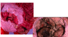
Oral melanomas. Most common in dogs. Around 90% of oral melanomas in dogs are malignant.
Smaller breed and oral pigmentation are predisposing factors.
Hematogenous and lymphatic routes

Amelanotic melanoma, dog
no melanin pigment; more invasive b/c cells are less differentiated

Oral melanoma, dog

Melanoma, dog – pulmonary metastases,

Squamous cell carcinoma, tongue, dog

Fibrosarcoma – palate and maxilla QH gelding, AVC

Congenital megaesophagus –persistent right aortic arch (vascular ring) - puppy
consequences:
- aspiration pneumonia
- vomiting without any digestion

Megaesophagus can also be acquired – most common in dogs. idopathic or myasthenia gravis

Choke,
foreign body stuck
when removed you will see the esophagus necrosis and degeneration

Reflux-esophagitis, horse
whitish color- keratin to protect against refluxed gastric acid
-hyperkeratosis primary with secondary erosins

Erosive-ulcerative esophagitis, BVD
or necrotizing

: Spirocerca lupi in dogs

Esophageal Osteosarcoma- S. lupi

Ruminal tympany or bloat- over distention of the rumen and reticulum by gases produced by fermentation
A. primary- often associated with new diets that promote the formation of stable foam
B. Secondary- caused by physical or functional obstruction of the esophagus resulting in failure to eructate

Bloat, cow-AVC

“Bloat line” – the most reliable post-mortem indicator of ante-mortem bloat -, cow, Atlantic Veterinary College, AVC.
blue=blood congestion

Foreign bodies
Traumatic reticulitis -cattle

chronic pericarditis and epicarditis is not an unsual complication; go through diaphram and into the heart

chemical rumenitis (lactic acidosis, grain overload) cattle

“stellate ulcers” (ruminal scars)
healing by fibrosis tissue from inflammation; from lactic acidosis

6-month-old heifer, history of grain overload. AVC-1998
Gastric dilation-volvulus (life-threatening condition)
Most common in large, deep-chested breeds of dogs; also in sows

Gastric dilatation-volvulus, dog

Dog, gastric dilatation, volvulus –Intraluminal hemorrhage, Rhodesian ridgeback dog, AVC-97
Abomasal displacement
Occurs most often in post-parturient dairy cows and calves
Left-sided is most common: generally non-fatal partial obstruction of abomasal flow
Right-sided: Represent ~15% of the abomasal displacement. 20% of these result in abomasal volvulus.
Abomasal displacements (left-sided or right-sided) lead to abdominal pain, elevated heart rate, anorexia, dehydration, depressed peristalsis with lack of feces and abomasal tympany (high-pitch ping elicited by percussion).

Gastric rupture
Most cases of gastric rupture in horeses are due to intestinal obstructions ileus; adynamic or mechincal ileus. Adynamic ileus results from inhibition of bowl motility more commonly caused by perinonitis
Since horses can’t vomit they get gastric ruptures secondary to gastric obstructions

Horse, chronic diaphragmatic hernia leading to gastric rupture and Death. UCVM-2008.
What is common to cause gastric impaction/rupture in horses?
Persimmons

Gastric ulcer, pig
Seen in pigs fed finely ground rations- melana gastric bleeding
What are possible causes?

Pig, gastric ulcer
Possible causes- finely ground feed, high copper, high starch and low protein diets, unsaturated fatty acids, hisatmine, stress
Pigs and horses- main complication- perferating ulcers
MDx and cause
MDx: multi-focal necrotizing ulcerative gastritis
Cause- NSAIDS
squamous is the esphagus
white=keratin

gastric ulceration; NSAIDS, horse
Causes ischemic necrosis
NSAID in equines- stomach and meduallary papilla of kidney

gastric ulcers in cats and dogs are idiopathic. Cutanous mast-cell tumors in dogs may lead to gastric ulceration.
Gastric ulcers:
- increased NSAID or seteriodal
- increased histamine levels ass with mast cell tumors or mastocytosis
- gastrin-secreting pancreatic islet cell tumors or gastrinomas

Right: Perforated gastro- duodenal ulcer in a dog with a mast cell tumor. Noah’s Arkives, UGA.
May occur in dogs with solitary cutaneous mast cell tumors.

Stomach, pig –thrombosis & hemorrhage (gastric venous infarction) secondary to endotoxemia or bacterial sepsis. Occasionally seen in ruminants and horses
Et: e. coli or salmonella
in pigs- e. rushtophatie, glasser’s disease

Uremic gastropathy (uremic gastritis, cat).

Uremic gastritis; 2 year old shi-tzu with familiar renal disease; Von-Kossa stain
CHF causes other locations- uremic gastritis, stomattis, left ventricular mural myocarditis, pneumonitis, thrombosis

Abomasitis lamb, brazy, Clostridium septicum
causes of death is endotoxemia

Abomastitis, necro-hemorrhagic with submucosal emphysema, Braxy Clostridium septicum

Mycotic abomasitis- often a sequel of long-term antibiotic therapy which destroys resident bacterial flora and promotes the growth of angio-invasive fungi such as Aspergillus, Absidia, Rhizopus, Mucor spp. . sepsis and other debilitating conditions are also predisposing factors
MDX: necro-hemorragic abomastits

Mycotic abomasitis, calf-

Mycotic vasculitis/perivascultis dogs with silver stain

Gasterophilus nasalis and intestinalis, horse

Erosive-ulcerative lesions caused by Gasterophilus intestinalis, horse

Abomasal folds. Proliferative (hyperplastic) abomasitis due to Ostertagia spp. “Moroccan leather” appearance of the affected abomasal mucosa.

Draschia megastoma (spirurid nematode) -brood pouch close to the margo plicatus (mucosal/submucosal nodule). Granulomatous gastritis, horse.

Haemonchus contortus is a tristrongylid nematode parasite that causes disease primarily in sheep and goats. Parasitic abomastitis leasds to blood loss, anemia, and hypoproteinemia.
Parasitic abomastitis, sheep- blood-suckign trichostronglyid parasite, can also affect goats and cattle
Lesion itself- superficial erosive gastritis
EDx: parsitic gastritis
“barber pole worm”

SCC
Horse, gastric squamous cell carcinoma
most common in horses;
mechanical obstruction- can’t eat

Peritoneal carcinomatosis. Horse, Gastric Squamous Cell Carcinoma, Noah’sArkives

Gastric lymphosarcoma, horse

LSA abomasum, cow

Multi-centric Lymphoma
What is the most common segmental anomaly of the intestine?
Atresisa coli

Segmental anomalies in the intestine are relatively common and can range from stenosis (incomplete occlusion of the intestinal lumen) to atresia (complete
occlusion/ obliteration of the intestinal lumen). Ischemia of a segment of gut during fetal development? It may be an autosomal recessive trait in Holstein calves.

Diagnosis: Atresia ani (imperforate anus) with concomitant recto-vaginal fistula – Right: Note prominent megacolon
Lethal white syndrome in foals
(congenital colonic aganglionosis – an autosomal recessive genetic disorder)
Most prevalent in the American Paint Horse. Usually (but not all) horses with a white-spotting pattern are carriers of this trait (heterozygus).


Microscopically there is absence of myenteric and submucosal parasympathetic ganglia in the wall of the ileum, cecum and colon leading to intestinal immotility and colic (colonic or ileocolonic aganglionosis).
Lethal white syndrom in foals

Enterolith, horse -
Composed of concentric lamellae of magnesium, ammonium phosphate (struvite) deposit around a “nucleus” foreign body such as a nail, wire etc. Vary in size, some may weight up to 10 kg.
Acquired obstructions
Trichobezoars
(hairballs) – cattle, Pathology Museum, Faculty ofVeterinary Science, University of Liverpool. Are not as heavy as enteroliths (located in the forestomach and
abomasum) . Also found in dogs and cats.
Phytobezoars
Phytobezoars or phytotrichobezoarscomposed mainly of plant material impregnated with Some phosphate salts may be found in the colon of horses.

Ascarids impaction, foal
more of a mechanical problem than a damage problem

Rectal stricture – Pig, chronic salmonellosis, (Salmonella typhimurium) Noah’s Arkives
Penetrating wounds (foreign bodies etc..) can also lead to stricture and stenosis within the GI tract.
Hernia:
The protrusion of an organ or part of an organ/tissue through an abnormal opening
Internal vs external hernia
Internal hernia: Displacement of intestine through a normal or
abnormal foramina within the abdominal cavity (rare).
Examples: incarceration (entrapment) of loops of the intestine within the slit-like epiploic foramen, or a rent (tear) in the omentum or mesentery.
External hernia VERY COMMON: Displacement of loops of intestine, omentum and occasionally other viscera (hernial contents) outside the abdominal cavity. Displaced contents are inside of a pouch (hernial sac, formed by the peritoneum and the skin) which protrudes through the hernial ring - an opening in the abdominal wall which could be acquired or natural (e.g.: vaginal ring of the inguinal canal).
If the displaced abdominal contents are not covered by parietal peritoneum or skin the lesion is referred to as “eventration”.

Mare. Eventration of the cecum secondary to trauma,

Mare. Eventration of the cecum secondary to trauma

Cat, diaphragmatic hernia due to trauma. It can also be congenital. Herniation of the liver and loops of the small intestine are more common.
Intestine in the thoraic cavity- happens after trauma i.e. HBC

Scrotal (inguinal) hernia - pig

Scrotal hernia, pig,

Incarceration of a piece of small intestinelead to venous infarction, sepsis, and death. Scrotal hernia, pig,

Umbilical hernia, pig, AVC

Strangulated loop of small intestine – umbilical hernia, pig, AVC

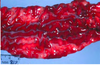
Torsion of the left colon -horse

Colonic torsion-horse

Intestinal volvulus - horse

Herniation of small intestine through the epiploic foramen (internal hernia) – horse, Cornell files

Intestinal strangulation by pedunculated lipomas.

Pedunculated lipomas
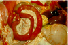
Intussusception


Intestinal intussusception, foal, Texas A&M, JEdwards.

Intestinal intussusception

Horse presented with severe colic unresponsive to palliative treatment to control the pain. Euthanized. Atlantic Veterinary College

Horse,cecocolonicintussusception,AVC

Viral Enteritis
BVD: Pestivirus, bison.
Most cattle develop a relative mild or subclinical Form of the disease.
MDx: ulcerative colitis
The most severe form of the disease (Mucosal disease – low morbidity but high mortality) occurs most commonly in persistently infected animals which subsequently become infected with a closely related CP strain; or when the persistent virus develops specific mutations.Affected
cattle are usually 6 months to 2 years
of age.

MCF
Gamma herpesvirus (genus rhadinovirus)
Affects a variety of ruminants, including bison and cervids.
In America we have tHe “sheep-Associated MCF” observed in bovids and deer in contact with sheep.
Fibrino-necrotizing vasculitis, with prominent lymphocytic perivascular infiltrates can be Found in several organs and tissues.
NO CLINICAL SIGNS IN SHEEP

Enteric coronaviral infections
Common cause of neonatal diarrhoea in calves; alone or in combination with other agents like rotavirus or Cryptosporidium
A&B: Transmissible gastro-enteritis (TGE) in piglets: Severe villous atrophy (blunting) and fusion. High mortality in young piglets.
LYMPHOCYTES AND PLASMA CELLS - FIP in cats

TGEV-Positive immunohistochemistry.
Small intestine, piglet. Note severe villus
blunting (villus attenuation/ atrophy).
can see through intestine
Cytokeratin- epithelial cells
Enteric rotaviral infections
Enteric rotaviral infections
Rotavirus can cause diarrhoea in young animals of any species
also causes damage to surface enterocytes resulting in variable degrees of villous atrophy.
Subclinical infections are common in piglets.

Parvovirus enteritis , puppy, Noah`s Arkives – CPV-2.
In cats the disease is caused by feline panleucopenia virus –bone marrow lesions dominate the clinical picture
CPV-2
Neuro-enterigic enterisits.
Panleukopenia, cat, TAMU. Segmental fibrino-hemorrhagic enteritis.

Parvovirus enteritis, puppy
In older cats a similar necrotizing enteritis can be caused by FeLV infection.

FIP
Bacterial Enteritis
E. coli
Important cause of enteritis in neonatal animals
Bacterial strains have different virulence factors which result
in different clinical syndromes
Virulence factors promote colonization or adhesion, metabolic dysfunction or death of enterocytes, affect the local or systemic vasculature, or promote invasion and septicemia.


Edema disease (enterotoxemic colibacillosis) in pigs.
Most common in pigs a few weeks after weaning.
Bacterial enterotoxin (verotoxin) that causes endothelial cell injury in arterioles resulting in fluid loss and edema. Affected animals may exhibit focal bilaterally symmetric encephalomalacia (Cerebrospinal angiopathy of swine).
Cholstridial enterotoxemia
Caused by Clostridium perfringens group (type) A to E. Type D is most common.
Affects the best nourished animals in the group.
Animals may be found dead or may exhibit bloody
diarrhea.
C. perfringens type D produces an angiotoxin (epsilon toxin) which in addition to intestinal lesions causes focal symmetrical encephalomalacia (FSE) in sheep.
You have to prove that the toxin is there, since the bacteria is normal in feces

Clostridial enteritis (Necrotic enteritis) in a chicken, AVC. Necrotic villi
are lined by Gram positive bacilli Clostridium perfringens type A.
Tyzzer`s disease
Tyzzer`s disease (Clostridium piliforme) – Occurs in multiple species. The main target is the liver but lesions also occur in the intestine (port of entry) and the heart.
Colitis X
Colitis X Typhlocolitis of horses: Probably the result of dysbacteriosis (often associated to antibiotic therapy or dietary changes) leading to proliferation of toxigenic clostridia, specially Clostridium perfringens type A and Clostridium difficile.
Salmonellosis
All Salmonella species are pathogenic
Clinical disease ranges from a localized enterocolitis to
septicemia
Stress factors are important in the onset of clinical disease
Zoonotic and nosocomial infection
Acute enteric lesions are those of an ulcerative and fibrino- necrotizing enterocolitis.
Intestinal contents are malodorous and contain mucus, fibrin and occasionally blood (feces: “septic tank odor”).

Horse, colon: edema and hemorrhage: Salmonellosis, Cornell

Fibrino-necrotizing entero-colitis, Horse, Salmonella, Cornell

Horse, Embolic mycotic pneumonia – Sequel of Salmonellosis, Cornell

Horse, Embolic mycotic pneumonia – Sequel of Salmonellosis, Cornell

Pig, chronic salmonellosis, button ulcers, Cornell files
fibrin
rectal strictures- are other manifestations of chronic salmonellosis; lead to fecal retention, megacolon, etc

Proliferative ileitis: Lawsonia intracellularis, Pig, Cornell files
The disease, currently named as Porcine Proliferative Enteropathy (PPE), causes different syndromes.

Necrotic enteritis: Lawsonia intracellularis, Pig. Cornell files

Proliferative hemorrhagic enteropathy, L. intracellularis, pig, AVC.
excessive crypts

Proliferative ileitis, Lawsonia intracellularis , pig, Warthin-Starry silver stain, AVC

Swine dysentery (spirochetal colitis ), caused by Brachyspira hyodysenteriae. The disease affects mainly pigs 8-14 weeks old and is characterized by large bowel diarrhea with mucous and blood in the feces.

Swine dysentery, Brachyspira hyodysenteriae
Rhodococcus equi
Causes an enterocolitis in young horses
Virulent factors allow the bacteria to survive within the
cytoplasm of macrophages and cause chronic disease
Typically R. Equi is associated with suppurative pyogranulomatous pneumonia in foals.
The enteric lesions are ulcerative and pyogranulomatous and are associated with prominent regional lymphadenitis.

Foal, pyogranulomatous colitis, Rhodococcus equi, Noah’s Arkives

McGavin. Pyogranulomatous colic lymphadenitis, Rhodococcus equi

Pyogranulomatous lymphadenitis, R. equi, foal. Noah’s

Foal, 6-week-old, Rhodoccocus equi pneumonia, Cornell

Mycobacterium avium, ssp. paratuberculosis
Johne`s disease (paratuberculosis) – Chronic disease of ruminants. Clinical disease, characterized by diarrhea, emaciation and hypoproteinemia, usually occurs in animals older than 19 months.

Granulomatous enteritis, Noah’s Archives
Granulomatous enteritis, Noah’s Archives
F03702, UGA, Bovine, Jejunum
Lesions can be found in the ileum, cecum and proximal colon. Ileocecal valve is usually affected.
lots of macrophages
ileal/cecal junction

Johne’s disease

Granulomatous lymphangitis with lymphangiectasia(when lymphnodess are grossly visible), Goat – Johne’s disease,

Granulomatous Colitis in Boxer dogs (Histiocytic Ulcerative Colitis) F09903 –F09904 –WSU.
The disease in Boxers appears to be associated with selective intramucosal colonization by specific strains of E. coli.
Similar findings have been reported in people with inflammatory bowel disease, particularly with Crohn’s disease.


Calf, Cryptosporidiosis, HE, Noah’s Archives
Diagnosis is made by the finding of numerous protozoal organisms attached to the apical surface of enterocytes.


Coccidial enteritis
A-D: Proliferative enteritis, goat kid, AVC.

Top: Proliferative, hemorrhagic & necrotizing enteritis, Coccidiosis, calf. Noah’s Arkive


Hookworms
Dogs: Ancylostoma caninum & Uncinaria stenocephala
Blood sucking parasites that may cause significant anemia and hypoproteinemia in puppies.
Larvae may be found in the colostrum.

Anaplocephala perfolita- hookworms in horses

Trichuris (Whipworm)
Trichuris spp. Parasitize the cecum and colon of domestic animals.
Most infections are subclinical
Severe infections may lead to bloody diarrhea, weight loss, dehydration and anemia.

Left: Ascaris suum infection, Pig, AVC
“Milk spotted liver”, pig, UCVM

Lymphosarcoma (LSA) in a pig. The tumor ruptured and led to fatal peritonitis. AVC
Neoplastic Diseases of the Intestine
Intestinal tumors are most common in dogs and cats.
Most primary tumors are carcinomas
Lymphosarcomas may arise in the intestine but in most cases they are a manifestation of multicentric lymposarcoma. The exception may be the cat
LSA is the most common neoplasm in cats and the “alimentary form” of lymphosarcoma has the highest incidence in this species.
