3.5 - 3.6 The Cytoskeleton Flashcards

1
Q

What are some of the functions of the cytoskeleton?

A
  • Mitosis - spindle
  • cell shape
  • cell migration and motility
  • intracellualr trafficking
  • supports membranes
  • mechanically links adjacent cells
  • muscle contraction
2
Q

What are the three main classes of filaments?

A
3
Q

What are intermediate filaments formed from?

A
  • Intermediate filaments are formed from multiple protofilaments which bind laterally to each other
  • Protofilaments of actin and tubulin assemble by head to tail binding of monomers
  • Cytoskeletal filaments are dynamic and adaptable
4
Q

What are some exampels of intermediate filaments proteins?

A
5
Q

What is the structure of intermediate filaments?

A
  • They are constructed from elongated protein subunits
  • The dimer has polarity
  • Tetramer is soluble and has no polarity
  • Protofilaments are antiparallel arrangement of dimers (extended alpha helix) and each end is identical so no polarity
6
Q

What gives the intermediate filaments their strength?

A
  • A rope like structure makes intermediate filaments strong when tetramers associate with each other
  • Multiple extended alpha helices form numerous hydrophobic interactions
7
Q

What are the roles of actin filaments in cells?

A
  • They change cell shape
  • Cell locomotion
  • Movement of organelles inside cell
8
Q

How do actin subunits assemble?

A
  • Actin subunits (G-actin) assemble head-to-tail creating polar actin filaments (F-actin)
  • The actin protofilament Globular (G) actin has ATP binding site
  • A protofilament comprises two parallel filaments of actin monomers assembled end to end (plus-end and minus end) giving polarity
9
Q

How are actin filaments organised into assemblies?

A
  • Actin filaments are organised into assemblies by cross linking proteins
  • Type of cross linking protein affects the type of assembly (meshwork vs bundles vs contractile)
  • Found in cells of the gut where parallel array of actin filaments are held in place by proteins
10
Q

What do bundles of F-actin form?

A
  • bundles of F actin form contractile rings in some cells
  • transmembrane proteins link to apical actin mesh work
11
Q

What is the role of F-actin bundles in processing of migrating cells?

A
  • Tight bundles in filopodia allow extension, they are tight paralell arrays of actin filaments
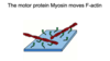
  • Stress fibles are contractile and exert tension, acted on by myosin motors to pull cell behind itself
  • Gel like network supports plasma membrane and allows broad extensions of cell (lamellipodia)
12
Q

Where is the network of F-actin found and what does it do?

A
  • A network of F actin is found beneath the plasma membrane of many cells (cortex) to support it
  • Actin binding proteins regulate gel-mesh formation of actin
  • Filamin allows for lamellipodia formation
    • it has bifold structure that arranges actin in mesh work
13
Q

How is the actin meshwork seen in red blood cells?

A

Spectrin regulates the actin meshwork in red blood cells so it can keep its shape as it squeezes through the capillary

14
Q

How does F-actin undergo polymerisation?

A
  • It self assembles from actin subunits
  • Faster at the plus end than minus because monomer needs to undergo conformational change before it can be added
  • At plus it will bind and initiate the conformational change
15
Q

What does depolymerisation of F-actin result in?

A
  • Depolymerisation results in the shortening of F-actin
  • If the concentration of monomers is insuffienct to replace the atp bound monomer then dynamic instability means the monomers come off
  • Faster dissociation at the plus end
16
Q

What happens at the critical concentration for F-actin polymerisation?

A
  • At Critical Concentration (CC):
    • The rate of subunits ON = rate of subunits OFF
  • Treadmilling
17
Q

How does the plus end of F actin compare to the minus end in polymerisation?

A

Plus end of F-actin grows (and shortens) more rapidly than minus end

18
Q

How can actin polymerisation be used to do mechanical work in cells?

A
  • Cells at the migrating edge be treadmilling
  • assembly of actin faster at positive end and losing at the minus end so it will grow in a certain direction
19
Q

What happens at the Critical concentration of G-actin?

A
  • Rate of G-actin addition = rate of G-actin loss
  • If the [G-actin] > CC then the filament grows
  • If the [G-actin] < CC then the filament shrinks
20
Q

How is F-actin growth controlled by regulating local available [G-actin]?

A
  • Thymosin binds G-actin and prevents addition to either plus or minus end of F-actin
  • Thymosin effectively reduces the local [G-actin] at bond ends
  • Concentrates G-actin where it needs to grow the actin
21
Q

How does profilin bind to G-actin?

A
  • Profilin competes with thymosin for binding to G-actin
  • Profilin-actin complex can be added to plus end, but not minus end of F-actin
  • Effectively increases the loacl [G-actin] at the plus end
22
Q

How does F-actin growth depend upon balance between thymosin and profilin?

A
  • Profilin competes with thymosin for binding to actin monomers and promotes assembly
  • Local concentration determines what happens to that filament
23
Q

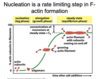
What is the rate limiting step in F-actin formation?

A
  • Nucleation is a rate limiting step in F-actin formation
  • Filament stability depends on the number of H bonds between subunits
  • Small filament assemblies are unstable, large assemblies more stable
  • Filament formation depends on formation of ‘nuclei’ of critical size
25
26
How can the lag phase of F-actin formation be abolished?
27
What is the pre-nucleus that nucleates F-actin growth?
* The ARP complex nucleates F-actin growth * ARP don't form filament themselves, there is no good minus end for the plus end of the actin filament to bind to
28
Which side of the actin filament promote nucleation on?
* ARP complex can promote nucleation from side of existing actin filament * Arp2/3 complex can bind to existing actin filament * Very effective polymerisation results in branching
29
What are some mechanisms of remodelling actin filaments?
* 100s of different actin binding proteins * Promote or inhibit assembly * Promote or inhibit disassembly * Stabilize filaments * Sever filaments and promote branching * Affect filament arrangement or membrane association
30
What moves F-actin?
31
32
How does Myosin form filaments in striated muscle cells?
* Myosin II forms thick filaments in striated muscle cells
33
What are the three major steps of cell locomotion?
1. Protrusion of filopodia - by actin polymerisation 2. Attachment - actin filaments connected by TM proteins to substratum 3. Traction - contraction by myosin-actin interactions move the rear of the cell forward Focal contacts need to be assembled and disassembled
34
What is it that pulls the cell body forwards?
* Actin-myosin interactions pull the cell body forwards * Myosin role at the trailing edge
35
How does actin polymerisation push the leading edge of migrating cells forwards?
* Plus ends associate with membrane at leading edge * Older filaments hydrolyse ATP and increasing dissembled (cofilin)
36
What are the three major forms of protrusion of actin polymerisation?
1. Filopodia 2. Lamellipodia 3. Pseudopodia
37
38
How do external molecules guide motile cells and axons?
* They bind to cell surface receptors * Bacteria produce chemoattractant, whuch binds cell surface receptors * Two pathways mutually antagonistic 1. Activates Rac → regulates actin polymerisation (G protein activates PIP3 lipid which faciliates actin polymerisation) → lamellipodial extension 2. Activates Rho → regulates myosin activity → actin filament bundling (stress fibres)
39
How do tubulin heterodimers assemble?
* Tubulin heterodimers assemble head to tail to create a polar filament * They have a plus and minus end (alpha tubulin and beta tubulin) * GTP binding site not ATP
40
What sort of proteins regulate the microtubules?
* Microtubules can be organised into regularly spaced bundles in cells * Microtubule associated proteins (MAP) regulate microtubule strability * Microtubule function in regulating cell shape and vesicle transport
41
Which Microtubule associated protein (MAP) is for where?
* Tau for axon * MAP2 for dendrites as it gives more widely spaced microtubules
42
What are cilia and flagella composed of?
* Cilia and flagella are composed of bundles of microtubules * Highly stabilised arrays of microtubules due to capping proteins and various other axonemal proteins * Arrangement regulated by structures called radical spokes
43
What end to microtubules self assemble from?
* Microtubules self-assemble from tubulin subunits * Microtubules grow faster at plus end (beta) than the minus end (alpha)
44
What phases do microtubules alternate between?
* Microtubules alternate between phases of rapid assembly and disassemly - dynamic instability * Random alterations in GTP cap (T-form of filament) can affect polymerisation rate and stability * If depolymerisation catches up with polymerisation then filament collapses (catastrophe)
45
What is the rate limiting step in microtubule formation?
* Nucleation is a rate limiting step in microtubule formation * Nucleation is unstable unless 4 subunits come together then tubule will form * Just like actin, initial polymerisation of microtubules is unstable unless there is a nucleation centre
46
What are microtubules nucleated by?
* Microtubules are nucleated by a gamma tubulin protein (ring) complex * N=Microtubules nucleate at the gamma tubulin ring complex (gammaTuRc) via their minus end (alpha) * Allows for rapid polymerisation and the plus end
47
What is the major microtubule organising centre in animal cells?
* The centrosome is tha major microtubule organising centre in animal cells * Centrosome can contain as many as 50 gammaTuRCs * Microtubule organising centre can be transported to various parts of the cell to initiate microtubule polymerisation at various sites
48
How do proteins bind to microtubule ends?
* Proteins that bind to microtubule ends can either stabilise or destabilise them * MAP stabilise microtubules and make them longer and less dynamic * Catastrophe pulls the microtubules out
49
What happens along stabilised microtubules?
50
Which cell transport pathway involves vesicles?
Vesicles move along MT tracks in the secretory-biosynthetic pathway
51
What are kinesins and dyneins?
* They are the microtubule motor proteins * attach via catalytic units to the microtubule and link via other proteins * Move using atp
52
How do kinesin and dynein generate force to move?
Kinesin and dynein generate force by coupling ATP hydrolysis to conformational changes
53
Which end of movement do kinesins and dyneins drive?
54