(1) Thoracic Wall And Cavities (Olinger) Flashcards
What are the components of the sternum?
(3 parts)
Manubrium
Body of the Sternum
Xiphoid process

Label the diagram


What are the three classifications of ribs?
(both names)
Vertebrocostal Ribs (True)
Vertebrochondral Ribs (False)
Vertebral (Floating) Ribs
What rib segments make up the following?
Vertebrocostal Ribs (True)
Vertebrochondral Ribs (False)
Vertebral (Floating) Ribs
Vertebrocostal Ribs (True) : 1-7
Vertebrochondral Ribs (False): 8-10
Vertebral (Floating) Ribs: 11,12
Which ribs are considered “typical”?
3rd-9th

What ribs are considered “Atypical” ribs?
1st, 2nd
10th-12th

Atypical Ribs:
Describe how the 1st rib is identified
Shortest
Broadest
Grooves for subclavian A. and V.
Scalene Tubercle

Atypical Ribs:
Descibe how the second rib is identified
Tuberosity of serratus anterior

What is this?

1st Rib
What is this?

Second Rib
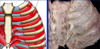
What are these?

Ribs 10, 11, 12
What is the difference b/w a simple vs a complicated rib fracture?
Simple: Break of the rib itself
Complicated: break of blood vessels, pleura, multiple rib fractures

What is this?

Typical Thoracic Vertebra
Label the diagram


Label the diagram


Label the diagram


Label the diagram


Describe how a rib articulates with the vertebral column:

Label the diagram


Label the diagram


During inspiration which direction does the thoracic cage move?
UPWARDS

During expiration, which way does the thoracic cage move?
Downward

Label the diagram

Synchondrosis
Identify the difference in location between a dislocation vs a separation