Week 3 - G - Neuropathology 2 - CNS demylination and dementia Flashcards
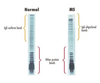
What do oligodendrocytes form to precipitate rapid slatatory conudction?
They form nodes of ranvier which greatly increase the speed of nerve impulse conduction
Do oligodendrocytes or schwann cells have a limited capacity to remyelinate after damage? Think about which is CNS and which is PNS
Oligodendrocytes have a limited capacity to remyelinate themselves after damage - this is the CNS Schwann cells have a greater remyelination capability - PNS
Demyelinating disorders can be classified as primary or secondary What is the main primary cause of demyelination? What is a primary cause that is rare and usually occurs in children, after a viral infection? symptoms can resemble the main primary cause
Main primary cause - Multiple sclerosis Acute disseminated encephalomyelitis - self limiting
Why does acute disseminated encaphlomyelitis occur?
Can typically occur after a minor infection such as a cold- the immune system becomes mis-programmed and activates immune cells to talk the healthy myelin covering the nerves
Acute disseminated encephalomyelitis is most commonly self limiting, what is the fatal form of ADEM where there is usually bleeding as it is a necrotizing vasculitis?
Acute haemorrhagic leukoencephalitis
Secondary demyelination can occur due to viral infection, such as JC virus This viral infection is seen as a side effect of certain MS treating drugs What can JC virus cause?
Progressive multifocal leukoencephalopathy (PML) is a rare (in the general population) but serious demyelinating disease of the brain, often resulting in severe disability or death, caused by lytic infection of oligodendrocytes by the JC polyomavirus (JCV).
When there is over-rapid therapeutic correction of hyponatraemia, triggering oligodendrocyte death… …demyelination can occur as a result. What is this ocndition known as? Is this primary or secondary demyelination?
Secondary demyelination due to rapid correction of hyponatraemia - known as central potine myelinosis
When is the peak incidence of multiple sclerosis and in which gender is it more common?
Peak incidence is 20-30years old, more common in females
What does mutiple sclerosis have a correlation with?
There is a well known correlation with latitude in that less sunlight means less vitamin D and therefore more northern countries have an increased rate of people with MS
Define multiple sclerosis
Autoimmune demyelinating disordr characterised by distinct episodes of neurological defecits, distinct in time and distinct neurological defects implicating two different sites of foci
What is needed for a diagnosis of MS?
Two distinct neurological deficits occurring at distinct times A neurological defecit implicating one neuro-anatomical site and an MRI scan appreciated defect at another neuro-anatomical site Mutliple dinstinct (usually white matter) lesions in the CNS
What can be seen on a lumbar puncture when checking for MS?
IgG Oligoclonal bands in the CSF
MS typically presents with the emergence of a focal neurological deficit What are the visual related neurological deficits that can occur in MS? - describe the sypmtom Is it usually unilateral or bilateral?
Unilateral (usually) optic neuritis - this is when there is pain on movmeent of the eye and a decrease in central vision (known as a central scotoma) INternuclear ophthalmopegia - usually bilateral in young patients with MS - if there is a lesion in the right medial longitdunial fasciculus (MLF) - the ipsilateral right eye will fail to adduct and the contralteral left eye will have nystagmus on abductio
There is a long list of clincial features of MS Name the optic nerve lesion problem? Spinal cord lesion problems? Brain stem lesions?
Optic nerve lesion - optic neuritis Spinal cord lesions - bladder dysfunction, spascticity, motor or sensory deficit in the limbs Brainstem lesiosn - ataxia, nystagmus, internuclear ophthalmoplegia
What are the different motor and snesory tracts in the spinal cord? Would a spinal cord lesion in these tracts cause contralateral or ipsilateral deficit What sensations do these tracts provide?
Dorsal column medial lemniscus tract - fine touch, vibration and proprioception - decussates in the medulla so ipsilateral if spinal cord lesion Spinothalamic tract - crude touch, pain and temperature - decussate in spinal cord (usualy about 2 levels above the dermatome level) - contralteral deficit if spinal cord lesion Corticospinal tract (pyramidal) - motor supply and decussate at medullary level so ipsilateral if spinal cord lesion
What are the different types of MS?
Relapsing remitting MS - 8out10 patients so most common Primary progressive MS - second most common Secondary progressive MS Progressive relapsing MS
Describe the different types of MS? Around half of the patinets with relapsing remitting MS will develop secondary progressive MS in how long? What is the most debilitating form of MS?
Relapsing remitting - around half of the patients with this will go on to develop secondary progressive MS within 15-20 years Progressive relapsing is the most debilitating form as patients will have a steady decline with superimposed MS attacks
What scan is used to aid in the diagnosis of MS? What is needed for diagnosis of MS remember?
MRI scan can be used Lesions disseminated in time and space - distinct focal neurological deficit implicating different neuroanatomical sites and distinct in time Can see the plaques on this T2 weighted scan
Is MS typically a white matter or grey matter disease and why?
Typically a white matter disease as this is where the axons from cell bodies (found in grey matter) are located hence the demyelination will occur where the myelin is
Are the shapes well demarcated or not?
he plaques are usually well demarcated
The plaques Can occur at any site in the CNS Lesions tend to be distributed in a non-symmetrical and non-anatomical manner. Where are the plaques often seen?
Often seen in Cranial nerve 2 - optic nerve Corpus callosum Cerebellum Brainstem (where the MLF - medial longitudinal fasciculus is found) Spinal cord lesions
In MS you can get acute (active) plaques and inactive (chronic) plaques Active plaques occur whilst demyleination is happening and inactive plaques are seen once the demylination has occured Describe what is seen histologically in the acute and inactive stage?
Acute plaques - ongoing demyleination, perivascular inflammation (12-24 hours you see red neurons, 24-48hours you see neutrophils, 48hours on theres is microglia) Inactive plaques - evidence of scarring and gliosis histologically is seen (after about a week of cell damage)
Environmental factors in multiple sclerosis Association with latitude Relationship with Vitamin D deficiency. Sunlight exposure What is the genetic factor haplotype linked to multiple sclerosis?
It is linked to the HLA DRB1 gene
Is multiple sclerosis multifactorial, genetic or environmental?
Multiple sclerosis is an autoimmune disease of multifactorial etiology The etiology of multiple sclerosis is linked to a variety of genetic and environmental factors.
Name two neurodegenerative diseases affecting the basal ganglia? Name a huge neurodgenerative broad category affecting the cerebral cortex?
Basal ganglia - Parkinson’s disease and Huntington’s disease Cerebral cortex - dememnita
Dementias are also characterised by progressive loss of neurons, typically affecting functionally related neuronal groups And often show symmetrical involvement of neurones, which contrasts with multiple sclerosis. Is dementia part of the normal ageing process?
Dementia is never part of the normal ageing process - it is always pathological
What are the four main primary types of dementia?
Alzheimer’s disease Lewy-body dementia Pick’s disease (fronto-temporal dementia) Huntington’s disease
What is the important type of secondary dementia?
This would be vascular dementia (multi-infarct dementia)
What is the most common type of dementia and what % of cases does it account for? What is the second and third most common causes of dmeentia?
Most common type - Alzheimer’s disease - 60-75% of cases Second most common - Vascular dementia Third most common - Lewy body dementia Fronto-temporal dementia is the fourth most common cause
Most common cause of dementia in elderly is Alzheimer’s disease When does the age of onset of Alzheimer’s usually occur?
Usually occurs after 60 years of age but early onset can be younger
Alzheimer’s disease has the potential to be familial What preotins have an asccoiated mutation in patients with Alzhiemer’s disease hence the familial aspect in a small number of cases ? What condition is there an increase in the incidence of AD?
Amyloid beta precursor protein - gene for this protein is found in chromsome 21 Or in the presenilin 1 (chromosome 14) and 2 proteins (chromosome 1) In Down’s syndrome, there is an increased incididence of AD, perhaps becuae there are now 3 chromosome 21 genes where the amyloid beta precusor protein is located and hence a higher chance of mutation
AD Becomes clinically apparent as an insidious impairment of higher intellectual function, with alterations in mood and behaviour. The onset can often be subtle, but once symptoms and signs are established, there is a steady progression What are some syptoms of more progressive AD?
Progressive disorientation Memory loss Aphasia (trouble with word finding)
While some atrophy does occur in ageing, the degree of atrophy in Alzheimer’s disease is far more marked… …underlying the fact that Alzheimer’s is a disease and not merely a more accelerated aspect of the ageing process What can happen macroscopically to the brain?
There is increased cortical atrophy resulting in the widening of the sulci in the brain and narrowing of the gyri
a term used to describe the increase in the volume of CSF, characterised on images as an enlargement of cerebral ventricles and subarachnoid spaces, caused by encephalic volume loss. What is this known as? It is seen when the ventricles enlarge in AD as well as other encephalic volume loss causing disease
This is known as hydrocephalus ex vacuo - compensatory enlargement of the CSF spaces often seen as a part of the ageing process but also seen pathologically
Which lobe of the brain is spared in Alzheimer’s disease?
Occipital lobe is spared
What are the microscopic pathological features seen in Alzheimer’s disease?
Loss of cortical neurones - Extensive neuronal loss with associated astrocyte proliferation - Simple neuronal atrophy - Gliosis Neurofibrillary tangles - Tau protein aggregates Neuritc plaques due to amyloid beta protein
Cerebral amyloid angiopathy also contributes to the amount of amyloid causing toxicity in the brain increasing the risk of Alzheimer’s disease How is amyloid angiopathy stained?
Use congo red staining and see a positive apple-green birefringence
What medication is given in the treatment of Alheimer’s disease?
Anticholineesterase inhibitors (AChE inhibitors) eg donepezil, rivastigmine, glantamine)
Acetylcholinesterase inhibitors are given in the treatment of mild to moderate dementia WHen the dementia progresses and the treatment stops working as well, what drug type can be tried?
NMDA receptor antagonists e.g. memantine used in moderate to severe In Alzheimer’s disease, excess glutamate can be released from damaged cells, leading to chronic overexposure to calcium, which can speed up cell damage. Memantine helps prevent this destructive chain of events by partially blocking the NMDA receptors that glutamate binds to (memantine is an anti-glutaminergic treatment). - NMDA-glutamate receptors allow for influx of CALCIUM, sodium and efflux of potassium
What is the drug treatment that can be given as a herbal remedy to treat Alzheimer’s dementia?
Ginkgo biloba - herbal remedy - interacts with warfarin
How does lewy body dementia typically present? How common is it?
Lewy body dementia is he third most common type of dementia - roughly 10% Presents with progressive dementia, along with hallucinations and fluctuating levels of attention/cognition
There is some overlap with Alzheimer’s and Lewy body dementia, which type typically affects memory later on? Fluctuation in severity of condition on a day-to-day basis What other disease features is there an association with in Lewy Body dementia that is important for diagnosis?
Lewy body dementia typically affects memory later in the course of the disease than Alzheimer’s disease There is the association with motor features of Parkinsonism, which are typically present at diagnosis, or emerge soon after
What are the clinically parkinsonism features of Lewy body dementia?
Loss of facial expression (hypomimia) Stooping and shuffling gait Rigidity - slow to intiate mvoement Bradykinesia Pill rolling resting tremor
All people with Lewy body dementia will go on to get parkinsonism What percentage of those with Parkinson’s disease will go on to get Lewy body dementia by 20 years? What causes Parkinson’s disease?
80% of patients after having Parkinson’s disease for at least 20 years will have gone on to have Lewy Body dementia Parkinson’s is caused by the degeneration of the dopaminergic neurons in the substantia nigra (pars compact)
What is seen macroscopically in Lewy body dementia?
Pallor in the substantia nigra where it usually black showing the absence of the dopaminergic neurons Substanita nigra is located in the midbrain
In a patient with Lewy body dementia, or Parkinson’s, what is the protein known as that forms the Lewy bodies and Lewy nitrites?
This is the alpha synuclein protein
What age does Huntington’s disease present in each gender? What is the mode of inheritance? What is the genetic mutation causing Huntingotn’s disease? Does Huntington’s disease cause a primary or secondary demenita?
Huntingtons disease presents in the 4th decade in women and 6th decade in men (30s and 50s) Autosomal dominant - cause by a triple nucleotide expansion , causing a CAG triplet repeat in the Huntingtin gene on chromsome 4, greater than 35 repeats causes the features of Huntingtin’s Huntingtin’s caused primary dementia
Clinical features: triad of emotional, cognitive and motor disturbance is seen in Huntington’s disease What are some of the symptoms?
Irritability, depression Chorea (dance like movements), slurred speech , apathy
What is usually the treatment options for Huntington’s disease? Which part of the basal ganglia is usually affected in Huntingotn’s disease?
Usually anti-depressants and exercise is proven to help in Huntington’s disease Typically the sriatum (caudate nucleus + putamen) (corpus striatum = caudate nucleus +putamen + globus pallidus)
Cortical atrophy occurs later in Huntington’s disease What is the compensatory mechanism seen in the lateral ventricles known as here?
Hydrocephalus ex vacuo
What is another name for fronto-temporal dementia? What age group does this dementia usually occur?
Pick’s disease Usually occurs between the ages of 50-60 years
Fronto-temporal dementia has the atrophy as seen in Alzheimer without the AD histology What are some of the symptoms of FTD ? What loves are spared from atrophy?
Pathologically there is extreme atrophy of the frontal and temporal lobes, with sparing of parietal and occipital lobes. Symptoms Characterised by progressive changes in behaviour, personality and social deterioration, leading to impairment of intellect, memory and language.
Is personality changes due to frontal or temporal lobe atrophy? Is language problems due to frontal or temporal lobe atrophy?
Personality hanges due to frontal atrophy Language problems due to temporal atrophy
What is seen histologically in Pick’s disease?
Pick’s proteins enriched with Tau potein are seen histologically Tau protein thereforre seen in both Fronto-temporal and Alzheimers dementia
Disorder involving a deterioration in mental functioning due to cumulative damage to the brain through hypoxia or anoxia (lack of oxygen) as a result of multiple blood clots within the blood vessels supplying the brain. What is this?
This is multi-infarct dementia (Vascular dementia)
Successive, multiple cerebral infarctions cause increasingly larger areas of cell death and damage. When a sufficient area of the brain is damaged, dementia results. What gender is it more common in? Where does this rank in the causes of dementia?
More common between men than in women And this ranks as the second most common form of dementia
Difficult to distinguish from Alzheimer’s disease, however there are clues to help What is the difference in disease onset and disease progression?
Alzheimers has an insidious gradual onset with a steadily progressing deterioration
Vascular dementia has an abrupt onset with a stepwise progression - reflecting the stepwise deterioration due to episodic vascular induced brain infarction
What is there likely to be a history of in a patient with vascular dementia?
History of hypertension, TIAs and/or stroke
Is it more common to have large vessel or small vessel infarcts in Multi-infarct dementia?
It is more common to have large vessel infarcts eg te cerebral arteries than small vessel eg lacunar (lenticulostriatal) artery infarcts in MID