Week 1 - C - Neuroanatomy 2 - Spinal Anatomy And Tracts Flashcards

1
Q

In the spinal cord there are how many paired nerves? What do the cervical and lumbar enlargement portions of the spinal cord supply?

A

31 paired spinal nerves Cervical supplies upper limb Lumbar supplies lower limb

2
Q

How is the anterior rami formed from the spinal cord?

A

The spinal cord gives off anterior rootlets that come together to make anterior roots The anterior and posterior roots then fuse together to form the spinal nerve at the intervertebral foramen which splits into the anterior and posterior rami

3
Q

What is contained into the dorsal root ganglion of the posterior root?

A

This contains cell bodies of sensory neurons

4
Q

The spinal cord terminates in a tapered cone-shape called the what? What vertebral level is this usually?

A

This is the conus medullaris L2

5
Q

What is the continuation of the spinal cord composed of pia mater that runs from the conus medullaris to the coccyx? What is the function of this cotninuation?

A

This is the filum terminale - this anchors the spinal cord

6
Q

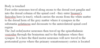
The spinal meninges are continuous with the cranial meninges via the foramen magnum. What are the three meningeal layers?

A

This would be the dura mater, arachnoid mater and pia mater

7
Q

Just as the filum terminale anchors the spinal cord, what is the ligament that attaches the lateral aspect of the spinal cord to the dura matter known as and helps anchor the spinal cord?

A

This is the denticulate ligament and is composed of pial and arachnoid tissue

8
Q

Is white or grey matter on the outside of the spinal cord? Which is H shaped?

A

The white matter is on the outside of the spinal cord with the grey matter in the H shaped inside

9
Q

A small central canal extends the length of the spinal cord. What does this open into rostrally?

A

This opens into the 4th ventricle

10
Q

The white matter is conventionally described as being made up of posterior, lateral (x2), and anterior funiculus, what is the smaller divisions of the funiculus known as? What is the grey matter of the spinal cord divided into?

A

Smaller divisions of funiculus are known as fasiculus It is divided into 4 horns left and right anterior and posterior horns hence the H shape

11
Q

How is it possible to tell apart the anterior and posterior horns if given an unlabelled spinal cord?

A

The posterior horns extend fully through the white matter of the spinal cord

12
Q

At spinal segments T1 to L2 there is also a smaller lateral horn What is contained in the lateral horns here?

A

The preganglionic sympathetic neurons for thoracolumbar outflow lie here

13
Q

The arterial blood supply to the spine contains 5 main arterial supplies What are they? and where are they derived from?

A

3 longitudinal arteries One anterior and 2 posterior that originate from the vertebral arteries Segemntal arteries derived from the vertebral, intercostal and lumbar arteries Radicular arteries derived from ventral and dorsal nerve roots

14
Q

What are the venous drainage veins of the spinal cord known as?

A

These are the longitudinal and sentimental veins

15
Q

What is the space between the dura mater and bone of the vertebra that can be used for anaesthesia? Why must you be careful when accessing this space?

A

This is the epidural space This is because in this space venous plexus’ exist and dont want to rupture one causing an epidural haemorrhage

16
Q

Where is the primary somatosensory cortex? Which love is it in?

A

It is located posterior to the central sulcus in the post-central gyrus It is in the postcentral gyrus of the parietal lobe

17
Q

Where is the primary motor cortex?

A

This is in he precentral gyrus located in the frontal lobe

18
Q

What is the major ascending spinal tract involved in fine touch and conscious proprioception? What is proprioception?

A

This would be the dorsal column/medial lemniscus spinal tract Proprioception is understanding ones movement in space

19
Q

Where do the fibres cross over in the dorsal column? What is crossing over also known as?

A

They cross over in the medulla It is known as decussation

20
Q

What is the word column in dorsal column also known as?

A

This is also known as the dorsal funiculus

21
Q

What does the dorsal funiculus contain?

A

Contains the fasciculus gracilis and fasciculus cuneatus

22
Q

Where do the axons for the fasciculus gracilis and the fasciculus cuneatus come from?

A

Fasciculus gracilis (graceful leg) - comes from the lower limb Fasciculus cuneatus - comes from the upper limb

23
Q

Does the fasciculus cuneatus or gracilis lie medial to one another?

A

The fasciculus gracilis lies medial to the fasciculus cuneatus

24
Q

In the dorsal column, explain the pathway if the axons came from eg the upper limb? (remember the 1st order, 2nd order and 3rd order neruones)

A

https://s3.amazonaws.com/classconnection/403/flashcards/11907403/png/picture1jpggifjpggif-15E344988D821EF1706.png

25
Q

Where do the synapses occur in the dorsal colum tract? Where does crossing over occur? What functions does the system help with?

A

Synapses occur in the cuneate/gracilis nucleus of the medulla Also in the thalamus Crossing over occcurs in the medulla where the second order neurones then follow the medial lemnsicus pathway Helps with fine touch, vibration&proprioception

26
Q

Now talked over the dorsal column/medial lemniscus pathway What is the other major sensory pathway?

A

This is the spinothalamic tract

27
Q

What sensations does the spinothalamic tract carry?

A

This tract carries deep pressure, pain (nociceptive) and temperature

28
Q

The dorsal column tract is clearly located in the dorsal column (funiculus) of the spinal cord Where does the spinothalamic tract lie?

A

The spinothalamic tract lies in the anterolateral region of the spinal cord

29
Q

What is another name for the spinothalamic tract?

A

The anteriolateral system

30
Q

Where do the first order neurones enter the spinothalamic tract?

A

They are carried along axons past the dorsal root ganglion and into a tract in the dorsal column called lissauer’s fasiculus

31
Q

What happens after the neurones reach lissauer’s fasciculus ? (say up to how they begin the spinothalamic tract from the anterolateral wall of the spinal cord)

A

The fibres travel in lissaeuers fasciculus from the white matter to the dorsal horn of the grey matter where they synapse in the substantial gelatinosa (usually a couple of vertebral levels above entry) and the second order neurones decussate to the anterolateral region of the spinal cord and travel up the spnothalamic tract in the axons

32
Q

Once the 2nd order neurones are in the spinothalamic vesiculus, hw do they end up in the postcentral gyrus?

A

They synapse again in the thalamus and the third order neurones travel to the primary somatosensory cortex in the postcentral gyrus of the parietal lobe

33
Q

Talk through from body touch to the postcentral gyrus for the spinothalamic tract?

A

https://s3.amazonaws.com/classconnection/403/flashcards/11907403/png/picture1jpggifjpggif-15E3473A5830E8DE02C.png

34
Q

The spinothalamic tract has the anterior and lateral spinothalamic tracts although they run alongside each other so are considered as one Which of the sensations do the anterior and lateral tracts carry? Where does decussation occur in the spinothalamci tract?

A
  • The anterior tractcarries pressure - The lateral tract carries temperature and pain - Synapses occur in - the substantia gelatinosa of the dorsal horn of they grey matter in the spinal cord, & in the thalamus -Decussation occurs in the spinal cord also instead of the medulla like the dorsal column tract
35
Q

What is the difference between upper and lower motor neurons?

A

Upper motor neurones originate in the motor region of the brain and travel down The lower motor neuron recieves information from the upper motor neurone (basically after the upper synaspes) and connects to the muscle/limb it supplies

36
Q

What is the main descending spinal cord tract?

A

This would be the corticospinal tract

37
Q

What is the motor function of the corticospinal tract?

A

It carries fine, precision movmeent and motor to limb muscles and axial muscles

38
Q

When the precentral gyrus (primary motor cortex) gives the signal for a movmeemnt, the neuron begins its travel Where does the neurone decussate for the lateral corticospinal tract?

A

The neuron travels past the thalmus and through the midbrain/pons to the medulla where the neurones decussaye to travel to the spinal cord

39
Q

What percentage of the neurones decussate in the medulla for the corticospinal tract? What out of the corticospinal tract motor functions will the lateral tract control?

A

85% decussate at the medulla and the lateral tract will give lower motor neurons to the limb muscles

40
Q

When the axial axons get to there desired levels by travelling down the naterior cortciospinal tract, where do they decussate?

A

They decussate to the anterior grey matter horn in the spinal cord at the appropriate level and supply the axial muscles

41
Q

Where do both the lateral and anterior corticospinal tracts synapse before giving lower motor nueornes to the muscles?

A

In the anterior horn of grey matter

42
Q

Why is the corticospinal tract sometimes reffered to as the pyramidal tract? What percentage of axons decussate at the medulla to become part of the lateral corticsopinal tract? Which muscles do the lateral and anterior tracts supply respectively? Where does the synapse occur?

A

This is because the tract forms visible ridges referred to as the ‘pyramids’ on the anterior surface of the medulla, so also called the pyramidal tract. 85% decussate at the medulla pyramids The lateral corticospinal tract supplies the limb muscles The anterior corticsopinal tract supplies the axial muscles The synapse occurs in the corticopsinal tract in the anterior horn of the grey matter in the spinal cord

43
Q

Which tract provides sensation for fine touch, vibration and proprioception? WHich tract provides motor control to the limbs and axial muscles? What tract provides sensation for deep pressure, temperature and pain?

A

This would be the dorsal column / medial lemnsicus tract The corticospinal tract - lateral corticospinal for limb muscles and anterior cortiocspinal for axial muscles Deep pressure, temp and pain would be the spinothalamic tract

44
Q

Would a lesion in the spinal cord cause contralteral or isilateral damage to the spinothalamic tract? WHat symptoms would be seen? Would a lesion in the spinal cord cause contralteral or isilateral damage to the DCML tract? What symptoms would be seen? Would a lesion in the spinal cord cause contralteral or isilateral damage to the corticospinal tract? If this is a hemisection of the spinal cord, what syndrome are these symptoms known as?

A

https://s3.amazonaws.com/classconnection/403/flashcards/11907403/png/picture1jpggifjpggif-15E388A626221A664D9.png

45
Q

What would happen if there was damage to the left corticospinal tract?

A

There would be hemiplegia of the right upper and lower limbs

46
Q

If there was a stroke or CVA occuring in the precentral gyrus, what tract would be affected?

A

This would affect the primary motor cortex casuing the affected tract to be the corticospinal tract

47
Q

Where are the primary motor and primary somatosensory cortex located? (lobe included)

A

Primary motor cortex - located in the precentral gyrus of the frontal lobe (just like anterior rootlets have motor axons in the spine) Primary somatosensory cortex - located in the postcentral gyrus of the parietal lobe (just like posterior rootlets have sensory axons in the spine) The precentral and postcentral gyrus - separated by central sulcus

48
Q

Regarding the homunculus, how would a lesion located in the logntiduinal fissure side of the primary somatosensory cortex differ from a lesion near the longitudinal fissure side of the primary motor cortex? (ie arms or legs)

A

It wouldnt differ, the legs would be affected If the lesions was further along the pre/post central gyrus then the arms would be affected

49
Q

There are a number of motor systems outside of the pyramidal tract and these are often referred to collectively as the “extrapyramidal system”. Which tract is thought to mediate reflex head and neck movement due to visual stimuli.? Where segemnt of the spinal cord does the axons input to mainly?

A

The tectopsinal tract is though to facilitate head and neck movements Its main input is to the cervical cord

50
Q

Where does the tectospinal tract begin and end?

A

The tectospinal tract begins in the tectum of the midbrain and ends in the anterior horns of mainly the cervical cords

51
Q

Reticular formation forms the central core of the brainstem. It has many nuclei and receives input from virtually all parts of the CNS. It has many functions, including the influencing of voluntary movement. Fibres originate in areas of the reticular formation in pons and medulla. In general is it fibres that originate in the pons that facilitate extensor movmements and inhibit flexor or the opposite?

A

In general it is fibres that originate in the reticular formation of the pons that facilitate extensor movements and inhibit flexor movements Fibres originating in the medulla inhibit extensor movements and facilitate flexor movements

52
Q

What is the descending motor tract that is ipsilateral?

A

This is the vestibulospinal tract - important for balance of the head when moving (vestibular - balance nerve)