The Thigh and Knee Flashcards

1
Q

Where is the lumbosarcal plexus located?

A

Abdomen and pelvis

2
Q

What does the lumbosacral plexus innervate?

A

The lower limb

3
Q

What spinal nerves form the lumbar plexus?

A

L1-L4

At each vertebral level, paired spinal nerves leave the spinal cord via the intervertebral foramina of the vertebral column. Each nerve then divides into anterior and posterior nerve fibres.

The lumbar plexus begins as the anterior fibres of the spinal nerves L1, L2, L3, and L4.

4
Q

What are the 6 major branches of the lumbar plexus?

A
  1. Iliohypogastric
  2. Ilioinguinal
  3. Genitofemoral
  4. Lateral cutaneous nerve of the thigh
  5. Obturator nerve
  6. Femoral nerve
5
Q

What spinal nerves form the lumbosacral trunk?

A

L4 and L5 - these then make connections with nerves from the sacral plexus. Together these form the lumbosacral plexus.

6
Q

What is the major nerve formed by the lumbosacral trunk?

A

Sciatic nerve

7
Q

What 2 parts does the sciatic nerve consist of?

A

Common fibular part and tibial part

8
Q

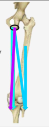
What are the roots of the obturator nerve?

A

L2, L3, L4

9
Q

What are the roots of the femoral nerve?

A

L2, L3, L4

10
Q

What are the motor functions of the obturator nerve?

A

Innervates the muscles of the medial thigh:

  • Obturator externus
  • Adductor longus
  • Adductor brevis
  • Adductor magnus
  • Gracilis
11
Q

What are the sensory functions of the obturator nerve?

A

Innervates the skin over the medial thigh.

12
Q

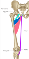
What are the motor functions of the femoral nerve?

A

Innervates the muscles of the anterior thigh – the illiopsoas, pectineus, sartorius and quadriceps femoris.

13
Q

What are the sensory functions of the femoral nerve?

A

Innervates the skin on the anterior thigh and the medial leg

14
Q

What forms the sacral plexus?

A

Formed by the anterior rami (divisions) of the sacral spinal nerves S1, S2, S3 and S4.

It also receives contributions from the lumbar spinal nerves L4 and L5 (lumbosarcal trunk)

15
Q

How does the sacral plexus begin? How does this then form the lumbosacral trunk?

A

The sacral plexus begins as the anterior fibres of the spinal nerves S1, S2, S3, and S4.

They are joined by the 4th and 5th lumbar roots, which combine to form the lumbosacral trunk –> this descends into the pelvis to meet the sacral roots as they emerge from the spinal cord.

16
Q

What are the 2 main destinations of the major nerves of the sacral plexus?

A
  1. Remain in pelvis to innervate structures here
  2. Leave the pelvis via the greater sciatic foramen
17
Q

What is the 1st major branch of the sacral plexus?

A

Superior gluteal nerve

18
Q

What are the spinal roots of the superior gluteal nerve?

A

L4, L5, S1

19
Q

Motor function of superior gluteal nerve?

A

Innervates the gluteus medius, gluteus minimus and tensor fascia lata

20
Q

Sensory function of the superior gluteal nerve?

A

None

21
Q

Route of the superior gluteal nerve?

A

The superior gluteal nerve leaves the pelvis via the greater sciatic foramen, entering the gluteal region superiorly to the piriformis muscle. It is accompanied by the superior gluteal artery and vein for much of its course.

22
Q

Route of the inferior gluteal nerve?

A

The inferior gluteal nerve leaves the pelvis via the greater sciatic foramen, entering the gluteal region inferiorly to the piriformis muscle.

It is accompanied by the inferior gluteal artery and vein for much of its course.

23
Q

Roots of the inferior gluteal nerve?

A

L5, S1, S2

24
Q

Motor and sensory function of the inferior gluteal nerve?

A

Motor Functions: Innervates gluteus maximus.

Sensory Functions: None.

25
Q

What are the roots of the sciatic nerve?

A

L4, L5, S1, S2, S3

26
Q

Motor functions of the sciatic nerve?

A

Tibial portion – Innervates the muscles in the posterior compartment of the thigh (apart from the short head of the biceps femoris), and the hamstring component of adductor magnus. Innervates all the muscles in the posterior compartment of the leg and sole of the foot.

Common fibular portion – Short head of biceps femoris, all muscles in the anterior and lateral compartments of the leg and extensor digitorum brevis.

27
Q

Sensory function of the sciatic nerve?

A

Tibial portion: supplies the skin of the posterolateral leg, lateral foot and the sole of the foot.

Common fibular portion: supplies the skin of the lateral leg and the dorsum of the foot.

28
Q

What is the bone of the thigh?

A

The femur

29
Q

What does the distal femur articulate with?

A

The proximal part of the large bone of the leg - the tibia - at the knee joint

30
Q

How does the femur lie? What is the purpose of this angulation?

A
  • The femur lies obliquely as the knee joint lies directly below the hip joint
  • This angulation of the femur returns the body weight to the midline
31
Q

Anterior view of right distal femur

A
32
Q

Posterior view of the distal femur. What are the 2 bony prominences highlighted?

A

Medial and lateral lips of the linea aspera (important for muscle attachment)

33
Q

Posterior view of right distal femur. What is highlighted in pink?

A

Medial and lateral supracondylar lines - sitting just proximal to the condyles

34
Q

What is highlighted in yellow (posterior view)?

A

Medial and lateral condyles of the femur

35
Q

What is highlighted here in black (posterior view)?

A

Intercondylar notch

36
Q

What is highlighted here in pink?

A

Adductor tubercle

37
Q

What is highlighted in blue (anterior view)?

A

Patellar surface - articulation with patella of knee cap

38
Q

Diagram of tibia and fibula

A
39
Q

Does the fibula participate in the knee joint?

A

No but is an important attachment site for one of the posterior thigh muscles

40
Q

Anterior view of right proximal tibia and fibula

A
41
Q

Posterior view of right proximal tibia and fibula

A
42
Q

How do the tibia and fibula lie in relation to each other?

A

Fibula lies laterally and tibia lies medially

43
Q

What is being pointed to?

A

Medial and lateral tibial condyles

44
Q

What is highlighted in pink?

A

tibial tuberosity

45
Q

What line is highlighted here?

A

Sharp anterior border of tibia - can be palpated anteriorly in leg

46
Q

What is highlighted here in blue?

A

Head, with the neck lying just underneath

47
Q

What is highlighted in yellow?

A

Tibial plateaus - flattened areas on proximal part of tibia that articulate with the femoral condyles

48
Q

What do the tibial plateaus articulate with?

A

The femoral condyles

49
Q

What is highlighted in blue?

A

Intercondylar tubercles

50
Q

What is highlighted in purple?

A

Soleal line

51
Q

What is circled in green?

A

Fibular head wih neck just underneath

52
Q

What is found between the fibula and tibia?

A

Strong interosseous membrane (has small holes in it to allow for passage of vessels through)

53
Q

What 3 bones are involved in the knee joint?

A
  • Distal femur
  • Proximal tibia
  • Patella
54
Q

What type of joint is the knee joint?

A

A synovial hinge joint

55
Q

What movements are allowed at the knee joint?

A

Flexion and extension

Very small degree of rotation

56
Q

What 3 articulations are found at the knee joint?

A
  • 2 femorotibial (medial and lateral sides)
  • Femoropatellar (between posterior aspect of patella and anterior aspect of distal femur)
57
Q

The joint surfaces in the knee are incongruent. What adds stability to the knee joint?

A
  • surrounding muscles
  • menisci (cartilage)
  • ligaments inside (intra-articular) and outside (extra-articular) the joint
58
Q

Label purple, yellow and blue - structures that aid stability

A

Purple - intra-articular ligaments

Yellow - extra-artciular ligaments

Purple - surrounding muscles

59
Q

What are the menisci?

A

C-shaped regions of fibrocartilage located on the medial and lateral aspects of the knee joint

60
Q

Function of the menisci?

A

Tibial plateau deepened by 2 plates of fibrocartilage – the menisci

Functions:

  • Increase joint congruency by creating a better fit for the femoral condyles
  • Distribute weight evenly across joint
  • Shock absorption
  • Assist in locking mechanism
61
Q

The external edges of the menisci are thicker. Why is this?

A

External edges attach to the fibrous capsule of the joint

62
Q

Menisci in cadaveric image

A
63
Q

Difference in stability of joint in flexion vs extension

A

Extension :

  • Most congruence
  • Most stable

Flexion:

  • Least congruence
  • Least stable
64
Q

What are the collateral ligaments?

A
  • Lateral/fibula collateral ligament
  • Medial/tibial collateral ligament
65
Q

What ligament is highlighted in purple?

A

Lateral/fibula collateral

66
Q

How does the fibular collateral:

  • Attach to the meniscus?
  • What is its shape?
  • What movement does it prevent?
A
  • Cord-like band
  • NOT attached to meniscus
  • Prevents adduction of the leg at the knee
67
Q

What ligament is highlighted in blue?

A

Tibial/medial collateral ligament

68
Q

How does the tibial collateral:

  • Attach to the meniscus?
  • What is its shape?
  • What movement does it prevent?
A
  • Flat band
  • Attached to meniscus
  • Prevents abduction of the leg at the knee
69
Q

What are the cruciate ligaments?

A

Found inside the knee joint –> intra-articular

70
Q

How many cruciate ligaments are there?

A

2 per knee:

  • Anterior (ACL)
  • Posterior (PCL)
71
Q

What is the function of the ACL?

A
  • Prevents anterior displacement of the tibia on the femur
  • Prevents hyperextension of the knee
72
Q

When is the ACL most often injured?

A

When the knee is hyperextended or force is applied anteriorly

73
Q

What is the function of the PCL?

A
  • Prevents posterior displacement of the tibia on the femur
  • Prevents hyperflexion
  • Main stabiliser of the flexed knee when weight bearing e.g. walking down hill
74
Q

When is the PCL most often injured?

A

Can be inured when landing on the tibial tuberosity with the knee flexed

75
Q

Posterior view of knee (with lots of tissues removed)

A
76
Q

What is highlighted in yellow?

A

Tibial plat

77
Q

What is highlighted in black?

A

Menisci (medial and lateral)

78
Q

What is highlighted in orange, green and blue?

A

Orange - Flat bandlike tibial/medial collateral ligament

Green - Posterior cruciate ligament (PCL)

Blue - Cordlike fibular/lateral collateral ligament

79
Q

Why are the tibial collateral ligament and the medial mediscus often injured together?

A

As they are connected

80
Q

What are the main stabilisers of the knee?

A

Muscles in the thigh that cross the knee

81
Q

What are the 3 compartments of muscles in the knee?

A
  1. Anterior
  2. Posterior
  3. Medial
82
Q

What are the muscles of the thigh separated by?

A

Fascial septae and enclosed by a fibrous sleeve called the fascia lata

83
Q

Where do the extensors of the knee lie?

A

In the anterior thigh (don’t get confused with the hip)

84
Q

Where do the flexors of the knee lie?

A

In the posterior thigh

85
Q

What is the action of the muscles in the medial compartment of the thigh?

A

Act upon the hip joint - adduction

86
Q

What are the muscles of the anterior thigh?

A
  • Iliopsoas
  • Quadriceps femoris
  • Sartorius
  • Tensor fascia lata
  • Pectineus
87
Q

What are the muscles of the anterior thigh innervated by?

A

Mostly by the femoral nerve

88
Q

What is the action of iliopsoas?

A

Flexes the hip –> is the major flexor of the hip

89
Q

Attachments of iliopsoas?

A

Iliacus: originates from the iliac fossa

Psoas major: originates from the lumbar vertebrae.

Both pass under the inguinal ligament, form a common tendon and insert onto the lesser trochanter

90
Q

Innervation of iliopsoas?

A

The psoas major is innervated by anterior rami of L1-3, while the iliacus is innervated by the femoral nerve.

91
Q

What is the action of pectineus?

A

Flexes and adducts the hip

92
Q

How does pectineus contribute to the femoral triangle?

A

The pectineus muscle is a flat muscle that forms the base of the femoral triangle

93
Q

Attachments of pectineus?

A

It originates from the pectineal line on the anterior surface of the pelvis, and attaches to the pectineal line on the posterior side of the femur, just inferior to the lesser trochanter.

94
Q

Innervation of pectineus?

A

Femoral nerve. May also receive a branch from the obturator nerve.

95
Q

What makes up the quadriceps femoris?

A

4 muscles, ‘the quads’:

  • Vastus Lateralis
  • Vastus Intermedius
  • Vastus Medialis
  • Rectus Femoris
96
Q

Actions of the quadriceps femoris?

A

Powerful extensors of the knee –> Extend the knee and stabilise the patella.

EXCEPT rectus femoris which is the only muscle of the quadriceps to cross both the hip and knee joints. It flexes the thigh at the hip joint, and extends at the knee joint.

97
Q

Innervation of quadriceps femoris?

A

All 4 innervated by the femoral nerve

98
Q

Attachments of rectus femoris?

A

Originates from the anterior inferior iliac spine and the area of the ilium immediately superior to the acetabulum. It runs straight down the leg and attaches to the patella via the quadriceps femoris tendon.

99
Q

What muscles is highlighted in pink?

A

Sartorius

100
Q

What are the attachments of sartorius?

A

ASIS –> medial tibia

Easily recognisable as is a bandlike thin muscle that takes an oblique course across the thigh

101
Q

Actions of sartorius?

A

Cross the knee and the hip:

At the hip joint, it is a flexor, abductor and lateral rotator. At the knee joint, it is also a flexor.

102
Q

Innervation of sartorius?

A

Femoral nerve

103
Q

What is highlighted here in blue?

A

Tensor fascia lata (is actually a gluteal muscle but can see when looking at the thigh anteriorly)

104
Q

What are the attachments of TFL?

A

Originates from the anterior iliac crest, attaching to the anterior superior iliac spine (ASIS). It inserts into the iliotibial tract (lateral side of thigh)

105
Q

Actions of TFL?

A

Flexes the hip and stabilises the knee

106
Q

What are the spinal roots of the femoral nerve?

A

L2-4

107
Q

How do all 4 muscles of Quadriceps Femoris converge?

A

Converge onto quadriceps tendon which runs over the patella and onto the patella tendon which itself attaches to the tibial tuberosity

108
Q

What is highlighted here in yellow? What are its attachments?

A

Vastus intermedius: originates from the anterior femur

109
Q

Name the highlighted muscles and their origins

A
  • Yellow –> Vastus intermedius, originates from the anterior tibia
  • Blue –> Vastus lateralis, originates from the greater trochanter and the linea aspera
    • N.B this is a posterior origin that then comes around anteriorly
  • Green –> Vastus medialis, originates from the intertrochanteric line and the linea aspera
  • Orange –> Rectus femoris, originates from the AIIS (so can contribute to hip flexion too)
110
Q

What are the boudaries of the femoral triangle?

A
  • Superior: inguinal ligament
  • Lateral: medial border of sartorius
  • Medial: medial border of adductor longus
111
Q

What forms the floor and the roof of the femoral triangle?

A
  • Floor: pectineus, iliopsoas and adductor longus muscles
  • Roof: fascia lata
112
Q

What branch of the femoral artery arises in the femoral triangle?

A

Profunda femoris

113
Q

What major superficial vein drains into the femoral vein in the femoral triangle?

A

Greater saphenous vein

114
Q

What are the structures in the femoral triangle from lateral to medial?

A

Femoral nerve, femoral artery, femoral vein

115
Q

What is being pointed at here? (femoral artery and vein have been removed)

A

Red: profunda femoris

Blue: point where great saphenous vein drains into femoral vein

116
Q

Where is the great saphenous vein formed?

A

The great saphenous vein is formed by the dorsal venous arch of the foot, and the dorsal vein of the great toe. It ascends up the medial side of the leg, passing anteriorly to the medial malleolus at the ankle, and posteriorly to the medial condyle at the knee.

As the vein moves up the leg, it receives tributaries from other small superficial veins. The great saphenous vein terminates by draining into the femoral vein immediately inferior to the inguinal ligament.

117
Q

The femoral nerve can be accessed in the femoral triangle. What would be purpose of this?

A
  • Anaesthetised with LA - ‘nerve block’
    • Femoral nerve innervates anterior thigh muscles, skin of anterior thigh and a branch called the saphenous nerve innervates the skin of the medial aspect of the leg and foot
118
Q

How do the medial thigh muscles act?

A

Adduct the hip joint

119
Q

What are the muscles of the medial thigh innervated by?

A

Obturator nerve (L2-4)

120
Q

What is the only muscle of the medial thigh that weakly flexes the knee too?

A

Gracilis

121
Q

What are the muscles of the medial thigh?

A

Adductor group:

  • Adductor longus
  • Adductor brevis
  • Adductor magnus

Others:

  • Obturator externus
  • Gracilis
  • (Pectineus)
    • N.B. this is generally considered an anterior thigh muscle that mainly flexes the hip but also contributes to adduction, innervated by the femoral nerve
122
Q

Attachments of adductor brevis?

A

Originates from the body of pubis and inferior pubic rami. It attaches to the linea aspera on the posterior surface of the femur, proximal to the adductor longus.

123
Q

Attachments of adductor longus?

A

Originates from the pubis, and expands into a fan shape, attaching broadly to the linea aspera of the femur

124
Q

Label

A

Green - pectineus

Blue - adductor brevis

Pink - adductor longus

Purple - gracilis

125
Q

Attachments of gracilis?

A

It originates from the inferior rami of the pubis, and the body of the pubis. Descending almost vertically down the leg, it attaches to the medial surface of the tibia, between the tendons of the sartorius (anteriorly) and the semitendinosus (posteriorly).

126
Q

What muscles of the medial thigh are more superficial? Which are deeper?

A

More superficial:

  • Adductor longus
  • Adductor brevis
  • Pectineus
  • Gracilis

Deeper:

  • Adductor magnus
  • Obturator externus
127
Q

What are the 2 parts of adductor magnus?

A
  • Adductor part
  • Hamstring part
128
Q

What are the attachments of the adductor part of adductor magnus?

A

Origin: inferior pubic ramus

Inserts: linea aspera of the femur and the gluteal tuberosity

129
Q

What is the innervation of the adductor part of adductor magnus?

A

Obturator nerve

130
Q

What are the attachments of the hamstring part of the adductor magnus?

A

Origin: ischial tuberosity

Inserts: adductor tubercle

131
Q

Innervation of the hamstring part of the adductor magnus?

A

Tibial nerve (adductor magnus has a dual innervation)

132
Q

Action of adductor magnus?

A

Both parts adduct the thigh. The adductor component also flexes the thigh, with the hamstring portion extending the thigh.

133
Q

What is the adductor hiatus?

A

Passage way through for femoral artery and vein into popliteal fossa –> can pass behind knee

134
Q

What are the attachments of the obturator externus?

A

Origin: margins of obturator foramen and obturator membrane

Inserts: trochanteric fossa

135
Q

Action of obturator externus?

A

Adduction and lateral rotation of the thigh.

136
Q

What are the spinal roots of the obturator nerve?

A

L2-L4

137
Q

What is the nerve of the medial thigh; muscles and skin?

A

Obturator nerve

138
Q

Where does the obturator nerve arise? How does it exit?

A

Arises in the pelvis and exits via the obturator foramen

139
Q

Inside the pelvis, where does the obturator nerve lie?

A

Location along the lateral wall of pelvis means it can be compressed by pelvic / ovarian masses

140
Q

If the obturator nerve is compressed in the pelvis, what is this associated with?

A

Signs and symptoms in the medial compartment of the leg

141
Q

What muscles are highlighted?

A

Purple - pectineus

Blue - adductor longus

Orange - gracilis

142
Q

What is highlighted?

A

Purple - pectineus (this has been removed)

Green - obturator externus

Blue - adductor brevis (adductor longus has been removed)

Pink - adductor magnus

Orange - gracilis

Black - obturator nerve

143
Q

What are the muscles of the posterior thigh known as?

A

the hamstrings

144
Q

What is the action of the hamstrings?

A

Extend the hip and flex the knee

145
Q

What are the hamstrings innervated by?

A

Tibial nerve of the sciatic nerve

146
Q

What are the 3 hamstring muscles?

A
  • Semimembranosus
  • Semitendinosus
  • Biceps femoris: long head

N.B. the hamstring part of adductor magnus can also extend hip but does not act on the knee

147
Q

Where do the hamstrings arise from?

A

Ischial tuberosity

148
Q

Attachments of semimembranosus?

A

Ischial tuberosity –> posterior aspect of medial tibial condyle

149
Q

Attachments of semitendinosus?

A

Ischial tuberosity –> medial aspect of superior tibia

150
Q

Attachments of biceps femoris long head and short head?

A

Long head: Ischial tuberosity –> fibular head

Short head: linea aspera –> fibular head

151
Q

Action of biceps femoris long head and short head?

A

Long head: flexes knee and extends hip

Short head: flexes knee but doesn’t extend hip

152
Q

Innervation of biceps femoris long head and short head?

A

Long head: innervated by the tibial part of the sciatic nerve

Short head: innervated by the common fibular part of the sciatic nerve.

153
Q

Label

A

Teal - semimembranosus

Purple - semitendinosus

Blue - biceps long head and short head

Pink - piriformis

Yellow - sciatic nerve

Yellow circle - popliteal fossa

154
Q

What vessels supply the muscles of the posterior compartment of the thigh?

A

Perforating branches from profunda femoris

155
Q

What is the popliteal fossa?

A

Diamond shaped fossa behind the knee

156
Q

What are the boundaries of the popliteal fossa?

A

Superomedially: Semimembranosus (teal), semitendinosus (purple)

Superolaterally: Biceps femoris (blue)

Inferior: 2 heads of gastrocnemius (calf muscle)

157
Q

How do the femoral artery and vein enter the popliteal fossa? What do they then become?

A

Enter via the adductor hiatus –> become the popliteal vessels (they are palpable here)

158
Q

What nerve runs through the popliteal fossa?

A

The tibial nerve - runs directly through and into the posterior aspect of the leg

159
Q

What are the 2 terminal branches of the sciatic nerve?

A

Tibial nerve and common fibular nerve

160
Q

Where does the sciatic nerve bifurcate?

A

Somehwere in posterior aspect of leg (around popliteal fossa)

161
Q

where does the common fibular nerve run?

A

Along lateral border of biceps femoris/popliteal fossa

162
Q

What are the motor functions of the tibial nerve?

A

the muscles of the posterior leg (calf muscles), and some of the intrinsic muscles of the foot

163
Q

What are the motor functions of the common fibular nerve?

A

the muscles of the anterior leg, lateral leg, and the remaining intrinsic foot muscles

164
Q

What muscle is responsible for rotation at the knee?

A

Popliteus

165
Q

How does rotation at the knee occur?

A
  • As joint approaches full extension, femur undergoes a few degrees of medial rotation on the tibia
    • This is called ‘locking’ the knee
  • Very stable position therefore thigh muscles can relax
  • The knee is ‘unlocked’ by the popliteus muscle which laterally rotates the femur (reverses medial rotation)
166
Q

Where is popliteus located?

A

The popliteus is located superiorly in the leg. It lies behind the knee joint, forming the base of the popliteal fossa.

167
Q

What artery supplies the thigh?

A
  • Femoral artery –> mainly the profunda femoris
    • Profunda femoris gives off perforating branches that travel through apertures in adductor magnus to supply the posterior thigh
168
Q

Describe the blood supply to the knee

A

Anastomoses around the knee formed by branches from the femoral, popliteal and posterior tibial arteries

169
Q

Normal knee xray AP

A

Pink - patella

Yellow - tibial plateaus

170
Q

Normal knee xray lateral

A
171
Q

How would you describe this injury? The patient is 20 years old – what does this tell you about the possible mechanism of injury?

A
  • Segmental fracture
  • Likely to be high level of trauma involved due to young age
  • Patient likely to have considerable blood loss or haemorrhage
  • Potential damage to nerves and vessels in the thigh
  • Other injures; pelvic? head? hip? lungs?
172
Q

This 67 year old was hit by a car. Can you see any abnormality here?

A
  • Lateral tibial condyle injury
173
Q

What’s happened here? More common in females – why?

A
  • Patellar disclocation
  • More common in females because women tend to have wider hips, causing the femurs to slant inward and join the knee at more of an angle
174
Q

What are the dangers of abnormal alignment at the knee joint?

A

Excessive wear and tear –> osteoarthritis

175
Q

What is genu varum?

A

Genu varum, is a varus deformity marked by (outward) bowing at the knee, which means that the lower leg is angled inward in relation to the thigh’s axis, giving the limb overall the appearance of an archer’s bow.

Tibia is adducted with respect to the femur

176
Q

What is genu valgum? How is the tibia in respect to the femur?

A

Genu valgum, commonly called “knock-knee”, is a condition in which the knees angle in and touch each other when the legs are straightened.

The tibia is abducted with respect to the femur

177
Q

How many bursae are found in the knee? What is their function?

A

4 - A bursa is synovial fluid filled sac, found between moving structures in a joint with the aim of reducing wear and tear on those structures.

178
Q

What are the 4 bursae of the knee?

A
  1. Prepatellar bursa
  2. Deep and superficial infrapatellar bursa
  3. Suprapatellar bursa
  4. Semimembranosus bursa
179
Q

What can cause bursitis in the knee? What are the 2 main types?

A
  • Prepatellar bursitis
    • Friction between the skin and the patella
      • Cause the prepatellar bursa to become inflamed, producing a swelling on the anterior side of the knee.
  • Suprapatellar bursitis
180
Q

Meniscal tears are another form of injury that can occur at the knee. What can these result from?

A

From injury or degenerative changes

181
Q

What are the dangers of meniscal tears?

A
  • Damage is associated with development of osteoarthritis
  • Displaced cartilage can become trapped during knee movements – pain or locking
182
Q

What is the treatment for meniscal tears?

A

Repair or resection

183
Q

What is the ‘unhappy triad’?

A

A combined knee injury affecting the:

  • Medial meniscus
  • Medial/tibial collateral ligament
  • ACL
184
Q

how does the ‘unhappy triad’ occur?

A

Excessive lateral twisting of the flexed knee or blow to lateral side of extended knee –> commonly seen in sports like football

185
Q

What causes all 3 aspects of the ‘unhappy triad’ to be affected?

A

As the medial collateral ligament is attached to the medial meniscus, damage to either can affect both structure’s functions. A lateral force to an extended knee, such as a rugby tackle, can rupture the medial collateral ligament, damaging the medial meniscus in the process. The ACL is also affected, which completes the ‘unhappy triad’.

186
Q

What is being pointed to? What is this an attachment site for?

A

AIIS - rectus femoris

187
Q

Which quadriceps muscle attaches to the greater trochanter?

A

Vastus lateralis

188
Q

What muscle attaches to the lesser trochanter?

A

Iliopsoas

189
Q

Where does vastus medialis take origin from?

A

The medial aspect of the intertrochanteric line

190
Q

Posterior aspect of proximal end of femur

A
191
Q

What bony ridge is being pointed to?

A

Gluteal tuberosity

192
Q

What ridge of bone is being pointed to? What muscle does it provide attachment for?

A

Pectineal line (just inferior to the lesser trochanter) - insertion point of pectineus

193
Q

What is being pointed to?

A

Medial and lateral lips of the linea aspera

194
Q

What is being pointed to?

A

Adductor tubercle - a tubercle on the lower extremity of the femur that provides attachment for part of adductor magnus

195
Q

What is being pointed to? What does this surface articulate with?

A

Surface that articulates with the patella

196
Q

What is being pointed to? (anterior view of tibia)

A

Condyle (medial and lateral)

197
Q

What is being pointed to?

A

Tibial plateaus

198
Q

What is being pointed to?

A

Intercondylar eminence - between the articular facets on the superior articular facet of the tibia, but nearer the posterior than the anterior aspect of the bone

199
Q

What is being pointed to? What is this an important attachment site for?

A

Tibial tuberosity - important attachment for quadriceps muscles

200
Q

What is being pointed to here? (posterior view of tibia). What is this an attachment site for?

A

Soleal line - an oblique line visible on the posterior surface of the tibia. Attachment site for soleus muscle (calf muscle)

201
Q

Labelled diagram of fibula

A
202
Q

Articulations of the knee joint in extension?

A
  • Posterior surface of patella articulates with the patella surface on the femur
  • Flattened tibial plateaus articulate with femoral condyles
203
Q

Why is the knee less stable in flexion?

A
  • Smaller area of femoral condyle is in contact with the tibia
    • Less stable
204
Q

What has happened here?

A

Fracture towards distal end of femur that has been left to heal on its own - bones now don’t align though as bone healing caused an anterior displacement of distal part and overlapping of bone fragments.

One leg likely to be shorter than other –> limp –> potential early arthritis

205
Q

What has happened here?

A

Fracture that been allowed to heal alone –> posterior displacement of distal end of femur

206
Q

What are the 3 main stages of bone healing?

A
  1. Inflammation
  2. Bone production
  3. Bone remodelling
207
Q

Describe the inflammation process during bone healing

A
  • When the bone is fractured there is bleeding into the area –> haematoma
  • This leads to inflammation and clotting of blood at the fracture site
  • This provides the initial structural stability and framework for producing new bone
208
Q

Describe the bone production phase in bone healing

A

Bone production begins when the clotted blood formed by inflammation is replaced with fibrous tissue and cartilage (known as “soft callus”). As healing progresses, the soft callus is replaced with hard bone (known as “hard callus”), which is visible on x-rays several weeks after the fracture.

209
Q

Describe the bone remodelling phase of bone healing

A

In remodeling, bone continues to form and becomes compact, returning to its original shape. In addition, blood circulation in the area improves. Once adequate bone healing has occurred, weightbearing (such as standing or walking) encourages bone remodeling.

210
Q

How can low levels of vitamin D affect your bones?

A

Vitamin D helps regulate the amount of calcium and phosphate in the body - helps your body effectively absorb these nutrients

211
Q

Where does the femoral nerve emerge?

A

Between iliacus and psoas major

212
Q

What muscle is being pointed to?

A

Tensor fascia lata - inserts onto the iliotibial tract

213
Q

What is being pointed to? Where does it insert? What is its function?

A

Iliotibial tract. Inserts on the medial aspect of the tibia and helps to stabilise the knee.

214
Q

What muscle of the anterior compartmen is attached to the ASIS? Where does this muscle insert?

A

Sartorius - attaches to the medial aspect of the tibia

215
Q

What muscle is being pointed to? What is its proximal attachment?

A

Rectus femoris - AIIS

216
Q

What is being pointed to?

A

Vastus lateralis

217
Q

Where do all the quadriceps muscles converge onto?

A

Onto the quadriceps tendon (common tendon)

218
Q

What vessels are being pointed to?

A

External iliac artery and vein

219
Q

What muscle is being pointed?

A

Iliopsoas

220
Q

What muscle is being pointed to?

A

Iliacus (in iliac fossa)

221
Q

What nerve is being pointed to?

A

Femoral nerve (between iliacus and psoas major)

222
Q

What muscle is being pointed to? What are its attachments?

A

Superior pubic ramus –> pectineal line (posterior femur just inferior to the lesser trochanter)

223
Q

What do iliopsoas and pectineus form?

A

The floor of the femoral triangle

224
Q

What is being pointed to?

A

Quadriceps tendon - this travels over the patella and then a patella tendon runs from the patella to the tibial tuberosity

225
Q

Where does the patellar tendon lie between?

A

Patella and tibial tuberosity

226
Q

What tendon is tapped in the knee jerk reaction?

A

Patellar tendon

227
Q

Where can the femoral nerve, artery and vein be accessed?

A

In the femoral triangle

228
Q

What is being pointed to?

A

Tensor fascia lata

229
Q

What is being pointed to?

A

Iliotibial tract

230
Q

What is the preferred site for a central line? Why might the femoral vein be used instead?

A
  • Jugular vein is preferred for a central line
  • If jugular vein is not able to be accessed (i.e. neck wound) then femoral vein can be used for temporary emergency access
231
Q

What vein is being pointed to? What does it drain?

A

Great saphenous vein - this is an important tributary of the femoral vein that drains the medial side of the foot and the medial aspect of the leg

232
Q

Where does the great saphenous vein drain into the femoral vein?

A

In the femoral triangle

233
Q

Where can the femoral pulse be palpated?

A

Just inferior to where the femoral artery crosses the inguinal ligament, it can be palpated to measure the femoral pulse. The femoral artery crosses exactly midway between the pubic symphysis and anterior superior iliac spine (known as the mid-inguinal point).

234
Q

When would the femoral nerve need to be blocked?

A

Nerve block to the leg –> LA

235
Q

What are the 3 boundaries

A
  • Inguinal ligament
  • Sartorius
  • Adductor longus
236
Q

What forms the floor of the femoral triangle?

A

Iliospsoas and pectineus

237
Q

What vessel is being pointed to?

A

Femoral artery

238
Q

What is the major arterial supply of the thigh?

A

Profunda femoris

239
Q

What is the only medial compartment muscle that crosses the knee?

A

Gracilis

240
Q

What muscle has the green string?

A

Adductor longus

241
Q

Where does adductor brevis lie?

A

Deep to adductor longus

242
Q

Where does adductor brevis insert? How does this compare to the insertion of adductor longus?

A

Adductor brevis inserts on upper 1/3 of linea aspera. Adductor longus inserts on middle 1/3 of linea aspera.

243
Q

What is the medial most muscle of the thigh?

A

Gracilis

244
Q

What are the actions of gracilis?

A

Flexes the knee and adduction of hip

245
Q

What muscle is being pointed to?

A

Adductor magnus

246
Q

What nerve is being pointed to?

A

Obturator nerve - passing through obturator canal

247
Q

What muscle is highlighted?

A

Obturator externus - inserts on the trochanteric fossa