Peripheral Lesions of the Upper Limb Flashcards
What does the level of a lesion dictate?
Dictates the extent of the motor and / or sensory deficits and the appearance of the affected limb –> All muscles downstream of the lesion that have not already been innervated will be affected.
Which muscles are innervated by the radial nerve?
All posterior compartment muscles:
- Triceps
- All forearm extensors (extensors of wrist, fingers, thumb, APL)
- No muscles in the hand
What muscles are innervated by the median nerve?
- Almost all anterior forearm flexor muscles (except flexor carpi ulnaris and 1/2 flexor digitorum profundus)
- Thenar eminence (NOT adductor pollicis)
What muscles are innervated by the ulnar nerve?
- Almost all intrinsic hand muscles
- Apart from lateral 2 lumbricals
- 1 and 1/2 of anterior forearm muscles:
- 1/2 flexor digitorum profundus
- Flexor carpi ulnaris
What spinal nerves contribute to the radial nerve?
C5-T1
What skin is innervated by the radial nerve?
Skin over parts of the posterior arm, forearm, ASB and dorsolateral hand
What nerve is affected in ‘wrist drop’? Is the lesion typically high or low?
Caused by a a high radial nerve lesion; injury to the nerve most commonly happens in the mid arm (around humeral shaft)

How does a high radial nerve lesion cause ‘wrist drop’? What muscles does it affect? What are the sensory disturbances?
- Paralysis of forearm extensors (posterior compartment)
- Unable to extend the wrist, fingers and thumb
- Sensory loss over the lateral dorsum
Why can the elbow still be weakly extended in ‘wrist drop’?
By this point the radial nerve has usually sent a branch to the long head of the tricep –> can still weakly extend the elbow
What is the effect of a low radial nerve lesion?
Sensory deficit only –> radial nerve doesn’t innervate any muscles of the hand

Why does a low radial nerve lesion cause sensory deficit only?
Injury to superficial radial branch close to the wrist gives sensory deficit only –> superficial branch is sensory only as contributes to the cutaneous innervation of the dorsal hand and fingers.
Describe the difference between the deep and superficial branch of the radial nerve
The radial nerve terminates by dividing into two branches: deep and superficial
Deep: (motor) – innervates the muscles in the posterior compartment of the forearm.
Superifical: (sensory) – contributes to the cutaneous innervation of the dorsal hand and fingers.
What spinal nerves does the median nerve carry fibres from?
C5-T1
What muscles does the median nerve innervate?
Arm: none
Forearm:
- Pronator teres, palmaris longus, flexor carpi radialis (superifical layer)
- Flexor digitorum superficialis (middle layer)
- Lateral half of flexor digitorum profundus, pronator quadratus, flexor pollicis longus (deep layer)
- Via anterior interosseous branch
Hand:
- Thenar eminence (via recurrent branch)
- Lateral 2 lumbricals (via palmar digital branch)
What is the sensory innervation of the median nerve?
Skin over lateral 1/2 of the palm of the hand
Where would a high median nerve lesion be? Mid? Low?
High: Cubital fossa (elbow) area
Mid: Mid forearm
Low: Wrist

What condition does a high median nerve lesion cause?
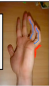
‘Bishop’s hand’ / ‘Hand of Benediction’ / ‘pointing finger’

Describe the fingers in ‘Bishop’s hand’ / ‘Hand of Benediction’?
- Unable to flex 2nd and 3rd digits
- Also unable to fully straighten 2nd and 3rd digits
- Can fully flex and extend 4th and 5th digits
- Thenar weakness
When does ‘Bishop’s hand’ / ‘Hand of Benediction’ become evident?
When the patient tries to make a fist
Why are the 2nd and 3rd digits unable to be flexed in a high median lesion?
FDP (and FDS) to these fingers is weak/paralysed (median nerve innervation)
Why are the 2nd and 3rd digits unable to be fully straightened in a high median lesion?
The lumbricals to these 2 fingers are paralysed/weak
Why can the 4th and 5th digits still be fully extended in a high median nerve lesion?
As FDP and lumbricals to these fingers are innervated by the ulnar nerve (lateral side)
Why is the middle finger variably affected in a high median nerve lesion? What condition does this lead to?
Middle finger is variably supplied by the ulnar portion of the FDP
If middle finger is predominantly supplied by the ulnar part of the FDP then it is just the index finger that cannot be flexed –> ‘pointing finger’
What type of lesion causes carpal tunnel syndrome?
Low median nerve lesion
What type of lesion causes ‘Simian hand’?
Low median nerve lesion
What characterises ‘Simian hand’?
- Thenar wasting
- Fine movements of 2nd and 3rd digitis impaired
- Sensory losses
- Inability to oppose thumb

What causes thenar wasting in a low median nerve lesion?
Recurrent branch of the median nerve that innervates the thenar eminence leaves the median nerve after it travels through the carpal tunnel
What causes impairment of the fine movements of the 2nd and 3rd digits in a low median nerve lesion?
Loss of lumbricals –> the median nerve innervates these
What spinal nerves does the ulnar nerve carry fibres from?
C8-T1
What muscles does the ulnar nerve supply in the arm, forearm, and hand?
Arm: none
Forearm:
- Flexor carpi ulnaris
- Medial half of FDP
Hand:
- Adductor pollicis
- Hypothenar eminence (ADM, FDM, ODM)
- Medial 2 lumbricals (to 4th and 5th digits)
- All interossei
What skin is innervated by the ulnar nerve?
Skin over medial ½ of the hand (palm and dorsum)
Unlike radial and median nerves, what do high and low lesions of the ulnar nerve both cause?
A deformity called claw hand - The claw hand looks slightly different depending on whether the nerve lesion is high or low
What does ‘claw’ hand refer to?
Combination of:
- Flexion at the IPJs of the 4th and 5th digits
- Hyperextension at the MCPJs of the 4th and 5th digits

How does the appearance of ‘claw’ hand differ between a high and low ulnar nerve lesion?
High lesion:
- Hyperextension at the MCPJs 4th and 5th but less flexion at the IPJs
- Claw doesn’t look as bad
Low lesion:
- Hyperextension at the MCPJs 4th and 5th but more flexion at the IPJs
- Claw looks worse
This is unsual as normally lower lesions are less severe
Is ‘claw hand’ evident present when the hand is at rest?
Yes - these deformities are fixed
Where is a high ulnar nerve lesion?
Elbow
Why are the 2nd and 3rd digits normal in a high ulnar nerve lesion?
As the median nerve is intact:
- FDP is functional and able to oppose the pull of ED
- Lumbricals functional and able to extend fingers and oppose FDP
What is the opposing muscle to FDP?
ED (with help from lumbricals)
Why is there only slight flexion at the IPJs at the 4th and 5th digits in a high ulnar nerve lesion?
-
Lumbricals and interossei to these fingers are innervated by the ulnar nerve
- Therefore cannot contribute to extension of these joints
- BUT –> FDP to these fingers is weak/paralysed (as medial half innervated by ulnar nerve)
- So cannot pull IPJs into signficant flexion
Why is there hyperextension at MCPJs of the 4th and 5th digits in a high ulnar nerve lesion?
- Medial half of FDP innervated by ulnar nerve
- Paralysed so unable to flex at MCPJ joint and oppose pull of ED
- Lumbricals lost = cannot flex at MCPJ and extend at IPJs
- Interossei lost = cannot flex at MCPJ and extend at IPJs
Unopposed pull of ED causes hyperextension at MCPJs
Describe the flexion at the IPJs between a low and high ulnar nerve lesion?
Low - significant flexion at IPJs/DIPJs
High - slight flexion at IPJs
Describe the hyperextension at the MCPJs of the 4th and 5th digitis between a high and a low ulnar nerve lesion?
High - significant
Low - significant
Describe the 2nd and 3rd digits in a high and a low ulnar nerve lesion?
High - normal
Low - normal
Why are the 2nd and 3rd digits normal in a low ulnar nerve lesion?
- Median nerve is still intact:
- Lateral half of FDP still functional and able to oppose the pull of ED
- Lumbricals are functional and able to extend fingers and oppose flexion
Why is there hyperextension at the MDPJs of the 4th and 5th digits in a low ulnar nerve lesion?
- Lumbricals lost –> cannot flex at MCPJ and extend at IPJs
- Interossei lost –> cannot flex at MCPJ and extend at IPJs
Unopposed pull of ED causes hyperextension at MCPJs
Where is a low lesion of the ulnar nerve found?
Wrist
Why is there significant flexion at the IPJs / DIPJs in a lower ulnar nerve lesion?
FDP to these 4th and 5th digits (medial side supplied by ulnar nerve) is STILL FUNCTIONAL as FDP has already been supplied by fibres by the time it reaches the wrist
- FDP still able to flex DIPJs
- Lumbricals and interossei lost –> cannot extend the fingers/oppose flexion by FDP
Why does the claw look worse in a lower ulnar nerve lesion?
- FDP to 4th & 5th preserved
- More flexion at the 4th and 5th
- Claw looks worse

In both upper and lower ulnar nerve lesions, the patient cannot straighten the 4th and 5th digits. Why?
As the lumbricals and interossei to the 4th and 5th digits have been taken out –> these are important in assisting extension at the IPJs
Describe the FDP in a higher ulnar nerve lesion
FDP to 4th & 5th weak / paralysed (cannot make a fist as cannot flex at the DIPJs): weakness / loss of flexion at the 4th and 5th = claw doesn’t look as bad (though more muscles are weak / paralysed)
Describe the FDP in a lower ulnar nerve lesion
FDP to 4th & 5th preserved causing greater degree of flexion at the 4th and 5th –> claw looks worse
Bishop’s hand (high median nerve injury) and claw hand (ulnar nerve injury) look similar. What are the important differences?
Bishop’s hand:
- Appears when patient tries to make a fist because they cannot flex the 2nd and 3rd digits
- No hyperextension the MCPJs
- Thenar weakness / wasting
Claw hand:
- •Patient can’t extend 4th and 5th digits
- Hyperextension the MCPJs
- Atrophy of interossei
