Leg and Ankle SDL Flashcards
How does the tibia articulate with the femur at the knee joint?
The medial and lateral tibial plateaus (condyles) articulate with the medial and lateral femoral condyles at the knee joint
What does the tibia articulate with distally at the ankle joint?
The talus
Where do the tibia and fibula articulate with each other?
The tibia and fibula articulate with each other at their proximal and distal ends
The tibia’s medial surface is extensive and superficial. What 2 clinical procedures is it therefore used for?
- Donor site for bone grafts
- Intraosseous (IO) access
When is IO access used?
In emergencies when the peripheral vasculature is shut down and the superficial veins cannot be cannulated
What type of joint is the IO membrane?
a form of fibrous joint which unites the tibia and fibula
What does the IO membrane separate?
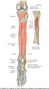
Separates the muscles of the anterior and posterior compartments of the leg (also acts as attachment site for muscles)
What separates the muscular compartments from each other in the leg?
The intermuscular septae

Do the intermuscular septae stretch?
No - are tough and inelastic
What is compartment syndrome?
When pressure builds in legs, intermuscular septae (formed by deep fascia) doesn’t stretch –> vessels and nerves are compressed
There is a foramen in the proximal part of the IO membrane – what passes through it?
Anterior tibial artery
What type of joint is the ankle?
Synovial hinge joint
What forms the ankle joint?
- The distal tibia, distal fibula and the talus
*

The ankle joint is very stable. Can you explain the anatomical basis for this stability?
- Good congruity
- Supporting ligaments
What movements are possible at the ankle?
- Flexion
- Extension
- Eversion
- Inversion
Describe flexion of the foot. What is it also called?
- Toes pointing towards floor
- Also called plantarflexion
- Performed by muscles of posterior compartment
Describe extension of the foot. What is it also called?
- Toes pointing upwards
- Also called dorsiflexion
- Performed by muscles of anterior compartment
In which position - flexion or extension – is the ankle is most stable? Explain your answer.
- In dorsiflexion / extension
- As the trochlear surface of the talus is wider anteriorly
- This forms a wedge that fits between the medial and lateral malleoli making dorsiflexion the most stable position for the ankle.
Where do inversion and eversion of the foot occur?
At the subtalar joint NOT the ankle joint
Is inversion or eversion of the ankle more common?
One of the most common injuries at the ankle is a forced inversion of the foot, which typically occurs when we fall over a kerb or step, or wobble over on the ankle when walking or running on uneven ground.
Which ligament complex is at risk in an inversion injury?
Lateral ligament complex
What can inversion injuries sometimes result in?
Fracture of lateral malleolus (distal fibula)
Which ligament is at risk from eversion injuries?
Medial ligament complex –> deltoid ligament
Why are ligament injuries typically quite slow to heal?
Ligaments are relatively avascular
Which blood vessels supply the ankle joint?
- Anterior tibial artery
- Posterior tibial artery
- Fibular (peroneal) artery
What are the muscles of the anterior compartment of the leg?
- Tibialis anterior
- Extensor digitorum longus (EDL)
- Extensor hallucis longus (EHL)
- (Fibularis/peroneus tertius)

As a group, what movements of the ankle and toes do the anterior compartment muscles allow?
- Extension / dorsiflexion of ankle
- Extension of toes (EHL and EDL)
Where does tibialis anterior originate and insert?
Origin: tibia, IOM membrane
Inserts: medial cuneiform and 1st metatarsal

Action of tibialis anterior?
Dorsiflexes ankle (and inversion)
Are EHL and EDL intrinsic or extrinsic muscles of the foot?
Extrinsic (arise in leg but tendons insert onto bones of foot)
Origin and insertion of EHL?
Origin: fibula and IO membrane
Insertion: distal phalanx of big toe
Action of EHL?
- Extends the big toe
- Dorsiflexes the ankle
Origin and insertion of EDL?
Origin: tibia and fibula
Insertion: distal phalanges of digits 2-5
Action of EDL?
- Dorsiflexes ankle
- Extends digits 2-5
Which nerve innervates all the anterior compartment muscles?
Deep fibular branch (of common fibular nerve)
What are shin splints?
Pain along the inside edge of the shinbone (tibia) due to inflammation of tissue in the area –> usually brought on by exercise
Which artery lies deep to EDL?
Anterior tibial artery
From which vessel does the anterior tibial artery arise? How does the anterior tibial artery gain access to the anterior compartment?
- Popliteal artery divides into anterior and posterior tibial arteries
- Anterior tibial artery passes through aperture in proximal IOM to reach anterior compartment
What does the anterior tibial artery continue in the foot as?
Travels over the anterior aspect of the ankle joint and into the foot as the dorsalis pedis artery

Which nerve accompanies the anterior tibial artery? From which larger nerve does it branch?
Deep fibular nerve - branch of the common fibular nerve
What condition can result if the common fibular nerve is injured at the neck of the fibula (e.g. due to fracture)?
Foot drop
What is ‘foot drop’, and why does it result from injury to the common fibular nerve? How do individuals with foot drop typically walk to compensate for this problem?
- Foot drop is a muscular weakness or paralysis that makes it difficult to lift the front part of your foot and toes.
- Damage to common fibular nerve –> damage to deep fibular branch
- Paralysis of extensors/dorsiflexors of the foot
- Individuals typically walk with a higher stepping gait to compensate
What are the muscles of the lateral compartment of the leg?
- Fibularis longus
- Fibularis brevis
What do the tendons of both fibularis longus and fibularis brevis pass posterior to to enter the foot?
The lateral malleolus

Origin of fibularis longus and brevis?
Fibula
Insertion of fibularis longus?
Travels under and into the sole of the foot –> attaches to:
- 1st metatarsal bone
- Medial cuneiform bone
Insertion of fibularis brevis?
5th metatarsal
What are the actions of fibularis longus and brevis?
Eversion of the ankle
Which nerve innervates the muscles of the lateral compartment of the leg?
Superficial branch (of the common fibular)
Which vessel provides the blood supply to the lateral compartment?
Fibular artery (branch of posterior tibial artery)
As it winds around the neck of the fibula, what two branches does the common fibular nerve divide into?
- Deep branch - innervates anterior compartment
- Superficial branch - innervates lateral compartment
How are the muscles of the posterior compartment of the leg arranged? What separates these layers?
- Into two layers, or groups: a superficial group and a deep group
- The transverse intermuscular septum separates these two groups from each other
What are the superficial muscles of the posterior leg?
- Gastrocnemius
- Soleus
- Plantaris
What is the most superficial muscle of the posterior leg?
Gastrocnemius

Describe the shape of soleus
Soleus is a large flat muscle that lies deep to gastrocnemius.

How do the tendons of the superficial posterior leg converge?
The tendons of these muscles converge onto the calcaneal (or ‘Achilles’) tendon, which inserts on to the calcaneum (heel bone).
Where does the calcaneal tendon insert?
On the calcaneum (heel bone)
What do the two large muscle bellies of gastrocnemius form?
The inferomedial and inferolateral boundaries of the popliteal fossa

Origin and insertion of gastrocnemius?
Origin: medial belly from the medial femoral condyle, lateral belly from the lateral femoral condyle
Insertion: common ‘achilles’ tendon which inserts on the calcaneum
Action of gastrocnemius?
- Plantarflexion/flexion of foot
- Inversion
origin and insertion of soleus?
- Origin: soleal line (posterior tibia)
- Inserts: achilles tendon
Action of soleus?
- Plantarflexion/flexion of foot
- Inversion
Apart from their actions on the ankle, what other important functions do these two large muscles have?
- Gastrocnemius is a powerful knee flexor (as crosses knee)
How is the calcaneal tendon (ankle jerk) reflex performed?
By holding the ankle in dorsiflexion and then tapping the Achilles tendon with a tendon hammer, just superior to its insertion point on the calcaneum –> usually more difficult to elicit than the knee jerk

What would you expect to see with a normal ankle jerk reflex?
Normal reflex is toe flexion (i.e. foot jerks towards its plantar surface)
Which peripheral nerve and which spinal nerves / cord segments are being tested?
- Peripheral nerve - posterior tibial nerve
- Cord segment - S1 being tested
The calcaneal (Achilles) tendon can rupture – either completely or partially. What is the usual mechanism of injury in calcaneal tendon rupture?
If the tension from a sudden, forceful contraction of the calf muscle becomes too great i.e. foreceful plantarflexion (often sport)
Signs and symptoms of Achilles tendon rupture?
- The feeling of having been kicked in the calf
- Pain, possibly severe, and swelling near the heel
- An inability to bend the foot downward or “push off” the injured leg when walking
- An inability to stand on the toes on the injured leg
- A popping or snapping sound when the injury occurs.
What are the deep muscles of the posterior leg?
- Popliteus
- Tibialis posterior
- Flexor hallucis longus (FHL)
- Flexor digitorum longus (FDL)

Action of popliteus?
It unlocks the extended knee by reversing medial rotation
Origin and insertion of tibialis posterior?
Origin: Tibia and fibula, IO membrane
Insertion: Navicular bone, medial cuneiform bone
Action of tibialis posterior?
- Plantarflexion of ankle
- Inversion
Origin and insertion of FHL?
Origin: fibula and IO membrane
Inserts: distal phalanx of big toe
Action of FHL?
- Plantarflexion of ankle
- Inversion of ankle
- Flexion of big toe
Origin and insertion of FDL?
Origin: tibia
Insertion: distal phalanges of digits 2-5
Actions of FDL?
- Flexion of digits 2-5
- Plantarflexion of ankle
- Inversion
What tendons pass posterior to the medial malleolus to enter the foot?
- Tibialis posterior
- FHL
- FDL
What is the order of tendons passing posterior to the medial malleolus?
From medial (closest to the medial malleolus) to lateral:
- Tibialis posterior
- is the most medial tendon, immediately behind the medial malleolus
- FDL
- FHL
Tom, Dick and Harry
Which artery and nerve also pass posterior to the medial malleolus? Which tendons do they travel between?
- Tibial nerve and posterior tibial artery
- Travel between tendons of FDL and FHL

Where can you palpate the posterior tibial pulse?
Behind the medial malleolus (not easy to palpate)

What vessels and nerves are at risk during fractures and dislocations of the ankle?
- Anterior and posterior tibial arteries
- Deep fibular and tibial nerves
What does the posterior tibial artery continue into the foot as?
Continues into the sole of the foot and gives rise to the plantar arteries (medial and lateral)
Describe the anterior and posterior tibial vein(s), and fibular vein(s)
Deep veins that accompany the arteries (and are usually paired, rather than single vessels)
Into which larger vein do the anterior and posterior tibial vein(s), and fibular vein(s) drain?
On the posterior surface of the knee, the anterior tibial, posterior tibial and fibular veins unite to form the popliteal vein
What are the important superficial veins found in the leg?
the great saphenous vein and the small saphenous vein.
What is the course of the great saphenous vein? Into which larger vein does it drain?
- It ascends up the medial side of the leg, passing anteriorly to the medial malleolus at the ankle, and posteriorly to the medial condyle at the kne
- Drains into the femoral vein in the femoral triangle
What is the course of the small saphenous vein? Into which larger vein does it drain?
- It moves up the posterior side of the leg, passing posteriorly to the lateral malleolus, along the lateral border of the calcaneal tendon
- Drains into the popliteal vein in the popliteal fossa
Identify the navicular, cuboid, medial, intermediate and lateral cuneiforms

The subtalar joint is an important inter-tarsal joint. What structures articulate at the subtalar joint? What movements occur here?
- Talus articulates with the calcaneum
- Eversion and inversion occur here
The calcaneum can be fractured, although it is a thick bone and considerable force is required. How are these fractures usually caused?
Calcaneal fractures are usually caused by a person falling from height and landing directly onto their feet (or foot).
Bunions and gout are two conditions that commonly affect the feet. WHat joint do they affect?
The first MTP joint
What is a bunion?
It’s a bony bump that forms on the joint where your big toe meets your foot – called the metatarsophalangeal (MTP) joint.
What is gout?
A type of arthritis - characterised by sudden, severe attacks of pain, swelling, redness and tenderness in the joints, often the joint at the base of the big toe.
How does gout occur?
- Urate crystals accumulate in your joint, causing the inflammation and intense pain of a gout attack.
- Urate crystals can form when you have high levels of uric acid in your blood.
- Your body produces uric acid when it breaks down purines — substances that are found naturally in your body.
- Sometimes either your body produces too much uric acid or your kidneys excrete too little uric acid –> urate crystals
How do the tendons of EDL and EHL travel? Where are they visible?
Travel over the dorsum of the foot to their insertions. These tendons, along with that of tibialis anterior, are usually visible under the skin of the foot.
