Posterior Thigh and Knee SDL Flashcards

1
Q

What are the bones of the leg?

A

Tibia and fibula

2
Q

Describe the location of the tibia

A

The tibia is much larger and lies medially

3
Q

Describe the location of the fibula

A

The fibula is a thin bone that lies laterally

4
Q

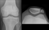
What forms the knee joint?

A

The proximal tibia articulates with the distal femur to form the knee joint - the fibula is not involved

5
Q

What 3 articulations is the knee joint composed of?

A
  • 2 femorotibial articulations (medial and lateral)
    • Medial femorotibial
    • Lateral femorotibial
  • A femoropatellar articulation between the distal femur and patella
6
Q

Bony landmarks of the anterior and posterior surfaces of the proximal tibia

A
7
Q

What important muscle group inserts on the tibial tuberosity and how do these muscles move the knee?

A

The quads –> these are powerful extensors of the knee

8
Q

What nerve winds around the neck of the fibula?

A

The common fibular nerve (also known as the common peroneal nerve). Here it is vulnerable to injury.

9
Q

Which nerve courses through the posterior thigh?

A

The sciatic nerve

10
Q

What is the sciatic nerve composed of?

A

2 separate nerves: the tibial nerve and the common fibular nerve which are bound together proximally but separate from each other in the posterior thigh and take different courses.

11
Q

What muscles are found in the posterior thigh?

A

The hamstrings: semitendinosus, semimembranosus and long head of biceps femoris

12
Q

Where do the hamstrings originate from?

A

The ischial tuberosity

13
Q

What is the common nerve supply of the hamstrings?

A

The tibial division of the sciatic nerve

14
Q

What are the actions of the hamstrings? What allows them to do this?

A

Because of their origin on the ischial tuberosity and their insertion on either the proximal tibia or fibula, they act in two ways: they extend the hip and flex the knee.

15
Q

Distal insertion of semimembranosus?

A

It attaches to the medial tibial condyle

16
Q

Distal insertion of semitendinosus?

A

the medial surface of the tibia.

17
Q

Distal insertion of the long head of biceps femoris?

A

Together, the long head and short head form a tendon, which inserts into the head of the fibula.

18
Q

Describe the long head of biceps femoris

A

The long head is a hamstring muscle, spanning the hip and knee joints and acting upon both.

19
Q

Describe the short head of biceps femoris

A

The short head is technically not considered a hamstring muscle, as its origin is from the posterior aspect of the femoral shaft, not the ischial tuberosity; for this reason it cannot extend the hip

20
Q

How does the short head of biceps femoris move the knee? What is its innervation?

A

Action: knee flexion

Innervation: common fibular part of sciatic nerve

21
Q

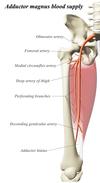
Where is adductor magnus found?

A

Adductor magnus is a large muscle of the medial compartment of the thigh.

22
Q

What are the 2 parts of adductor magnus?

A
  1. Adductor part
  2. Hamstring part
23
Q

Origin and insertion of adductor part of adductor magnus?

A

Origin: Inferior pubic ramus

Inserts: along the linea aspera

24
Q

Action of adductor part of adductor magnus?

A

Adducts the hip

25
Q

Which nerve innervates the adductor part of adductor magnus?

A

Obturator nerve (L2-4)

26
Q

Diagram of adductor magnus

A
27
Q

Where does the hamstring part of adductor magnus insert? What does this tell you about the action that this muscle has?

A

Inserts on the adductor tubercle on the femur –> can only act on the hip; extends the thigh

28
Q

Innervation of hamstring part of adductor magnus?

A

Tibial nerve

29
Q

Which vessel(s) supply the posterior compartment of the thigh?

A

Perforating branches of profunda femoris

30
Q

Where is the adductor hiatus located? What travels through it?

A

A hiatus (gap) between the adductor magnus muscle and the femur. This allows the passage of the femoral vessels from the anterior thigh to the posterior thigh and then the popliteal fossa.

31
Q

What is the popliteal fossa?

A

This is a diamond-shaped depression located behind the knee.

32
Q

What muscles form the boundaries of the popliteal fossa?

A
  • Semimembranosus
  • Semitendinosus
  • Biceps femoris
  • Gastrocnemius – medial and lateral heads (a muscle in the posterior leg)
33
Q

What structures form the floor and roof of the popliteal fossa?

A
  • Floor: posterior surface of the knee joint capsule, popliteus muscle and posterior femur
  • Roof: popliteal fascia and skin
34
Q

The popliteal artery is the direct continuation of which vessel?

A

The femoral artery

35
Q

What does the popliteal artery then divide into?

A

An anterior and a posterior tibial artery - which supply the leg and the foot

36
Q

What can atherosclerotic disease of the popliteal artery or injury to the popliteal artery lead to?

A

Can seriously compromise the perfusion of the leg and foot and may lead to irreversible ischaemia and loss of the foot +/ - the leg.

37
Q

What type of vein is the popliteal vein?

A

The popliteal vein is a deep vein which drains the leg and foot and travels alongside the popliteal artery.

38
Q

After the popliteal vein leaves the popliteal fossa, how does it enter the anterior thigh?

A

Via the adductor hiatus (alongside the popliteal artery)

39
Q

After leaving the popliteal fossa, what does the popliteal vein continue proximally as?

A

The femoral vein

40
Q

What can a thrombus (DVT) in the popliteal vein lead to?

A

A thrombus in the leg may travel ‘upstream’ to the lungs, causing hypoxia and infarction of the lung tissue. A large thrombus may occlude the pulmonary trunk leading to death.

41
Q

Which superficial vein drains into the popliteal vein in the popliteal fossa?

A

Small saphenous vein

42
Q

What type of joint is the knee?

A

A synovial hinge joint

43
Q

What movements are possible at the knee joint?

A

Flexion and extension

44
Q

The articular surfaces of the joint are incongruent. What does stability depend on?

A

largely depends on the surrounding soft tissue structures – cartilage, ligaments and muscles.

45
Q

What is the patella?

A
  • The patella is a triangular sesamoid bone that develops within the quadriceps tendon.
  • It has a base, an apex, and two surfaces – an anterior and an articular surface.
  • The articular surface has a medial and lateral facet.
46
Q

What does the patella articulate with?

A

The distal femur

47
Q

What does the medial facet of the patella articulate with?

A

articulates with the medial condyle of the femur.

48
Q

What does the lateral facet of the patella articulate with?

A

articulates with the lateral condyle of the femur.

49
Q

What are the 2 main functions of the patella?

A
  1. Leg extension – Enhances the leverage that the quadriceps tendon can exert on the femur, increasing the efficiency of the muscle.
  2. Protection – Protects the anterior aspect of the knee joint from physical trauma.
50
Q

What is a bipartite patella?

A

A congenital condition (present at birth) that occurs when the patella (kneecap) is made of two bones instead of a single bone. Normally, the two bones would fuse together as the child grows but in bipartite patella, they remain as two separate bones.

51
Q

Why is patella dislocation more common in females than males?

A

Females have wider pelves relative to the length of their femurs; in females the femurs lie at relatively more oblique angles than in males and thus a strong quadriceps contraction is more likely to displace the patella laterally in females than in males.

52
Q

What happens during a patella dislocation?

A

The patella bone is displaced out of the patellofemoral groove

53
Q

How do most patella dislocations occur?

A

Most occur laterally and are caused by high force impact on the patella or forceful sudden twisting of the knee (e.g. sports)

54
Q

Why do most patella dislocations occur laterally?

A

Because of the powerful superolateral pull of rectus femoris, vastus intermedius and vastus lateralis.

55
Q

What 2 mechanisms counteract this tendency towards lateral displacement?

A
  1. The vastus medialis, when it contracts, pulls the patella medially at a more horizontal angle (compared to the superolateral/vertical pull of the other three quads)
    1. This counteracts the lateral movement of the patella
  2. The lateral femoral condyle projects more anteriorly than the medial
    1. This limits the movements of the patella
      1. N.B. if this bony feature is underdeveloped, recurrent dislocation of the patella may occur.
56
Q

What specialised cartilages does the knee contain?

A

The menisci

57
Q

What intra-articular ligaments does the knee contain?

A

The anterior and posterior cruciate ligaments (ACL and PCL)

58
Q

What are the menisci and cruciate ligaments both vital for?

A

Stability at the knee

59
Q

Where are the menisci located?

A

They are C shaped and attached at both ends to the intercondylar area of the tibia. In addition to the intercondylar attachment, the medial meniscus is fixed to the tibial collateral ligament and the joint capsule.

60
Q

What is the function of the menisci?

A

They have 2 functions:

  1. To deepen the articular surface of the tibia, thus increasing stability of the joint.
  2. To act as shock absorbers by increasing surface area to further dissipate forces.
61
Q

Where are the tibial and fibular collateral ligaments located?

A
  • Tibial (medial) collateral ligament:
    • Wide and flat ligament
    • Found on the medial side of the joint
    • Proximally, it attaches to the medial epicondyle of the femur, distally it attaches to the medial condyle of the tibia.
  • Fibular (lateral) collateral ligament
  • Thinner and rounder than the tibial collateral
  • This attaches proximally to the lateral epicondyle of the femur, distally it attaches to a depression on the lateral surface of the fibular head.
62
Q

Function of the tibial and fibular collateral ligaments?

A

They act to stabilise the hinge motion of the knee, preventing excessive medial or lateral movement

63
Q

Why are the medial meniscus and the tibial collateral ligament commonly injured together?

A

As the medial meniscus is attached to the tibial collateral ligament

64
Q

Following an injury, why is the meniscus relatively slow to heal?

A

As relatively avascular structure with a limited peripheral blood supply

65
Q

What is the intercondylar area?

A

Lies between the medial and lateral tibial plateaus (not involved in articulation of the knee joint)

66
Q

What is the intercondylar area an important attachment site for?

A

ACL and PCL

67
Q

What does the patella tendon insert on?

A

The tibial tuberosity

68
Q

Attachments of the ACL?

A
  • Anterior intercondylar area on the tibia
  • Medial aspect of lateral femoral condyle
69
Q

Function of ACL?

A

Prevents anterior displacement of the tibia on the femur

70
Q

Attachments of the PCL?

A
  • Posterior intercondylar area on the tibia
  • Lateral aspect of the medial femoral condyle
71
Q

Function of the PCL?

A

It prevents posterior displacement of the tibia on the femur and stabilises the flexed knee when weight-bearing (i.e. when walking downstairs / downhill).

72
Q

Which ligament stabilises the flexed knee when weight-bearing (i.e. when walking downstairs / downhill)?

A

PCL

73
Q

Pathology of cruciate ligaments

A
74
Q

How can injuries to the cruciate ligaments affect the knee joint?

A

Injuries to the cruciate ligaments destabilise the knee joint

75
Q

How is the ACL typically injured? What types of forced movements are typically to blame?

A

Can be torn by hyperextension of the knee joint, or by the application of a large force to the back of the knee with the joint partly flexed.

Also involved in the ‘unhappy triad’ (medial meniscus, tibial collateral ligament, ACL)

76
Q

How is the PCL typically injured? What types of forced movements are typically to blame?

A

The most common mechanism of PCL damage is the ‘dashboard injury’. This occurs when the knee is flexed, and a large force is applied to the shins, pushing the tibia posteriorly. This is often seen in car accidents, where the knee hits the dashboard. The PCL can also be torn by hyperextension of the knee joint, or by damage to the upper part of the tibial tuberosity.

77
Q

What are the major contributors to the stability of the knee joint?

A

The tendons and muscles around the knee koint

78
Q

What other movement is possible at the knee joint, but only to a minor degree?

A

Rotation

79
Q

How does the knee undergo roatation?

A

The knee ‘locks’ and ‘unlocks’ - as the knee reaches full extension, there is a small degree of medial rotation of the distal femur on the tibia – this is ‘locking’.

80
Q

How does ‘locking’ of the knee affect its stability?

A

it increases the stability of the extended knee

81
Q

What is ‘unlocking’ of the knee? What muscle performs this?

A

When the knee is then flexed from full extension, the first thing that happens is ‘unlocking’ – a small degree of lateral rotation of the femur on the tibia; achieved by the contraction of popliteus.

82
Q

As the popliteal artery descends down the popliteal fossa, what branches does it give rise to? What do these supply? How do these join with branches from the femoral artery?

A

Several small genicular arteries –> these supply the knee joint

Branches also descend to the knee joint from the more proximal parts of the femoral artery (and its branches), thus creating an anastomotic network of vessels around the joint.

83
Q

Why are anastomoses around the knee joint clinically important?

A

The genicular anastomosis provides collateral circulation to supply the leg –> allows perfusion of knee if there is an occlusion in a vessel

84
Q
A