The Heart Flashcards
(29 cards)
The Mediastinum
Everything between the lungs
- Heart
- Blood vessels coming off or coming into to the heart
- Esophagus (sits behind the heart)
Covers with loose connective tissue
heart sits in 2nd and 6th rib

Serous Membrane
Simple squamous epithelium (mesothelium) on loose areolar connective tissue
-Line body cavities that are closed to the exterior
Produces a thin serous fluid - pericardial fluid (case of the heart), oily type substance - reduce fiction when heart is contracting and relaxing. between surfaces of the heart and serous membrane surround it
Folds on itself - parietal layer visceral layer
- directly against the heart (visceral percardium)
- Parietal percardium out piece against the chest wall
And in between and there’s a space filled with percardial fluid to help reduce friction when the heart’s contracting or expanding

Pericadium
- 3 Layers:
- Fibrous Pericardium (outermost) - dense connective tissue - anchor the rest of the percardium in the heart within the thorracic cavity - connects to hold the heart in place
- Parietal Pericardium
Pericardial cavity space where the percardial fluid in the serous fluid
- Visceral Pericardium (innermost layer)/ epicardium also known as the outer most layer of the heart
- Serous fluid located in pericardial cavity

Clinical Correlate: Pericarditis

Inflammation/roughening of the serous lining of the pericardial cavity
swelling between three layers and rubbing against eachother and friction
- Often associated with a viral infection or autoimmune disease, respiratory infections
- Retrosternal pain - pain behind the sternum
-Can be treated with anti inflammatory
• Severe cases: cardiac tamponade - pericadium cavity starts to fill with blood or inflammatory fluid eventually rupture the blood vessels and blood will come out. Pressure on the heart so the heart has to contract (squeezes) and limit how much the blood can fill in chambers and less blood to enough blood to the body (can be life threatening)
- treatment percadriocentesis - needle and draw out the blood
Layers of the Heart
- Epicardium/visceral percadium - innermost layer
- Myocardium - Muscle layer, thickest layer of heart when contracting occurs
- Endocardium - Endothelium, simple squamous epithelium and loose areolar connective tissue. Purpose is to line the cavities in the heart and have a smooth surface to reduce friction as blood is moving through
continous with innermost endothelim, the tunica intima of our blood vessels. It covers the values of the hearts
-Outermost layer of the heart will have fat interculated within there. Fat increases linearly no matter what you do you’ll have an increase in fat within your epicardium, however after 40 it seems the amount of fat is related to the amount of obesity we have increased adipose tissue within the epicardium
How is cardiac muscle different from skeletal muscle?
Skeletal muscle - multi nucleate, voluntary
Cardiac muscle - uni nucleate, short cylindrical, striatians due to actin and myosin, involuntary, intercalated discs
Cellular Organization of the Myocardium
- -intercalated discs - (look like egg cartons) two adhacent cells will interdigitate and link up. We also see fascia adherens simiar to desmosomes link those intermediate filaments (contrete pillars) very tough to pull apart
- Gap junctions so cells can communicate with eachother. important for ions move back and forth and critical for electrical signaling between the cells and beat in synchrony
- Plasma membrane are called the Sacrolemma t-tbules, z-discs
- Cardiomyocytes have more mitochondria than skeletal muscle fibers so the type 1 muscle fibers (slow oxidative muscles, never fatigue and low level of force, bc it has low mitochondria)
- Each cardio muscle fibers is wrapped in an endomysium with loose connective tissue
Surface Anatomy and Orientation
- 4 chambers in heart: Right Atrium, Right Ventricle, Left Artium, Left Ventricle
- Apex is point to the left hip
- We don’t see the atrias from the outside bc they’re covered by auricles (mean ear) Muscular pouches they will increase the capacity of the atria so more blood can flow into them - more blood flow into the ventricles to pump to the body. - Expansion pack for the atria (right/left auricle)
- (atria receiving blood from outside the heart)
Left ventricle is more posterior to the heart
(septum means wall)
- Interventricular septum - Interior muscular wall separating wall from the right and left artium
- Interatrial septum
- Anterior interventricular sulcus - indidation where the wall sits also one in the posterior side
- Coronary suclus - running around the heart running horizontally
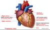

- Base of the heart is mostly made of left atrium
- Anterior surface (front of heart) coming out is the right ventricle
- heart sits right on top of the diaphragm
-Right atrium is over on the right pulmonary surface
(pulmonary - lungs)
right ventricle has a cresant shap and surrounds the left ventricle (if we’re poking the heart we’ll mostly touch the right ventricle)
left ventricle is thicker in terms of muscle; pumping blood to the body to put gravity to head and to the toes
right ventricle thinner bc it pumps blood to the lungs
Part of the left ventricle will sit on the diaphragmatic surface
Inditation - anterior interventricular sulcus -anterior side of the coronry arteries run within that groove
Inferior View

Chambers of the Heart
if we removed the fat we removed the epicardium, the fat intercalates within the epicardium.
Chambers of the Heart

Right Atrium - receives deoxygenated blood from the body from the atrium will allow that blood to go down to the right ventricle (thinner bc it’s pumping blood to lung) at lungs blood will become oxygenated. The oxygen we breath in will hop onto red bloodcells - oxygenated blood will return to heart into the left atrium and from there it will go into the left ventricle will pump the blood out to the entirety of the blood to make sure all the tissues have good oxygenation and nutrients

- Right atrium receives deoxygenated blood from (blue)
the superior (receives deoxygenated blood above the diaphragm) and inferior vena cavae (lower half of body, leg pelvis,lower abdominis), as well as the coronary sinus (heart needs it’s own blood supply)
- Right ventricle receives deoxygenated blood from the right atrium
- Right ventricle pumps deoxygenated blood into the pulmonary trunk which splits to left/right pulmonary arteries (lungs)
- Blood becomes oxygenated at the lungs.
What carries the oxygen in blood? red blood cells - protein hemoglobin
Carbon dioxide is also released at the lungs which is by product of cellular respiration of cellular metabolism when cells are breaking down sugars to produce energy, a byproduct is the production of carbon dixide and blow that off when we exhale. Carbon Dioxide is higher in deoxygenated blood
- Pulmonary veins bring oxygenated blood back to the left atrium
In general any blood vessel that carrying blood away from the heart is called an artery
Any blood vessel brings blood to the heart - vein
In general blood moving away from heart is oxygenated.
the only time we don’t see that are Pulmonarary arteries are carrying deoxygenated blood away from the heart to the lungs and plumonary veins are bringing oxygentated blood from the lungs back to the heart
- Oxygenated blood flows out of the left atrium and into the left ventricle.
- The left ventricle then pumps oxygenated blood into the aorta (all the blood supply will branch off of)
- Oxygenated blood circulates throughout the body
- Tissues use oxygen from blood, which then becomes deoxygenated.
- Deoxygenated blood enters into veins and ultimately into the superior and inferior vena cavae.
Continous cycling
Right Atrium
- Receives deoxygenated blood from superior vena cava, inferior vena cava, and coronary sinus there’s gonna be a little coronary orifice betwn valve that exits the right atrium and the inferior vena cava where that coronary sinus brings in the deoxygenated blood from the heart muscle itself.
- Covered by right auricle that allows for the expansion
- Posterior part of right atrium is smooth
- Anterior part rough appreance is lined with pectinate muscles which help assist w/ emptying blood from the right atrium (only find that in the two atria)
- Anterior and posterior parts separated by the crista terminalis (ridge/dividing line)
Right Ventricle
- Forms most of anterior surface of the hea rt
- Receives deoxygenated blood from the right atrium
- Special Features:
- Trabeculae carneae - flesh pillars, large pillars of muscle that keep the ventricle open and prevent collapse of the ventricle
- Papillary muscles - attached to the chordar tendanae (heart strings)
- Chordae Tendinae (heart strings) attached to the valves that separate the atria from the ventricles
Left atrium

Left atrium makes up most of the base of the heart (posterior surface)
- Makes up most of heart’s posterior surface/base
- Receives oxygenated blood from 2 left and 2 right pulmonary veins
- Contains some pectinate muscles in the auricle part which helps with emptying the left atrium during atrial contraction
Left Ventricle

Left Ventricle only difference is how thick the muscle is as it’s sending blood to the systemic circulation
- Most of the inferior surface of the heart
- Receives oxygenated blood from the left atrium
- Pumps blood into systemic circuit
- Special Features:
- Trabeculae carneae
- Pappillary muscles
- Chordae tendinae
Mini-Review: What are the structural and functional differences between the 4 chambers of the heart?
Right/left atrium - pectinate muscles within the auricles that help with emptying whereas the ventricles have the trabeculae carneae, which helps to prevent collapse. The ventricles are going to much thicker (left). The ventricles and blood is flowing from the atria into the ventricles. The right side of the heart is all deoxygenated blood, left side oxygentated
Right atrium receives blood from superior and inferior vena cavae, and coronary sinus.
The left receives from four surfaces the two left and two right pulmonary veins
Ventricles have papillar muscles attached to the chordae tendinae
Clinical Correlate: Heart Failure
Failure to keep up with body’s oxygen demands
- Often due to weakened ventricles
- Can affect left ventricle, right ventricle, or both
Due to myocardial infraction - heart attack if the heart attacks causes damage to the muscle of the left ventricle remember the muscle can’t replicate - long term heart failure
Also, Atherosclerosis narrowing of our arteries bc of inflammation and build of fats (obseity)
That’s why we get our blood pressure checked bc high blood pressure is a strong signal for long term heart failure.
What symptoms might you see in a patient?
- left ventricle pumps blood to the body so if it’s failing individual will have pain in distal extremidites
- if blood is receiving normal amount of blood - blood can back up into the pulmonary surface and have patient will have difficulty breathing
With right ventricle failure, the right ventricle has a hard time pumping to the lungs if there are problems have pulmonary embolism (blocking one of the blood vessels in the lungs) or chronic smoker then the right ventricle has to pump aginst the higher pressure and will fail overtime
See swelling in the feet both sides will be affected
Valves are Critical for Movement of Blood through the Heart

Valves separate each chamber of the heart and prevent blood from flowing backwards (one way valves)
- Atrioventricular valves = Tricuspid and Bicuspid (Mitral) valves seperate the atria from the ventricles Tricuspid (right side of the heart) Bicuspid (left side)
- Semilunar valves = Pulmonary (separating the right ventricle pulmonary trunk) and Aortic valves (separating the left ventricle with the aorta
Operations of the AV valve

Blood is being returned to the right atrium we have blood returned to left atrium. Constantly moving through all chambers of the heart
- As the blood comes in it’ll put pressure against the AV valves (the tricuspid and bicuspid valves) and it puts pressure on it the valves will open up and flow down to the ventricles as the ventricles fill right before the ventricles contract the atria contracts and squeeze out a little bit of additional blood.
Papillary muscles are attached to the heart string j. ust before the ventricles contract the paillary muscles will contract pull on the chordae tendinae which pull on the AV valves then
- the ventricles contract to force blood out
The purpose of the paillary muscles and the chordae tendineae is to hold the valves and prevent them from opening in the wrong direction
Opearations of the Semilunar Valve

As blood comes up Valves are forced to open due the pressure within the ventricles and when the ventricles relaxes as they are in diastole (relaxation) the intraventricular pressure will fall and as a result there isn’t as much force pushing the blood up up into the aorta and the pulmonary arteries so blood, due to the force of gavity will come back bc how the cusps are shaped they’re pushed open, the blood will catch in those cusps and then hold them closed.
Sytole when ventricles are contracting
Lub-Dub sound is due the closing of the valves of the heart. The “Lub” is due to the closing of AV valves and the “Dub” is due to closing of the semilunar valves
Fibrous “Skeleton” of the Heart
Dense connective tissue
- 4 Fibrous rings + membranous part of interventricular septum
- Anchors the valves of the heart - fibrous connective tissue and covered with endocardium
- Attachment sites for the myocardium
Myocardial Insertion into Cardiac Skeleton
Why is the myocardium oriented in a circle?
-If we are going to have efficient squeezing, we want the muscle orientated in kind of a circle so it’s like a drawstring bag that as you tighten it closed as the muscle shortens

