Skin Flashcards
Keratinocytes are of what embryological tissue origin? What are they?
Ectoderm
They are keratinizing epidermal cells (they produce keratin) that are the main cells in all 5 layers of the skin
What do keratinocytes produce?
- In spinosum - keratin
- Blue & in stratum granulosum
- Keratohyalin granules (filaggrin), which organize keratin into keratin-filaggrin protein complexes
- Lipid-rich lamellar bodies/granules
Keratohyalin granules turn the cell what color on an h&e stain?
Blue!
Recall: Hematoxylin is a basic dye that stains acidic structures (nucleus, keratohyalin granules, calcified material)
Formation of a water-protective layer of skin requires
- Cells undergo apoptosis and extrude their nucleus as well as their lipid-rich lamellar granules, creating the water-protective barrier
- The cells become stratified squamous (keratinized) epithelium: flattened bags of keratin-filaggrin complexes surrounded by a cell membrane
In stratum granulosum
Stratum spinosum holds the cells that
Are filled with keratin bundles called tonofilaments that converge into small, spiny cellular extensions and insert on desmosomes to strengthen cell-to-cell adhesion
Stratum granulosum cells make
Keratohyaline granules and Lamellar granules ; purplish-blue
Stratum corneum cells
have no organelles; they are cornified (filled with keratin, extruded organelles, and flattened)
Stratum basale
One-cell thick layer of cells bound to the basement membrane below by hemidesmosomes
Contains stem cells
Melanocytes are from neural crest cells. Where are they? How do you recognize them?
Sits on the basal layer alongside keratinocytes; have a pale cytoplasm

Melanin funciton
Protect keratinocytes from UV radiation
Langerhans cells - what is their function and where are they? Origin?
- What: Engulfs foreign antigens, goes to lymph nodes, and presents antigen to an active lymphocyte
- Where: Stratum spinosum
- Origin: monocytes in bone marrow
Merkel corpuscle
- Merkel cell + nerve ending
- Tactile discrimination
- In the stratum basale, but not seen in an H& E

Epidermolysis Bullosa is a dysfunction in what cell junciton?
Hemidesmosomes anchor basal cells to the basal lamina
Friction can cause the epidermis to separate and fluid builds up between the dermis & epidermis –> blister

Keratin tonofibrils made by stratum spinosum keratinocytes insert onto..
Desmosomes

Outer layer of the dermis
Papillary layer: sticks up with dermal papillae into the epidermal ridge
- Loose connective tissue, lots of collagen 3, blood vessels

Deeper layer of the dermis - what is it called? what type of tissue is it/describe its characteristics
Reticular layer
- Dense irregular connective tissue
- Fewer cells, more fibers such as collagen1 and elastic fibers

What are these wavy dark lines?
Elastic fibers of the reticular dermis

Hypodermis/subcutaneous tissue is made of what kind of connective tissue? What does it have a lot of?
Loose connective tissue that loosely binds skin to adjacent organs to allow for movement
Has a lot of blood vessels
Melanocyte produciton of melanin - what are the two steps and where does is occur?
- Tyrosinase oxidizes tyrosine to DOPA
- DOPA turns into melanin
Occurs in premelanosomes: membrane-bound structures derived from golgi
How do mealnocytes “donate” their melanin pigment to keratinocytes?
The premelanosomes mature into melanosomes, which eventually become colored pigments. They move to the tip of the melanocytes, where a keratinocyte will engulf it along with part of the cell membrane (cytocrine secretion)
Hair production is just like keratin production, it’s just a harder keratin.
Hair follicles are associated with what other two structures?
- Sebaceous gland: Produces sebum to lubricate the skin and hair; protect against infections
- Arrector pili: autonomically innervated smooth muscle; when cool, it causes the hair to stick straight up

Oblique and cross section of hair follicle - which part is continuous with teh surface of teh skin?
External root sheath is the only part continuous with the surface of the skin
Note the dermal papilla in the center giving the hair follicle nutrients

Nail matrix makes the keratin and then gets pushed out

Sebaceous gland
Holocrine secretion of sebum

Eccrine sweat glands
- All over your body; note how the duct opens onto the skin’s surface
- Innervated by cholinergic nerve endings
- Small secretory portions & ducts
- Can modify their products on their way to the surface of the skin (sebaceous & apocrine don’t)

How are apocrine sweat glands visibly different from eccrine sweat glands? Where are they? What do they produce? How are they innervated?
- Bigger lumen
- Only located in the skin of axillary and perineal regions
- Produces pheromones
- Innervated by adrenergic nerve endings

Meissner corpuscles - location, composition, and detection
Location: Dermal papilla & papillary layer of fingertips, palms, soles
Composition: modified Schwann cells with a nerve axon exiting at its base
Detection: light touch

Pacinian Corpuscles are where?
Look like onions in the hypodermis
Touch, pressure, vibration

Free nerve endings are unencapsulated. Can you see them in an H&E stain?
NO.

Ruffini corpuscles have ___ fibers sticking through it, so as collagen gets pulled it, it stimulates these to respond to stretch & torque.
Krause end bulbs can respond to low frequency vibration because they’re penetrated by a ___ fiber.
Ruffini, collagen fiber
Kraus, sensory fiber
What layer of skin is this?

Hypodermis


Reticular layer of dermis
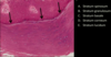
What is the region highlighted in yellow?

Stratum granulosum
What’s the function of this structure?

Pacinian corpuscle - to respond to vibration stimuli
What layer of the integument thas a decent amt of vasculature within the tissue (choose all that apply)?
- Epidermis
- Papillary layer of dermis
- Reticular layer of dermis
- Hypodermis
- Subcutaneous tissue
Everything but the epidermis.
Epidermis is avascular

The ducts of both eccrine and apocrine sweat glans use what kind of epithelial cells?
Stratified cuboidal .
(Note: myoepithelial cells are in the secretory part of only eccrine sweat glands)
Identify the region indicated by the arrows
Granulosum

Epidermal ridge
Ehlers’ danos syndrome (defect in collagen production) would affect which the most?
- Dermis
- Hypodermis
- Arteriovenous shunts
- Hair follicles
- Sweat glands
Dermis - connective tissue supporting epithelium; collagen, elastin, reticular fibers

Bulb of hair follicle
What gland is this?

Apocrine sweat gland, secretory portion; because it has a big lumen
Decrease in tyrosinase would do what to your skin?
Decrease skin pigment becasue tyrosinaes is crucial for melanin produciton
Which is most prevalent on the surface of a melanocyte?
- Gapjunctions
- hemidesmosomes
- tight junctions
- microvilli
- cilia
Hemidesmosomes holds it down
What sensation stimulate a ruffini corpuscle?
Stretch or torque
What skin layer would most likely be to the origin of skin cancer?
Stratum basale
What are the 3 components of the pilosebaceous unit?
- Hair follicle: downgrowths of epidermal epithelium
- Sebaceous glands: outgrowths of the external root sheath
- Arrector pili: originates in the subcutaneous layer and contracts to expel sebaceous gland secretions

What is this region?

Papillary dermis
Ichthyosis
Filaggrin normally binds keratin fibers to create an effective water barrier.
Ichthyosis: filaggrin mutations causes scaly skin and hyperkeratosis from compensatory repair mechanisms causing increased cell proliferation
Albinism
Genetic disorder that involves a defect in tyrosinase or in melanin production rate
Which part o fthe hair follicle will become cornified (hard keratin)?
Internal root sheath

What part of the hair follicle separates the connective tissue from the follicle?
Glassy membrane

Which part of the nail gives rise to the nail

The secretory part of the ECCRINE sweat gland vs the APOCRINE sweat gland is composed of what cell types?
Eccrine - clear, dark, myoepithelial cells
Apocrine- simple cuboidal or columnar cells
Which sweat gland is more involved in the physiological response to increased body temp and stress?
Eccrine sweat glands
Kraus end bulbs respond to what? Where are thye?
Low frequency vibration in skin of penis and clitorus

Describe the transformation of keratinocytes from the basal to the superficial layers, ultimatelyr esulting in keratinization
- Stratum basale: Keratinocyte is a basal cell
- Stratum spinosum: Keratinocyte starts making keratin and are are attached to each other by desmosomes (looks spiny)
- Stratum granulosum: Filaggrin is released from keratohyalin granules and lipid rich organelles are extruded to form a water barrier
- Stratum corneum: Lost nuclei and desmosome attachments; “keratinized”
Where in the body are melanocytes?
In the skin and the iris
Cutis laxa
Impaired elastic recoil becuase elastic fibers don’t form properly
hair follicles, hair, sweat glansd, sebaceous glands, nails, and mammary glands project from the dermis but
They are epidermal derivatives - they originate from ectoderm
The outermost papillary layer has
____ anchoring the intermediate filaments to the basal lamina
____ anchoring actin to the basal lamina
Hemidesmosomes anchor IF to the basal lamina
Focal adhesions anchor actin to the basal lamina
Panniculus carnasus
STriated muscle deep to the subcutaneous fascia
Subutaneous tissue contains ___ and ___ so drugs are absorbed rapidly
Loose connective tissue of fat cells and numerous blood vessels (drugs absorbed quickly)

Blood vessels that nourish teh skin come from two plexi where?
Between the hypodermis and dermis
Papillary layer vs deeper reticular layer of the dermis contains what kind of tissue?
- Papillary layer: Loost connective tissue (type 1 & 3 collagen)
- Blood vessels, nerve processes, some elastic fibers
- Reticular layer: Irregular dense connective tissue (type I collagen)
- More fibers, fewer, cells and ELASTIC FIBERS
