Hormonal Regulation of Testes Flashcards
(61 cards)
GnRH is a ___ hormone synthesized by neuroendocrine cells whos cell bodies reside in the ____ and ___ nuclei.
Released into the ____ in the median eminance to go bind gonadotrophs and stimulate synthesis & release of FSH & LH
GnRH is a peptide hormone synthesized by neuroendocrine cells whos cell bodies reside in the arcuate and preoptic nuclei.
Released into the primary capillary plexus in the median eminance to go bind gonadotrophs and stimulate synthesis & release of FSH & LH
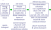
Describe testosterone regulation
Hypothalamus GnRH > Ant pituitary LH > Leydig cell testosterone
-
Testosterone neg feedbacks itself:
- inhibits hypothalamic GnRH
- inhibits anterior pituitary’s LH
- Estrogen from sertoli cells inhibits testosterone production from Leydig cells

LH activates ___ cells to secrete ___
FSH activates ___ cells to secrete ___
LH activates Leydig cells to secrete testosterone

FSH activates Sertoli cells to secrete estrogen, ABP, growth factors, and inhibin B to induce & regulate spermatogenesis
Inhibin B from ___ does what?
Inhibin B from Sertoli cells neg feedbacks anterior pituitary to inhibit FSH production

Activin from __ does what?
Activin from anterior pituitary antagonizes inhibin B to increase FSH synthesis

What hormone is responsible for the size of the testis?
FSH
Because it controls sertoli cell proliferation and seminiferous tubule growth
What hormone is most important in the initiation of spermatogenesis during puberty?
FSH because it controls Sertoli cell proliferation, seminiferous tubule growth, production of ABP, and development of blood-testis barrier
___ levels correlate with total sperm count and testicular volume, and can be used as an index of spermatogenseis
Inhibin B
Androgens are produced by the ___, then they diffuse intot he ___ where it’s converted to ___
Produced by leydig cells, diffuses across basement membrane into the seminiferous tubule where Sertoli cells turn it into estrogen

Describe how testosterone circulates
~50% is bound to albumin
~44% is tightly bound to sex hormone-binding globulin (SHBG) - not bioavailable
2-3% are free

When T is transferred to target tissues, what can happen to it?
What happens if it does not get fixed to tissues?
In the tissues, it can remain as T, or converted to DHT or estrogen
T that doesn’t get fixed to tissues is rapidly converted by the liver into inactive products for excretion in gut or urine
Testosterone is the pinrcipal circulating androgen and nearly all is produced by the testis.
Binds to its receptor, ____
androgen receptor
T, DHT, or weak androgens all bind to this receptor that regulates gene expression. PSA tumor markers (PSA is a gene) are really markers for androgen receptor activity
Functions of testosterone
High local levels of T in the testes -> spermatogenesis
Peripheral testosterone in th ebody -> masculine characteristics (either as T, DHT, or estrogen)
How do testosterone levels change from fetal life to childhood to puberty?
- Fetal: hCG from placenta stimulates testes to produce T
- Childhood: no T
- Puberty: Pulsatile GnRH secretion -> FSH & LH -> T

In puberty, plasma levels of FSH & LH increase primarily during ___.
___ and ___ are responsible for normal pubertal growth.
sleep -> GH & T, but this diurnal rhythm of FSH & LH is lost after puberty (~16-18yo)
growth hormone & testosterone are responsible for normal puberty growth
Aging & testosterone
- After age 40, T declines every year with increased SHBG (less bioavailable T)
- Decreased T:E
- Decreased LH pulse frq
- Decreased DHT in reproductive tissues
Hypergonadism - how does it present before or after puberty?
Before puberty - precocious puberty
After puberty - early hair loss
Causes of hypergonadism
- Hypothalamic, adrenal, or Leydig cell tumors
- LH receptor mutations
- Congenital adrenal hyperplasia
- Pinealoma - tumor that destroys pinealocytes so melatonin cant inhibit GnRH release
Melatonin regulation of GnRH
Melatonin inhibits the GNRH pulse generator (functioning since birth) throughout childhood
At puberty, body mass increases -> decreased [melatonin] -> GnRH pulse generator re-activated
Hypogonadism types
-
Primary hypogonadism - testes is messed up
- low T, high LH
- Also called hypergonadotropic
- Damage, cryptorchidism, gonadal dysgenesis, enzyme defects, LH receptor defects, etc
-
Secondary hypogonadism - hypothalamic/pituitary messed up
- low FSH, LH, and T
- Also called hypogonadotropic
- genetic defects; adrenal hypoplasia; mutations of GnRH receptor; etc
To differentiate between primary and secondary hypogonadism, you look at levels of
LH
- Primary* - low T / high LH / normal FSH
- Secondary* - low T, LH, and FSH
How can you differentiate between hypothalamic or pituitary dysfunction in secondary hypogonadism (hypogonadotropic)?
Give low-dose GnRH stimulation/priming, then a high dose GnRH injection
If the hypothalamus is the problem, you see increased LH
If pituitary is the problem, this has no effect on LH
Intratesticular vs excurrent ducts
- Intratesticular:
- straight tubules
- rete testis
- Excurrent:
- efferent ductules
- ductus epididymis
- vas deferens
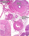
Straight tubules / tubuli recti
very short terminal section of the seminiferous tubules

Initial segments lined with sertoli cells
Near their termination, they narrow and become lined with simple cuboidal epithelium