Muscle Flashcards
All muscle tissues are involved in what two functions?
Movement
Heat production
All muscle tissue contractions depend on myofilaments, which are composed of
Thin filaments - actin
Thick filaments- myosin
Satellite cells
Unfused prescursor myoblasts/ stem cells that persist in muscle; They fuse with present muscle fibers to support growth with additional nuclei
Why do skeletal muscles have limited regeneraitive capacity?
Skeletal muscle cells DO NOT undergo mitosis; they have satellite cells with limited capability to divide & repair
Organization of skeletal muscle
Fascicle > Muscle fibers > Myofibrils > Myofilaments > Thin & Thick filaments
Muscle fibers (aka muscle cells) are bound by
Connective tissue (epimysium, perimysium, endomysium)- not cell junctions

Fascicle
Group of muscle cells held together by perimysium

Visible characteristics of skeletal muscle
- Polygon shaped cross-section; Rectangular longitudinal section
- Peripheral nuclei
- Striated

Epimysium- what is its composition and function?
Dense irregular connective tissue that ensheaths the entire muscle and carries vascular and nerve supply

Endomysium wraps around
Each individual muscle fiber/ cell
Thick filaments
-
Myosin II: 2 heavychains, 4 light chains
- Light chains contain actin, ATP binding sites, ATPase, and motor activity
- Lined up tail-to-tail to form bipolar thick filaments

What’s the black stuff?

Mitochondria
Thin filaments- F-actin, tropomyosin, troponin
Tropomyosin masks myosin-binding sites on F-actin
Ca+ binds torponin C to pull off tropomyosin, exposing the myosin-binding sites for the thick filament
A sarcomere (contractil eunit of skeletal muscle) is either
Distance between 2 adjacent Z lines
I band + A band
A band composition
Full length of thick filaments with some overlap from thin filaments

I band composition
Only thin filaments
H band composition
Only thick filaments; appears lighter because it lacks thin filaments

M line function
Holds thick filaments in place and links them to one another
What happens in muscle contraction
Sarcomere shortens, so
- Z lines brought closer
- I band shortens
- H band shortens
- A stays teh same*
Sliding filament theory
- ATP-myosin hydrolyzes -> binds actin
- Pi is released -> conformational change (cock)
- ADP is released -> power stroke
- New ATP binds -> release actin
Transverse tubules
Invaginations of the plasma membrane at A-I band junctions that propagate impulses down into all levels of the muscle cell
Serves as voltage-sensor proteins

Sarcoplasmic reticulum
Terminal cisternae; stores Ca2+ via gated Ca2+ release channels
Two of these and 1 T-tubule form a triad together

Dystrophin
Links actin to endomysium through dystrophin-associated glycoprotein complex
Ensures that shortening of myofibers is transmitted to surrounding connective tissue, resulting in muscle contraction
Rigor mortis
Lack of ATP –> can’t release actin, so there’s no detachment and muscles stiffen
Motor unit of skeletal muscle
Motor neuron (from ventral horn of spinal cord) + all the muscle fibers it innervates
Less muscle fibers, finer movement
Neuromuscular junction (motor-end plate)
Where synapse occurs between motor nerve and muscle fiber; point of contact between axon & muscle fiber

Initiation of contraction
- Action potential arrives
- Synaptic transmission at neuromuscular junction
- Propagation of AP along sarcolemma
- Hyperpolarization of T-tubules
- Conformational change of voltage-sensor proteins
- Gated Ca++-release channels open
- Ca++ released into sarcoplasm from SR
- Troponin binds Ca++
- Myosin-actin interaction
Myasthenia gravis
Autoimmune disorder attacking Ach receptors on post-synaptic sarcolemma. (The body tries to fix this by digesting affected receptors, but replaces them with less responsive receptors.)
–> ptosis
Fiber types of skeletal muscle
- Type I : slow oxidative; fatigue resistance; red
- Type IIa: fast oxidative; glycolytic; intermediate
- Type IIb: fast glycolytic; fatigue-prone; white

Sprinting is associated with what type of fiber?
Type IIb
Fast glycolytic; fatigue prone
Contrasting red (type I) vs white (type II) fibers- Vascularity, size, SR mitochondria, myoglobin, etc
Type I is smaller and has less SR, but is otherwise more rich in everything else

Myotendinous junction
Where the connective tissues surrounding skeletal muscle fibers become continuous with the dense collagenous (Type I collagen) tissue of tendon

Sensory Receptors vs Golgi Tendon Organs
Sensory receptors penetrate the muscle spindle itself to detect stretch/tension of extrafusal muscle fibers and relay it to the CNS –> maintain posture & regulate opposing muscle groups
GTO: Encloses sensory axons penetrating among the collagen bundles in tendons at the myotendinous junction to send info about stretch and tension to the CNS

What is the cursor pointing at?
Muscle spindles are located in the connective tissue island between muscle fascicles

Visible characteristics of cardiac muscle
- Branching
- Intercalated discs
- Central nuclei

Cardiac muscle is held together by
Gap junctions
demosomes: intermediate filaments for cell adhesion
Adhering junctions to actin filaments of sarcomeres

How to differentiate between skeletal and cardiac muscle on a cross section?
Cardiac = centrall located nuclei

Contracitle apparatus of smooth muscle
-
Myosin filaments (side-polar) and actin filaments
- But no troponin, no specific patterns (like sarcomeres)
-
Dense bodies- bind to intermediate filaments (vimentin, desmin), actin, and the membrane
- Similar to Z-discs
-
Caveolae: dyad
- Similar to T-tubule system
- Gap junctions
Actin anchors to dense bodies, which also attach IF to the sarcolemma

Regeneration of smooth muscle
High proliferative capacity (hyperplasia)
Arises from vascular pericytes
Side-polar thick filament of smooth muscle

3 ways to initiate smooth muscle contraction
Mechanical (stretching)
Depolarization (neuronal)
Chemical (vasopressin, angiotensin II , etc)
Visible characteristics of smoth muscle
- longitudinal
- fusiform, small
- no striations
- corkscrew-shape dnucleus when contracted
- central nucleus
- cross-section
- central nucleus with little cytoplasm
- variable nuclear profile
Regulation of smooth muscle contraction by calcium
Note: no troponin involved

How to differentiate between smooth muscle and dense irregular connective tissue?
Smooth muscle has more cells & nuclei; whereas, in dense irregular connective tissue has more collagen, less cells

White matter vs gray matter of spinal cord
White matter of spinal cord - no neurons
Gray matter of spinal cord - neurons
Muscle spindles
Encapsulated proprioreceptors that help control body posture and coordinate the action of opposing muscles; consists of
- Connective tissue capsule
- Intrafusal muscle fibers
- Sensory nerve fibers penetrating those fibers
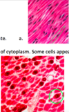
What kind of muscle is this

Nuclei are peripheral -> skeletal
