palpable masses Flashcards
Why is knowledge of embryological routes useful in mass determination?
Give an example of a mass that is embryological in origin?
Give another condition that is related to this embryology
- Knowledge of the route of travel of an embryological tissue can help explain certain masses in which the movement to the adult position has not occured.
-
Example: Lingual thyroid
- Thyroid gland starts at foramen caecum, a hole at the midline of sulcus terminalis of the tongue
- It should descend down into the neck
- Lingual thyroid is a normal thyroid gland which has not descended
- Thyroglossal cysts are cysts that form in the thryoglossal duct which is the embryological route into the neck
- Normally thyroid gland elevates with swallowing, not always with tongue protrusion
- Thyroglossal cysts form anywhere in the midline and will move with swallowing and tongue protrusion.

What relevant history questions should be asked in a patient presenting with a lump?
- Duration –> long development likely to be cancer, acute likely infectious
- exacerbating/ alleviating factors –> come and go (hernia) or remain?
- Pain in swelling location? Pain anywhere else?
- History of trauma
- Previous treatment/ intervention/ surgery?
- site dependent questions
- neurological disturbance or distribution
- Temporally associated systemic symptoms
What other factors may be taken into account when forming a diagnosis of a mass?
- Age (cancer)
- sex (sex- specific problems e.g. herniation types)
- social history (activity and herniation)
- occupation (activity and herniation, exposure to carcinogens etc.)
- medical history
How would you examine a lump?
what would you look for?
WIPE (Wash/ introduce/ patient details/ explain)
–> informed consent
–> explain what will happen, why you are checking certain things and what they can expect
1st –> inspect and observe mass (colour, size, evenness, hair etc).
2nd –> palpate (hard or fluid filled?)
3rd –> Percuss
4th –> auscultate (bowel sounds or bruits)
explain findings to patient
thank and end consulation
What are we looking for when examining lumps?
SPACE PIT
S - Size, shape, surface
P - position
A - attachments/ auscultate (on ligament/ tendon?)
C - colour, consistency
E - edge (even or not?)
P - pulsation/ thrills/ fluctuation (i.e fluid filled is it movable/ compressible?)
Inflammation
Transillumination
What are the methods for determining the origin/ content of a lump?
- Transilluminate –> fluid filled?
- Fluctuance –> Fluid containing –> pressing fingers into one region, does it rise in another region? (I.e pushing fluid into another region).
- Auscultation –> bruits? bowel sounds?
How would a ring shaped mass be described?
How would a curved mass be described?
- Ring shaped described as Annular
- Curved mass described as arcuate
What is a nodule or papule?
- Nodule / papule =palpable mass of specific size
what is a macule?
what is a pustule?
- Macule is a flat region of surface colour change
- pustule = small pocket of puss
How do you determine which layer a lump is in?
- The layer a lump is in and any attachments needs to be determined –> relies on knowledge of structures and tissue layers.
- Lumps that are within the skin can be moved with the skin:
- e.g. epidermoid cyst (benign cyst found on skin, common)
- papilloma –> wartlike growth on mucous membrane or epidermis, often benign
- If the lump is under the skin it may still move but the skin will be able to move freely over it.

If the mass is sub-epidermal:
What techniques can be used to determine the tissue layer location of a mass?
- Bone masses are usually immobile and hard. They will move with the bone.
- muscle and tendon masses can be moved by or have their movement limited by muscle contraction e.g. ganglion cysts (fluid filled cyst of tendon).
- neural masses only tend to move medial to lateral
- pressing on neural mass can cause pain/ tingling/ sensory loss (sensory loss will be in the distribution of that nerve).

What key factors do we look for with skin tumours?
What are three common examples of skin cancer?
-
ABCD:
- Asymmetry
- Borders
- Colour
- Dimensions
- Examples:
- basal cell carcinoma –> UV induced, often small, slow growing, waxy, dark, bleed easily. Least risky of skin cancers. Cancer of basal cells in lowest layer of epidermis.
- squamous cell carcinoma –> second most common skin cancer, in squamous cells of the skin. UV induced.
- melanoma –> cancer of melanocytes

How can lymphadenopathy present?
What are causes of lymphadenopathy?
- lymphadenopathy can present as a palpable and relatively non mobile mass
- Enlargement can be unilateral during cancer or infection
- causes–> either primary or tumour metastases, infectious (often red, hot, inflamed).

What is virchow’s node?
What is trosier’s sign?
What conditions are associated with this?
What conditions are associated with the contralateral node?
Virchows node= left side supraclavicular node
receives lymph drainage from the pelvic and abdominal cavities –> can signify gastric, ovarian, testicular, renal cancer
Troiser’s sign = enlarged hardened virchows node
Differentials –> breast cancer, lymphoma, infection
Right supraclavicular lymphadenopathy -> thoracic malignancy, e.g. oesophageal cancer, and Hodgkin’s lymphoma.
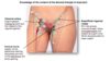
What is a sister mary joseph nodule?
- Sister mary joseph nodule refers to palpable mass at the umbilicus as a result of metastasis of a malignant cancer from the pelvic or abdomen
- Embryology provides potential clue of the route of cancer spread –> via urachus (remnant of the allantois) which attaches bladder to the umbilicus or via falciform ligament attaching liver to anterior abdominal wall.

What should you be aware of in a male patient if a lump appears in the groin or scrotal region?
- Consider first if you can get above the swelling –> If no it is likely to be a hernia, if you can it cannot be a hernia.
- If hernia is suspected can you reduce it? Test by the cough impulse
- Is the mass solid or fluid filled?
- where is the lump relative to the testicle? where is the testicle? Does the testicle look / feel like a bag of worms? (think varicocele).
- If there is pain assume it is testicular torsion until proven otherwise. (Testicular torsion –> testicle twists on its own blood supply.)
What should you consider in terms of a scrotal mass?
What are the differential diagnoses? What tests can be done to confirm/ refute diagnosis?
- Blood supply of the testicle (testicular arteries) is contained within the spermatic cord, which travels via the inguinal canal.
-
Differentials:
- Inguinal hernias emerge from the abdominal wall and descend down towards the testicle –> there are indirect hernias in which the loop of bowel passes through both the deep inguinal ring, inguinal canal and superficial ring into the scrotum. Due to a patent process vaginalis. —> And Direct hernias in which the bowel loop passes through superficial inguinal ring, more likely older patients due to weakness in posterior abdominal wall in region of Hesselbach’s triangle.
- Hydrocele –> fluid that completely surrounds the testicle, will be able to feel the spermatic cord above but not the testicles. Transillumination should reveal fluid filled.
- Varicocele –> venous swellings occuring around the spermatic cord ( looks like a bag of worms within the scrotum).
- Testicular cancer –> feel normal regions of the testicle with hard lumps within
- Spermatocele or epididymal cyst –> fluid filled bag superior to the testicle, testicle will feel normal.

A 10 year old boy presents with a swelling in his scrotum. His mother tells you it is not there in the morning but appears during the day, then disappears if he lies down. Examination reveals the swelling transilluminates, but you can’t get your hand above the swelling. What is this?
- It transilluminates which points toward a hydrocele, however unable to get hands above the swelling –> points towards a herniation.
- Cannot be a herniation as it transilluminates indicating it is fluid
- Therefore is it fluid accumulation due to patent process vaginalis -> when the testicles descend to the scrotum they pull with them a loop of peritoneum, which should fibrose. If this doesnt occur it is called a patent process vaginalis.
- Fluid from the abdomen is able to enter scrotum with gravity.

Define a hernia
What types of hernias are there?
- Protrusion of a tissue or organ through a structure or tissue that normally retains it.
- Classic hernias:
- Inguinal
- Femoral
- Incisional or postoperative
- umbilical
- lumbar (superior and inferior).
What is an inguinal hernia?
What are the two types?
How do they present differently in anatomy?
- Inguinal hernia = abdominal contents enter into inguinal cancal
- Direct inguinal hernia (20%) –> bowel enters inguinal canal “directly” through weakness in posterior wall of the canal termed Hesselbach’s triangle.
- Direct hernias occur more commonly in older patients secondary to abdominal wall laxity or increase intraabdominal pressure
- Indirect inguinal hernais (80%) –> bowel enters the inguinal canal via deep inguinal ring and superficial inguinal ring.
- Indirect inguinal hernias arise from incomplete closure of the processus vaginalis, outpouching of the peritoneum that allows testicular descent and normally fibroses.
- In surgery differentiated by position relative to inferior epigastric vessels:
- indirect inguinal hernias will be lateral
- direct inguinal hernias will be medial
What are the clinical features of an inguinal hernia?
- Lump in the groin that will initally disappear with pressure or when patient lies down (reducible hernia)
- If the hernia becomes trapped it will become painful, tender and red,
- Patient may also present with bowel obstruction, strangulation of the bowel can occur. –> irreducible and tender tense lump, pain out of proportion to clinical signs.
What is involved in the examination of an inguinal hernia?
- Reducible? Observe patient lying down and standing (should reduce with lying down). Also with minimual pressure.
- Observe the site and direction of the hernia –> inguinal or femoral? Inguinal = superomedial to the pubic tubercle, femoral hernias are inferolateral to pubic tubercle.
- make other regional observations
- compare the sides
- test the cough impulse –> reducible hernia will reappear, irreducible hernia will not
- pressure over the alternate inguinal rings
- ausculate
- if it has entered the scrotum can you get above it? is it separate from testis?
What are three causes of groin swelling in females?
- Femoral hernia
- canal of nuck = female equivalent of processus vaginalis
- bartholin gland cyst
what are two bony landmarks that mark the femoral triangle?
What forms the borders of the femoral triangle?
What forms the roof and the floor?
What are the contents?
where do the feoral vessels and saphenous nerve enter?
Where does the saphenous vein go and what is the clinical relevance?
- bony landmarks –> ASIS and pubic tubercle
- Superior border = inguinal ligament
- lateral border –> medial portion of sartorius
- medial border –> medial border of adductor longus
- floor –> partially iliacus, psoas major, pectineus, adductor longus
- roof –> superficial fascia, deep fascia, skin
- Contents from medial to lateral:
- femoral canal containing lymph nodes (cloquets node)
- femoral vein
- femoral artery
- femoral nerve
- Note the femoral vessels and saphenous nerve enter the subsartorial/ adductor canal and pass into the popliteal fossa
- the saphenous vein pierces the roof of the femoral triangle and can dilate here creating a saphena varix