Eyes- orbit, movement and reflexes Flashcards
What is the accomodation reflex?
Describe the three action that occur
The pathway that it involves
What region of the brain needs to be intact for this to occur?
- The accomodation reflex allows us to focus on a near object and involves a fattening of the lens which focuses the image on the retina –> process known as accomodation.
- Accomodation reflex involves:
- convergence of the eyes
- lens accomodation (fattening)
- pupillary constriction
- Accomodation reflex pathway:
- Light hits the back of the retina –> primary visual cortex
- Primary visual cortex –> frontal eye field
- Frontal eye field:
- CN iii nucleus –> medial rectus (vergence)
- Edinger westhphal nucleus —> ciliary ganglion —> Sphincter pupillae (pupil constriction) and Ciliary body (Lens fattening).
- Contraction of the ciliary muscles relaxes the suspensory ligaments, making the lens fatter
- Pupillary constriction prevents light from wide angles entering the eye, allowing clear image.
- Midbrain needs to be intact for accomodation reflex.

What is argyll roberston/ prostitute’s pupil?
- Argyll robertson/ prostitutes pupil describes pupils that are constricted but are unable to constrict further when exposed to light (Loss of pupillary light reflex). They are still able to accomodate.
- Due to tertiary syphilis which affects the pre-tectal nucleus, and sometimes diabete neuropathy.
- Loss of pre tectal nuclei which have communicative bilateral fibres leads to loss of pupillary light reflex.
- Pre- tectal nuclei are unable to communicate to Edinger Westphal nuclei.
- However Edinger westphal nuclei are both still intact, meaning the accomodation reflex can occur.
- Accomodation reflex requires intact CN ii (afferent pw) and intact CN iii (efferent pw), and midbrain.

How would damage to the Edinger Westphal nucleus present?
What commonly causes damage to this nucleus?
- Loss of direct and consensual pupillary light reflex on the affected side.
- Pupils remain dilated and unreactive.
- EWP receives from pretectal nucleus, loss of the EWP means parasympathetic preganglionic fibres cannot hitchike along CN iii and reach post ganglionic fibres in the ciliary ganglion.
- Therefore no postganglionic innervation to sphincter pupillae muscles, no pupillary constriction on affected side.
- Other side with intact EWP nucleus will have direct reflex and consensual reflex (Shining light in eye with EWP lesion still leads to pupillary constriction of other eye.).
- Common causes: vascular lesion, tumour, brainstem lesion.

Describe the pupillary light reflex pathway
- Light hits back of the retina, afferent p/w via CN II
- Carried to Pre tectal nucleus which has bilateral communicative fibres to the other pre tectal nucleus
- Pre tectal nucleus sends impulse to edinger westphal both ipsilaterally and contralaterally (PTN in midbrain close to superior colliculus)
- EWP nucleus has parasympathetic pre ganglionic efferent fibres that synapse with post ganglionic fibres in the ciliary ganglion. (EWP in midbrain)
- These parasympathetic fibres hitchhike along CN iii
- Ciliary ganglion post ganglionic efferents innervate sphincter pupillae muscles, leading to constriction of the pupil.

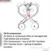
How would CN III compression present?
What would occur if there was a vascular lesion to CN III?
- No direct or consensual pupillary light reflex on the affected side (parasympathetics cannot reach ciliary ganglion).
- Dialted and non reactive pupil
- CN III compression = loss of all CN III functions:
- loss of innervation to all extraocular muscles except superior oblique and lateral rectus –> down an out eye
- Full ptosis
- Dilated pupil
- CN III vascular lesion –> sparing of pupillary functions
What is a Marcus Gunn Pupil?
How is it tested for?
What does this indicate?
- Reduced pupillary constrictor response, detected by swinging flashlight test
- Patients pupils constrict less (Reduced consensual and direct reflex) on the affected side when light is shone into that eye, when light is shone into the unaffected eye both pupils constrict the expected amount (normal consensual and direct reflex).
- Indicates afferent limb pathology:
- lesion in optic nerve, disrupting communication from retina –> pretectal nucleus
- Retinal disease

What three muscles act on the eyelid?
What are their actions?
What are their innervations?
- Levator palpabrae superioris –> innervated by CN III, action is to raise eyelid
- Superior tarsal muscle —> Innervated by sympathetic fibres, action is to enhance eyelid raise during flight/ fight response
- Obucularis oculi –> closes the eye, innervated by CN VII

If each of the three muscles acting on the eyelid stopped working what would be the effect?
- Loss of obicularis oculi –> loss of ability to close the eye, and flush debris and lacrimal fluid across the eye from lateral to medial. Dry, painful, irritated eyes.
- Loss of superior tarsal –> helps to raise the eyelid, partial ptosis
- Loss of levator palpabrae superioris –> full ptosis
What is the golden rule when it comes to innervation of extraocular muscles?
- All extraocular muscles are innervated by CN III except:
- Lateral rectus –> innervated by abducens nerve , CN VI
- Superior oblique –> innervated by trochlear nerve, CN IV

Label each of the extraocular eye muscles


What neurovascular structures pass through the superior orbital fissure?
Where do extraocular muscles originate from?
- CN III
- CN IV
- CN Va
- CN VI
- Opthalmic vein
The rectus muscles originate from a common tendinous ring at the posterior aspect of the orbit.
Inferior oblique arisus from antero medial floor of the orbit, acts like a hammock sitting under the eye.
Superior oblique originates from superior medial portion of the eye.

In which axis can the eye move?
The eye can move in three perpendicular axis:
1) Left and right
2) torsion -> twisting inward and and outward
3) up and down

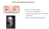
How can eye movements be tested?
What might clinicians use?
Eye movements can be tested using the H test, it involves moving the eye to a different starting position each time which isolates a specific muscle movement.
This testing pattern isolates the movements of specific muscles and so not all clinicians will use it. Many clinicals use a diagonal pattern of testing and with compare movements of the L and R eye in 6 different directions.

What are the actions of the medial and lateral rectus muscles?
- Medial rectus adducts the eye, lateral rectus abducts the eye

How do we test the functions of Superior oblique, inferior oblique, superior rectus and inferior rectus individually?
- The H test aligns these muscle tendons in a way that allows them to bring about their movement but minimises the effects of other muscles.

What are the actions of the superior and inferior rectus muscles?
How are their actions tested clinically?
- Superior rectus originates from superior part of common tendinous ring at back of the orbit and attaches the superior and anterior portion of the sclera.
- Main action elevates eyeball (also helps adduct/medially rotate).
- Inferior rectus originates from inferior portion of common tendinous ring and attaches to the inferior anterior aspect of the sclera.
- Main action is to depress the eyeball, also helps adduct and laterally rotate.
- During the H test their actions are tested by 1st getting the patient to look laterally (lateral rectus). Then:
- look up tests superior rectus
- looking down tests inferior rectus

What are the actions of the superior and inferior oblique muscles?
How can we test their function clinically?
- Superior oblique: arises from superior medial portion of the orbit (body of sphenoid bone), travels through trochlear and insert on superior aspect of the eyeball
- Depresses, abducts and medially rotates the eyeball
- Inferior oblique: arises from orbital floor and attaches to sclera of the eye, posterior to lateral rectus.
- Elevates, abducts and laterally rotates the eyeball
- Function tested again with H test:
- patient looks medially
- Then looks superior - tests inferior oblique
- then inferior - tests superior oblique
- Diagonal movements in and up and in and down test both medial rectus + oblique muscle involved.

What is Hering’s law and what is its importance when testing the cardinal positions of gaze?
Hering’s law states that extraocular muscles have equal and simultaneous innervation. This means that when testing the cardinal positions of gaze, both eyes should move by the same amount, at the same time and in the same direction

What symptoms might a patient complain of if their extraocular muscles do not obey Hering’s law?
- Neck pain as the patient tries to compensate for the affected eye to prevent diplopia by turning their head.
What does CN VI supply?
What symptoms would a patient with CN VI lesion/ nucleus damage complain of?
- Abducens nerve supplies the lateral rectus muscle which abducts the eye.
- Lesion of CN VI or its nuclei leads to ipsilateral inability to abduct the eye, which rests in an adducted position –> convergent squint
- This leads to horizontal diplopia which is worse when the patient looks towards the affected side
How would a CN III lesion present?
- CN III supplies 4/6 extraocular muscles, x 1 that opens the eyelid and the sphincter pupillae and ciliary body
- CN III lesion presents with:
- complete ptosis on affected side
- down and out position of the eye –> divergent squint
- horizontal and vertical diplopia
- dilated pupil on the affected side thats unreactive todirect or consensual light
- consensual pupil light reflex intact in contralateral (unaffected eye)>

What does CN IV carry innervation to?
What would a CN IV lesion present with?
- CN IV innervated the superior oblique muscle which depresses, and medially rotates the eye.
- Lesion presents with:
- Ipsilateral upward deviation and outward rotation of the affected eye (deviation and extorsion).
- Vertical diplopia worse when descending stairs/ reading paper
- Torsional diplopia -> double vision where images are twisted apart from each other
- Patient tilts head away from the lesion to help prevent diplopia (Counteracts extorsion by inferior oblique), complain of neck ache.

The eyes can move in several different ways:
What are three main movements they can do?
- Tracking target –> smooth pursuit of a moving target
- Stabilise target –> target is stable, you are moving
- Saccadic movements, scan target to target –> fast movement
What centres control eye movements?
- Multiple brain centres control eye movements:
- Vestibular nuclei –> reflex movement of eyes with head movements
- Parapontine reticular formation –> coordination of eye movements, horizontal gaze and saccadic movements
- frontal eye field –> control of visual attnetion, voluntary eye movements, saccadic movements
- Several saccade centres
- Visual association area –> allows us to relate past/ present visual information and recognise/ evaluate what is seen


