Introduction to CNS anatomy Flashcards
What does the nervous system enable the body to react to? What are the two main divisions of the nervous system?
The nervous system enables to body to continously react to changes to both the internal and external environment. To main divisions are the central and peripheral NS.
What further divisions do the central and peripheral NS’s split into?
Central NS: Brain, spinal cord cranial nerve II (optic nerve) and the retina.
Peripheral NS: Spinal nerves and cranial nerves (except CN II) , autonomic NS (parasympathetic and sympathetic divisions) somatic motor and somatic sensory nerves (part of the spinal nerves).

How do spinal nerves leave the spinal cord?
Paired, via intervertebral foramen.
What are the two main cell types of the nervous system?
Neurons and neuroglia. Neurons work in harmony with neuroglial cells (also called glial cells).
What are the main characteristics of neurons?
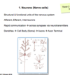
Main characteristics:
- Structural and functional units of the NS
- Afferent (sensory) and efferent (motor)
- Interneurons (patellar reflex)
- Allow rapid communication via synapses and Neurotransmitters.

Describe general neuron structure.
Neurons have a general structure:
Dendrites: projections sensitive to NT’s at gap junctions
Cell soma/ body: metabolic centre of the neuron
Axon: Long projection from cell body to other neuron
Axon terminal: portion of the axon which communicates with other neurones/ body tissues via SYNAPSES.

What are the main characteristics of neuroglial cells?
- Non- neural/ non exictable
- Supporting cells, nourish and insulate neurons.
- x5 more abundant
What are the types of neuroglial cells in the Central NS?
4 types of neuroglial cells:
1) Oligodendrocytes- myelinate and provide structural framework
2) Astrocytes- Maintain BBB, recycle NT’s
3) Microglial cells- clear debris and pathogens via phagocytosis
4) Ependymal cells- Line ventricles of brain and central spinal canal, produce CSF.

What are the main types of neuroglial cells in the Peripheral NS?
1) Schwann cells - myelinate axons of peripheral nerves
2) Satellite cells- regulate nutrient and NS levels in the ganglia.

What are spinal nerves? What are the divisions of each (i.e what type of information is involved in each branch?)
They are mixed motor and sensory nerves. Sensory information can be: 1) visceral sensory (organs and vessels 2) somatic sensory (tissues) 3) special sensory (taste) Motor information can be: 1) somatic motor - skeletal muscle 2) Visceral/ autonomic motor 3) Branchiomotor- provide motor innervation to pharyngeal arches

Describe the formation of the neural tube:
Where does the nervous system develop from? what week does this happen?
What happens to form the neural tube?
What are the open ends of the neural tube called?
When do they close?
- The nervous system forms from the ectodermal plate at the beginning of week 3.
- The edge of the ectodermal plate thickens and becomes folds.
- These folds start to lift away from the rest of the ectodermal plate and fuse within the midline- forming the neural tube.
- The fusion first begins in cervical region and then continues towards the head and caudal regions.
- The open ends of the neural tube form the cranial and caudal neuropores.
- The cranial neuropore closes at day 25 and the caudal neuropore at day 27.

What can go wrong with neural tube folding?
What key prenatal supplement can prevent this?
- If the cranial neuropore does not close (day 25) it can result in anencephaly (absence of the brain) or mesoanencephaly ( herniation of brain tissue through partially formed cranium).
- If the caudal neuropore does not close (day 27) is results in spina bifida.
- Spina bifida ranges in severity - milder form = spina bifida occulta.
- Spina bifida occulta= failure to close neural arches of lumbar/ sacral vertebrae.
- Folic acid is a key prenatal supplement

Describe the development of the adult brain:
how many primary brain vesicles form and at what week?
what are they called?
How many secondary brain vesicles come from these?
what are they called?
What week does this occur?

What do the secondary brain vesicles go on to form?
They go on to form the adult brain, vesicles, brainstem and cerebellum.
- Telencephalon- cerebral hemispheres
- Diencephalon- thalamus and hypothalamus
- Mesencephalon- midbrain
- Metencephalon- cerebellum and pons
- Myelencephalon- medulla

What develops between the cerebral hemispheres and the thalamus/ brainstem?
If this didnt develop what could occur?
A flexure develops between the cerebral hemispheres and the thalamus/ brainstem.
This means the superior portion of the cerebral hemispheres can be described as dorsal.
The inferior portion of this flexure can be described as ventral.
If this flexure didnt develop our eyes would point upward.

What divides the two hemispheres of the brain?
What could a raised portion of the cerebral hemispheres be described as?
what could depressions/ folds in the cerebral hemispheres be described as?
What divides the temporal lobe from the rest of the brain?
The longitudinal fissure divides the L and R sides of the brain.
Raised portions described as a gyrus.
depressions = sulci.
The lateral / sylvian fissure divides the temporal lobe from the rest of the brain/

What is the name of a distinct white matter tract that connects the R/L hemispheres?
What forms the brainstem?
What structure sits below the corpus callosum and above the brainstem?
What structure comes of inferiorly to this?
What are these two structures collectively known as?
- The corpus callosum is a distinct white matter tract.
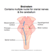
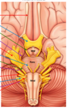
- Brainstem: Midbrain, pons. medulla.
- The thalamus sits below the corpus callosum and above the brainstem.
- The hypothalamus comes off inferiorly to the thalamus, ending in the pituitary gland inferiorly.
- Hypothalamus plus thalamus = Diencephalon

Label the image

What other structure does the corpus callosum intersect with?
What does this structure do?
- The corpus callosum (connecting R and L hemispheres) intersects with another white matter tract called the corona radiata.
- The corona radiata carries ascending and descending tracts between the cerebral cortex, thalamus and spinal cord.

What sulcus divides the frontal and parietal lobes?
What lies immediately anterior to this sulcus?
And posteriorly?
What sulcus divides frontal lobe from temporal?
How many gyri are there on the temporal lobe?
- The central sulcus divides the frontal and parietal lobes.
- Anteriorly to central sulcus is the precentral gyrus which is the primary motor cortex of the frontal lobe.
- Posteriorly is the post central gyrus which is the primary somatosensory cortex of the parietal lobe.
- Lateral sulcus/ sylvian fissure divides the frontal lobe from the temporal lobe.
- There are 3 gyri on the temporal lobe: 1) superior temporal gyrus 2) middle temporal gyrus 3) inferior temporal gyrus

what lobe is underneath the lateral sulcus/ sylvian fissure?
What are its functions?
- The insular lobe lies underneath the lateral sulcus.
- Involved with higher pain processing, emotion and language processing.

What divides the parietal lobe from the occipital lobe?
What sulcus is on the occipital lobe that has a key function?
- The parieto-occipital sulcus divides the parietal lobe from the occipital lobe.
- On medial aspect of the occipital lobe is the calcarine sulcus that forms the primary visual cortex.

What is the cingulate gyrus?
what system is it a part of?
where can it be found?
- The cingulate gyrus is an integral part of the limbic system
- It can be found superior to the corpus callosum on the medial aspect of the cerebrum.
- Has important functions in higher autonomic control such as HR, RR and pain processing.

What areas divide different regions of the cerebral cortex?
what are these regions based on?
- Brodmann’s areas separate different regions of the cerebral hemispheres
- These regions are based on histological structure of the cortex.


