Larynx Flashcards
(34 cards)
what is the primary function of the larynx?
protect the tracheobronchial tree
What is speech articulation controlled by?
Whereas the vocal cords within the larynx are responsible for phonation, the pharynx, soft palate, tongue and lips control speech articulation
Where does the larynx begin (vertebral level) and where does it end?
The larynx is suspended from the hyoid bone above around C2. It extends from the hyoid bone to the cricoid cartilage- vertebral level C6/C7.
What are the main components of the larynx? (Bony and cartilaginous structures?)
Describe its structure, where these components are in relation to each other and their attachements.
The hyoid bone most superior, thyroid cartilage immediately inferior to this (largest of the three cartialges) and cricoid cartilage inferiorly to this (shaped like a signet ring which broad arch posterior and narrower arch of tissue anteriorly). The epiglottis is a leaf shaped cartilage- attaches to the anterior wall of the thyroid cartilage and projects posterosuperiorly.

What might the larynx allow us to regulate in the thorax and abdomen?
The larynx allows us to regulate the pressure within the the thorax by closing off the airways and increasing thoracic/abdominal pressure e.g. valsalva manouvre as in childbirth/defecation.
Which cartilage is more prominent in men than women?
The thyroid cartilage is formed by two laminae, the right and left. These converge and join anteriorly, forming the laryngeal prominence (Adam’s apple). Angle between two laminae more prominent in men than women so laryngeal prominence more apparent in men than women.

There are no open gaps in the larynx due to the presence of multiple membranes between the cartilage.
Describe these membranes in the larynx.
1) the thyrohyoid membrane extends from the thyroid cartialge up to the hyoid bone
2) the cricothyroid membrane projects from the cricoid cartialge inferiorly to the thyroid cartialge superiorly

What might the cricothyroid membrane be used for in an emergency?
Cricothyroid membrane can be used to access the airway during an emergency - procedure called cricothyroidotomy.
Used to access the airway in the event there is an upper respiratory blockage e.g. in cases of anaphylaxis, trauma and mass bleeding involved the mouth/pharnyx, laryngospasm, uncontrollabled emesis, clenched teeth, foreign body obstruction.

What two structures project off the thyroid cartilage?
describe how the thyroid cartilage articulates with the cricoid cartilage, what joint exists and what does this joint allow?
What does movement of the thyroid cartilage allow us to alter?
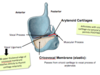
The thyroid cartilage has two horns on its posterior surface, the superior and inferior horn.
The inferior horn articulates with the superior surface of the cricoid cartilage on the posterior aspect of the larynx, a synovial joint exists between them which permits movement.
The thyroid cartilage is a highly mobile unit and the vocal cords articulate with the anterior aspect of this thyroid cartilage. Change of shape of this cartilage allows us to change the shape of the vocal cords and therefore pitch.
Why might a hyoid bone fracture be fatal?
Hyoid bone fractures often associated with strangulation, car accidents/ trauma.
Fatality due to the bleeding and swelling that occurs over the top of the airways leading to suffocation.
What two accessory cartilages are in the larynx that articulate with the cricoid cartilage underneath and allow us to alter our vocal cords?
What joint exists here and what does that permit?
The arytenoid cartilages articulate with the cricoid cartilage inferiorly and a synovial joint exists here which allows rotational movement- PIVOTING. This alters the shape of our vocal cords and therefore allows us to alter pitch.

What is the shape of the arytenoid cartilages?
What processes come off the arytenoid cartilages, where are they located and what attaches to these points?
What is the action provided by these attachements?
The arytenoid cartilages are shaped like pyramids.
One each of the paired arytenoid cartilages there are two processes 1) the vocal process 2) the muscular process
The vocal process is anterior and allows the attachment of the vocal ligament which projects from this anterior tip of the arytenoid to the thyroid cartilage anteriorly.
The muscular process is posterior and lateral and allows the attachment of muscles that will move this arytenoid process at its synovial joint, either adducting or abducting the vocal ligaments.

What membrane passes from the cricoid cartilage and to the vocal ligament?
What actually forms the vocal ligament?
What are its attachment points?
The cricovocal membrane is an elastic membrane that extends from the cricoid cartialge inferiorly and projects upward to the vocal ligament. Its free, upper thickened edge actually forms the vocal ligament, it inserts posteriorly on the vocal process of the arytenoid cartilage and anteriorly to the thyroid cartilage.
How many strong membranes does the larynx contain and what kind of mucosa covers them?
What other structures does this kind of mucosa line?
What type of epithelium is respiratory mucosa?
The larynx contains two strong membranes covered in respiratory mucosa.
Respiratory mucosa lines most of the larnyx. laryngopharynx and oropharynx.
The respiratory mucosa is made up of psuedostratified ciliated columnar epithelium.
What kind of mucosa covers the vocal ligaments and how does this differ from the rest of the larynx?
The larynx is covered in respiratory mucosa (pseudostratified ciliated columanr epithelium) whereas the vocal cords are covered by stratified sqaumous epithelium to resist the abrasion caused by the banging of the vocal cords together.
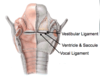
What are the two strong membranes contained within the larynx and what do their borders form?
1) aryepiglottic membrane which projects from the apex of the arytenoid cartilage to the epiglottis and then extends infeiorly.
Its free lower border forms the vestibular ligament.
2) the cricovocal membrane which projects from the cricoid cartilage and inserts onto vocal process of the arytenoid and the anterior portion of the thyroid cartilage.
Its free upper thickened edge/ border forms the vocal ligament.

What exists between the vestibular ligament and vocal ligament (vestibular folds and vocal folds)?
What are the two parts of this called?
What does it allow?
A mucosal pouch exists inbetween the vestibular and vocal ligaments and is formed of two parts 1) the ventricle 2) the saccule
Ventricle and saccule form this 1 mucousal pouch
The ventricle and saccule sit inbetween the vocal ligament and vestibular ligament and allows the vocal cords to slam together to produce phonation independently of the membrane above (the arytenoid membrane above the cricovocal membrane). Allows vocal cords to slam together quite aggressively without moving the rest of the larynx.
What are the vestibular and vocal ligaments called when covered in mucosa?
Vestibular ligament ——> vestibular fold
Vocal ligament ——-> vocal fold

On an endoscopic view of the larynx what membrane fold forms a posterior rim?
What cartilage is seen on the anterior of an endoscopic view of the larynx?
What is the space enclosed by the vocal folds called?
What is another name for the vestibular folds and what is their function?
Where would the ventricule and saccule be found?
The aryepiglottic fold forms a rim around the top of the larynx posteriorly.
The epiglottis cartialge is seen on the anterior of a superior endoscopic view of the larynx
The space enclosed by the vocal folds is called the rima glottidis
The vestibular folds are also called false vocal cords and their function is to protect the airway and support the vocal folds in phonation.
Inbetween the vocal folds and vestibular folds are the ventricule and saccule.
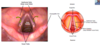
what pathology is shown in this image?
What effect would this have on the patient?

Vocal cord nodules on the vocal folds - patient would have difficulty with phonation and protection of airways
Where is the aryepiglottic muscle found and what is its function?
The aryepiglottic muscle is found below the aryepiglottic fold (formed by the aryepiglottic membrane that projects from the apex of arytenoid to epiglottis superiorly and then inferiorly).
When this muscle contracts it closes the top of the larynx- reducing the size of the laryngeal inlet.

What nerve supplies the motor and sensory innervation to the larynx?
The vagus nerve
What branches does the vagus split into to innervate the larynx?
What does each branch do?
The vagus nerve splits into 1) superior laryngeal nerve 2) recurrent laryngeal nerve
The surperior laryngeal nerve further splits into the external laryngeal nerve and internal laryngeal nerve.
The external laryngeal nerve projects down over the larynx until it reaches the cricothyroid muscle which it provides motor innervation to.
The internal laryngeal nerve enters the larynx and does all the sensory innervation to the larynx above the vocal folds.
The recurrent laryngea nerve is a branch that moves back in the thorax, enters the larynx to innervate all the other muscles of the larynx (except cricothryoid muscle) and provides sensory innervation below the vocal folds.

Fill the blanks:
All nerve supplies in the larynx are __________.
E.g. the right recurrent laryngeal nerve supplies the ________ side of the larynx.
All nerve supplies in the larynx are ipsilateral.
E.g. the right recurrent laryngeal nerve innervates the right side of the larynx.


