Cardiac embryology Flashcards
(33 cards)
During development the heart is a _________ structure that rotates to the ______.
Left sided structures sit mostly __________.
Right sided structures sit mostly __________.
During development the heart is a midline structure that rotates to the left.
Left sided structures sit mostly posteriorly.
Right sided structures sit mostly anteriorly.

Where do fetal blood vessels start to develop from?
Describe the development of the fetal circulation.
What week does this occur?
- Fetal blood vessels develop from the extraembryonic mesoderm that surrounds the chorionic cavity and embryo.
- Blood vessels develop in this layer called the chorion.
- The extraembryonic mesoderm also forms the connecting stalk which will become the umbilicus
- Fetal blood vessels in the extraembronic mesoderm connect to the embryo by the umbilicus.
- Umbilical arteries carry deoxygenated blood from the embyro to the chorion
- Umbilical veins carry oxygenated blood back into the embryo
- Embryonic blood vessels of the chorion contact maternal blood vessels
- Uternine maternal blood vessels supply maternal blood to the trophoblastic lacunae.
- Occurs by the end of week 3.

Where does the heart develop from?
What process occurs and what is formed early on?

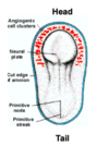
- From angiogenic cell clusters at the head end of the embryo and close to the neural plate
- These angiogenic cell clusters come together to form rods
- These rods cannalise and from two endocardial heart tubes.
What happens after the formation of the two endocardial tubes?
When does the heart start beating?
When is blood first pumped through?
What other cells are involved?
- Lateral folding causes fusion of the two endocardial tubes.
- Heart starts beating at days 22-23
- Blood first flows through the heart at the end of week 4.
- Neural crest cells are involved in the process.
What is strange about the positioning of the developing heart/mouth/brain in the embryo?
What is above the heart region? What does the heart develop into?
What corrects the abnormal positioning of the embryonic regions?
The develoing heart is intially siutated above the head of the embryo.
- The heart develops above the prochordal plate which forms the mouth region.
- The heart develops in a region with an open cavity above it which will form the pericardial cavity
- The heart develops into this cavity becoming wrapped in the pericardium.
- The prochordal plate is originally above the ectoderm which will form the brain
- Order at that point in the embryo is heart, mouth, brain.
- reversal occurs during longitudinal folding, brain develops quickly and grows over mouth and heart regions
- The heart is moved into the future thoracic region.

What is the septum transversum?
Where is it originally located in the embyro?
What does it form?
The septum transversum is a region in the embyro that originally overlies the heart primitive tissue. It will go on to form the fibrous pericardium and part of the diaphragm. During longitudinal folding it shifts beneath the heart.

Fill the blanks


What does the heart become surrounded by?
How does this happen?
- The heart becomes surrounded by the pericardium
- This happens as the endocardial tube grows into the pericardial cavity overlying it becoming completely surrounded by it
- The pericardium molds to the developing heart
- Points of reflection of the visceral peritoneum becomes continuous with the parietal peritoneum forming the pericardial cavity.

What regions does the heart tube dilate into?
What does each of these regions become?
From head to tail:
- Truncus arteriosus- becomes the pulmonary trunk and aorta
- Bulbus cordis- becomes the right ventricle
- Primitive ventricle- forms the left ventricle
- Primitive atrium- forms the rough part of both atria
- Sinus venosus- becomes the smooth wall of the right atrium

Fill in the blanks


What is the atrioventricular canal?
What forms in this region?
The atrioventricular canal is the narrow connection between the common atrium and common ventricular region.
The endocardial cushion will develop here and from this the atrioventricular valves will form.
What region connects the primitive ventricle and common arterial outflow (truncus arteriosus)?
What does this region then become to connect to the truncus arteriosus?
What is the difference in the wall texture between these two regions?
The bulbus cordis connects the primitive ventricle and common arterial outflow (the truncus arteriosus).
The bulbus cordis then develops into the conus cordis which leads from the future ventricles into the truncus arteriosus.
The conus cordis remains smooth walled and associated with the outflow tracts. The proximal third of the bulbus cordis and ventricular region become trabeculated.

What needs to occur after the dilated regions of the heart tube have formed?
The heart tube needs to develop its inter atrial and inter ventricular septi that will split the heart into L and R sides.
Describe the process of atrial septation
What does this allow in the embryo?
- Septum primum starts to grow off the roof of the common atria towards the endocardial cushion in the AV canal.
- Septum primum grows down to meet it, the foramen inbetween the endocardial cushion and septum primum is known as foramen primum
- Septum primum contacts endocardial cushion closing foramen primum but holes form higher up that merge to form foramen secundum.
- To the right of septum primum, a 2nd septum develops called septum secundum.
- Septum secundum grows over foramen secundum and towards the endocardial cushion but doesnt contact it however.
- The hole that remains is foramen ovale.
- Septum secundum is rigid and tough whereas septum primum is floppy and acts like a valve.
- Allows right to left shunting of blood which bypasses the fetal lungs which are fluid filled and non functional. Allows oxygenated blood from the placenta to enter the systemic circulation.

Describe how the heart tube is split into L and R regions?
What is different between R and L sides of the heart?
What needs to develop on the L side of the heart?
What does this mean for wall texture?
- Septal formation doesnt happen exactly down the midline but occurs after the sinus venosus (which will form part of the right atrium) and splits the right atrium from left atrium.
- The right atrium is then joined to the right ventricle region and has both venous inflow (at the sinus venosus) and arterial outflow.
- The left atrium is joined to its ventricle and arterial outflow but has no venous inflow.
- The pulmonary veins actually form from the Left atrium.
- Originally as a single pulmonary vein that then branches into 4 pulmonary veins
- The left atrium remodels and grows out to contact the the 4 branches of the pulmonary veins, absorbing the tissue before it- it forms the smooth part of the left ventricle.

What forms the smooth part of the right atrium?
What forms the rought part?
The smooth part forms from the right side of the sinus venosus.
The rough part is formed from the embryological atrium.

What forms the smooth part of the left atrium?
What forms the rough part of the left atrium?
The smooth part of the left atrium forms from outgrowth of the budding pulmonary vein which gets absorbed into the wall of the remodelling L atrium.
The rough part froms from the embryological atrium.

Describe heart tube folding
The heart tube begins to fold due to growth in specific regions that occurs at a faster rate than in others.
The venous inflow remains at the bottom and is relatively fixed in position.
The arterial outflow/ aortic arches are also fixed and remain superior.
The bulbus cordis grows rapidly and pushes anterior and inferior to the right. (Forms the right ventricle).
The atria are pushed superiorly and posteriorly.
The ventricle region is pushed towards the left. (Forms the left ventricle).
Starts to mimic the positions of the adult heart. Day 22-24.

What occurs if the heart tube folds the wrong way?
what is this caused by?
A condition called Dextrocardia where the heart hangs to the right instead of the left.
Caused by the Bulbus Cordis moving downward and left and the ventricle moving to the right.

why does shunting of blood occur from right to left in the embryo?
What happens post natally?
- In utero: R to L shunting occurs because:
1) Lungs are fluid filled, high pulmonary vascular resistance to flow.
2) With little pulmonary circulation there is low venous return to the L side of the heart.
3) Left sided pressure less than right
4) Septum primum pushed towards the left from the right. - Post natal: L sided pressure overcomes R sided.
1) Baby takes its first breath
2) fluid filled lungs are drained and become functional
3) pulmonary vascular resistance decreases
4) pulmonary circulation increases
5) Venous return to the left side increases as does pressure
6) Septum primum pushed against rigid Septum secundum.

What is it called when the foramen ovale remains open?
Are there any consequences to this?
This is called a probe-patent foramen ovale.
Under normal physiological conditions there are no consequences to this.
During the valsalve manouvre R sided P can transiently increase above the L. (E.g. straining during childbirth).
This can lead to risk of emboli moving from R to L and increase risk of transient ischaemic attack and stroke.
What test can be used to visualise a probe patent foramen ovale?
The microbubble test. Ultrasound probe used to introduce micorbubbles into R atrium. If probe patent foramen ovale bubbles enter L atrium, viewed via ultrasound.

What is an atrial septal defect?
What are the consequences of one?
Where can it occur?
An atrial septal defect refers to a hole between the two atria.
The consequence of an isolated ASD is not a congenital cardiac defect:
Some are asymptomatic, some close during growth.
ASD’s can be seen at:
1) Foramen/Septum secundum.
2) Foramen/ Septum primum
3) Endocardial cushion
What week/s does atrial septation occur?
Weeks 4-5


