Musculoskeletal, Skin, and Connective Tissue - First Aid Flashcards
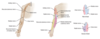
Arm Abduction

Rotator Cuff Muscles
Shoulder muscles that form the rotator cuff (SItS):
-
Supraspinatus (suprascapular nerve)
- abducts arm initially (before the action of the deltoid)
- most common rotator cuff injury (trauma or degeneration and impingement → tendinopathy or tear)
- assessed by “empty/full can” test
-
Infraspinatus (suprascapular nerve)
- externally rotates arm
- pitching injury
-
teres minor (axillary nerve)
- adducts and externally rotates arm
-
Subscapularis (upper and lower subscapular nerves)
- internally rotates and adducts arm
Innervated primarily by C5-C6.

Overuse Injuries of the Elbow:
- repetitive flexion (forehand shots)
- idiopathic
Medial Epicondylitis
(golfer’s elbow)
Overuse Injuries of the Elbow:
- repetitive extension (backhand shots)
- idiopathic
Lateral Epicondylitis
(tennis elbow)
Wrist Region:
So Long To Pinky, Here Comes The Thumb.
- Scaphoid
- Lunate
- Triquetrum
- Pisiform
- Hamate
- Capitate
- Trapezoid
- Trapezium

- Scaphoid (palpable in anatomic snuff box) is the most commonly fractured carpal bone, typically due to a fall on an outstretched hand.
- Complications of proximal scaphoid fractures include avascular necrosis and nonunion due to retrograde blood supply.
- Fracture not always seen on initial x-ray.
- Dislocation of lunate may cause acute carpal tunnel syndrome.

Wrist Region:
- also called boxer’s fracture
- common fracture caused by direct blow with a closed fist (eg. from punching a wall or individual)
- most commonly seen in 4th and 5th metacarpals
Metacarpal Neck Fracture
Wrist Region:
- entrapment of median nerve in carpal tunnel (between transverse carpal ligament and carpal bones)
- nerve compression → paresthesia, pain, and numbness in distribution of median nerve
- thenar eminence atrophies but sensation spared, because palmar cutaneous branch enters hand external to carpal tunnel
- suggested by ⊕ Tinel sign (percussion of wrist causes tingling) and Phalen maneuver (90° flexion of wrist causes tingling)
- associated with pregnancy (due to edema), rheumatoid arthritis, hypothyroidism, diabetes, acromegaly, and dialysis-related amyloidosis
- may be associated with repetitive use
Carpal Tunnel Syndrome

Wrist Region:
- compression of ulnar nerve at wrist
- classically seen in cyclists due to pressure from handlebars
Guyon Canal Syndrome
Common Pediatric Fractures:
- incomplete fracture extending partway through width of bone following bending stress
- bone fails on tension side
- compression side intact
- bone is bent like a green twig
Greenstick Fracture

Common Pediatric Fractures:
- axial force applied to immature bone → cortex buckles on compression side and fractures
- tension side (other side of cortex) remains intact
Torus (buckle) Fracture

Hand Muscles
- Thenar (median)
- Opponens pollicis
- Abductor pollicis brevis
- Flexor pollicis brevis
- superficial head (deep head by ulnar nerve)
- Hypothenar (ulnar)
- Opponens digiti minimi
- Abductor digiti minimi
- Flexor digiti minimi brevis
- Dorsal interossei (ulnar)
- abduct the fingers
- Palmar interossei (ulnar)
- adduct the fingers
- Lumbricals (1st/2nd, median; 3rd/4th, ulnar)
- flex at the MCP joint
- extend PIP and DIP joints

Both groups perform the same functions (OAF).:
- Oppose
- Abduct
- Flex
DAB = Dorsals ABduct
PAD = Palmars ADduct

Upper Extremity Nerves
- Axillary (C5-C6)
- Musculocutaneous (C5-C7)
- Radial (C5-T1)
- Median (C5-T1)
- Ulnar (C8-T1)
- Recurrent Branch of Median Nerve (C5-T1)

Upper Extremity Nerves:
- Causes of Injury:
- fractured surgical neck of humerus
- anterior dislocation of humerus
- Presentation:
- flattened deltoid
- loss of arm abduction at shoulder (> 15°)
- loss of sensation over deltoid muscle and lateral arm
Axillary (C5-C6)

Upper Extremity Nerves:
- Causes of Injury:
- upper trunk compression
- Presentation:
- loss of forearm flexion and supination
- loss of sensation over lateral forearm
Musculocutaneous (C5-C7)

Upper Extremity Nerves:
- Causes of Injury:
- compression of axilla, eg. due to crutches or sleeping with arm over chair (“Saturday night palsy”)
- midshaft fracture of humerus
- repetitive pronation/supination of forearm, eg. due to screwdriver use (“finger drop”)
- Presentation:
- wrist drop: loss of elbow, wrist, and finger extension
- ↓ grip strength (wrist extension necessary for maximal action of flexors)
- loss of sensation over posterior arm/forearm and dorsal hand
Radial (C5-T1)

Upper Extremity Nerves:
- Causes of Injury:
- supracondylar fracture of humerus (proximal lesion)
- carpal tunnel syndrome and wrist laceration (distal lesion)
- Presentation:
- “ape hand” and “pope’s blessing”
- loss of wrist flexion, flexion of lateral fingers, thumb opposition, lumbricals of 2nd and 3rd digits
- loss of sensation over thenar eminence and dorsal and palmar aspects of lateral 31⁄2 fingers with proximal lesion
Median (C5-T1)

Upper Extremity Nerves:
- Causes of Injury:
- fracture of medial epicondyle of humerus “funny bone” (proximal lesion)
- fractured hook of hamate (distal lesion) from fall on outstretched hand
- Presentation:
- “ulnar claw” on digit extension
- radial deviation of wrist upon flexion (proximal lesion)
- loss of wrist flexion, flexion of medial fingers, abduction and adduction of fingers (interossei), actions of medial 2 lumbrical muscles
- loss of sensation over medial 1 1/2 fingers including hypothenar eminence
Ulnar (C8-T1)
Upper Extremity Nerves:
- Causes of Injury:
- superficial laceration of palm
- Presentation:
- “ape hand”
- loss of thenar muscle group: opposition, abduction, and flexion of thumb
- no loss of sensation
Recurrent Branch of Median Nerve (C5-T1)

Humerus fractures, proximally to distally, follow the _____.
ARM
Axillary → Radial → Median
Brachial Plexus
Randy Travis Drinks Cold Beer:
- Roots
- Trunks
- Divisions
- Cords
- Branches

Brachial Plexus Lesions
① Erb palsy (“waiter’s tip”)
② Klumpke palsy (claw hand)
③ wrist drop
④ winged scapula
⑤ deltoid paralysis
⑥ “saturday night palsy” (wrist drop)
⑦ difficulty flexing elbow, variable sensory loss
⑧ decreased thumb function, “pope’s blessing”
⑨ intrinsic muscles of hand, claw hand
Brachial Plexus Lesions:
- Injury:
- traction or tear of upper trunk
- C5-C6 roots
- Causes:
- Infants—lateral traction on neck during delivery
- Adults—trauma
- Muscle Deficit:
- deltoid, supraspinatus
- infraspinatus
- biceps brachii
- Functional Deficit:
- abduction (arm hangs by side)
- lateral rotation (arm medially rotated)
- flexion, supination (arm extended and pronated)

Erb palsy (“waiter’s tip”)
Brachial Plexus Lesions:
- Injury:
- traction or tear of lower trunk
- C8-T1 root
- Causes:
- Infants—upward force on arm during delivery
- Adults—trauma (eg. grabbing a tree branch to break a fall)
- Muscle Deficit:
- Intrinsic Hand Muscles:
- lumbricals
- interossei
- thenar
- hypothenar
- Intrinsic Hand Muscles:
- Functional Deficit:
- Total Claw Hand:
- lumbricals normally flex MCP joints and extend DIP and PIP joints
- Total Claw Hand:

Klumpke palsy
Brachial Plexus Lesions:
- Injury:
- compression of lower trunk and subclavian vessels
- Causes:
- cervical rib
- Pancoast tumor
- Muscle Deficit:
- same as Klumpke palsy
- Functional Deficit:
- atrophy of intrinsic hand muscles
- ischemia, pain, and edema due to vascular compression

Thoracic Outlet Ssyndrome
Brachial Plexus Lesions:
- Injury:
- lesion of long thoracic nerve
- roots C5-C7
- Causes:
- axillary node dissection after mastectomy
- stab wounds
- Muscle Deficit:
- serratus anterior
- Functional Deficit:
- inability to anchor scapula to thoracic cage → cannot abduct arm above horizontal position

Winged Scapula
Distortions of the Hand
- At rest, a balance exists between the extrinsic flexors and extensors of the hand, as well as the intrinsic muscles of the hand—particularly the lumbrical muscles (flexion of MCP, extension of DIP and PIP joints).
- “Clawing”—seen best with distal lesions of median or ulnar nerves. Remaining extrinsic flexors of the digits exaggerate the loss of the lumbricals → fingers extend at MCP, flex at DIP and PIP joints.
- Deficits less pronounced in proximal lesions; deficits present during voluntary flexion of the digits.
- Atrophy of the thenar eminence (unopposable thumb → “ape hand”) can be seen in median nerve lesions, while atrophy of the hypothenar eminence can be seen in ulnar nerve lesions.

Knee Exam
LAMP:
- Lateral femoral condyle to anterior tibia: ACL
- Medial femoral condyle to posterior tibia: PCL

Knee Exam:
- bending knee at 90° angle, ↑ anterior gliding of tibia (relative to femur) due to ACL injury
- Lachman test also tests ACL, but is more sensitive (↑ anterior gliding of tibia [relative to femur] with knee bent at 30° angle)
Anterior Drawer Sign

Knee Exam:
bending knee at 90° angle, ↑ posterior gliding of tibia due to PCL injury
Posterior drawer sign

Knee Exam:
knee either extended or at ∼ 30° angle, lateral (valgus) force → medial space widening of tibia → MCL injury
Abnormal Passive Abduction

Knee Exam:
knee either extended or at ~ 30° angle, medial (varus) force → lateral space widening of tibia → LCL injury
Abnormal Passive Adduction

Knee Exam:
- During flexion and extension of knee with rotation of tibia/foot:
- pain, “popping” on external rotation → medial meniscal tear (external rotation stresses medial meniscus)
- pain, “popping” on internal rotation → lateral meniscal tear (internal rotation stresses lateral meniscus)
McMurray Test

Common Hip and Knee Conditions:
- Inflammation of the gluteal tendon and bursa lateral to greater trochanter of femur
- treat pain with NSAIDs, heat, stretching
Trochanteric Bursitis
Common Hip and Knee Conditions:
- common injury in contact sports due to lateral force applied to a planted leg
- classically, consists of damage to the ACL, MCL, and medial meniscus (attached to MCL); however, lateral meniscus injury is more common
- presents with acute knee pain and signs of joint injury/instability

“Unhappy Triad”
Common Hip and Knee Conditions:
- inflammation of the prepatellar bursa in front of the kneecap
- can be caused by repeated trauma or pressure from excessive kneeling (also called “housemaid’s knee”)
Prepatellar Bursitis
Common Hip and Knee Conditions:
popliteal fluid collection in gastrocnemius-semimembranosus bursa commonly communicating with synovial space and related to chronic joint disease (eg. osteoarthritis, rheumatoid arthritis)
Baker Cyst
Ankle Sprains
-
Anterior TaloFibular ligament
- Always Tears First—most common ankle sprain overall, classified as a low ankle sprain
- due to overinversion/supination of foot
- Anterior Inferior Tibiofibular ligament
- most common high ankle sprain

Lower Extremity Nerves
- Iliohypogastric (T12-L1)
- Genitofemoral(L1-L2)
- Lateral Femoral Cutaneous (L2-L3)
- Obturator (L2-L4)
- Femoral (L2-L4)
- Sciatic (L4-S3)
- Common Peroneal (L4-S2)
- Tibial (L4-S3)
- Superior Gluteal (L4‑S1)
- Inferior Gluteal (L5-S2)
- Pudendal (S2-S4)
Lower Extremity Nerves:
- Sensory—suprapubic region
- Motor—transversus abdominis and internal oblique
- Cause of Injury:
- abdominal surgery
- Presentation:
- burning or tingling pain in surgical incision site radiating to inguinal and suprapubic region
Iliohypogastric (T12-L1)
Lower Extremity Nerves:
- Sensory—scrotum/labia majora, medial thigh
- Motor—cremaster
- Cause of Injury:
- laparoscopic surgery
- Presentation:
- ↓ anterior thigh sensation beneath inguinal ligament
- absent cremasteric reflex
Genitofemoral (L1-L2)
Lower Extremity Nerves:
- Sensory—anterior and lateral thigh
- Cause of Injury:
- tight clothing
- obesity
- pregnancy
- pelvic procedures
- Presentation:
- ↓ thigh sensation (anterior and lateral)
Lateral Femoral Cutaneous (L2-L3)
Lower Extremity Nerves:
- Sensory—medial thigh
- Motor—obturator externus, adductor longus, adductor brevis, gracilis, pectineus, adductor magnus
- Cause of Injury:
- pelvic surgery
- Presentation:
- ↓ thigh sensation (medial) and adduction
Obturator (L2-L4)
Lower Extremity Nerves:
- Sensory—anterior thigh, medial leg
- Motor—quadriceps, iliacus, pectineus, sartorius
- Cause of Injury:
- pelvic fracture
- Presentation:
- ↓ thigh flexion and leg extension
Femoral (L2-L4)
Lower Extremity Nerves:
- Motor—semitendinosus, semimembranosus, biceps femoris, adductor magnus
- Cause of Injury:
- herniated disc
- posterior hip dislocation
- splits into common peroneal and tibial nerves
Sciatic (L4-S3)
Lower Extremity Nerves:
- Superficial:
- Sensory—dorsum of foot (except webspace between hallux and 2nd digit)
- Motor—peroneus longus and brevis
- Deep:
- Sensory—webspace between hallux and 2nd digit
- Motor—tibialis anterior
- Cause of Injury:
- trauma or compression of lateral aspect of leg
- fibular neck fracture
- Presentation:
- loss of sensation on dorsum of foot
- Foot Drop—inverted and plantarflexed at rest, loss of eversion and dorsiflexion; “steppage gait”
Common Peroneal (L4-S2)
PED = Peroneal Everts and Dorsiflexes; if injured, foot dropPED
Lower Extremity Nerves:
- Sensory—sole of foot
- Motor—biceps femoris (long head), triceps surae, plantaris, popliteus, flexor muscles of foot
- Cause of Injury:
- knee trauma
- Baker cyst (proximal lesion)
- tarsal tunnel syndrome (distal lesion)
- Presentation:
- inability to curl toes and loss of sensation on sole
- in proximal lesions, foot everted at rest with loss of inversion and plantarflexion
Tibial (L4-S3)
TIP = Tibial Inverts and Plantarflexes; if injured, can’t
stand on TIPtoes
Lower Extremity Nerves:
- Motor—gluteus medius, gluteus minimus, tensor fascia latae
- Cause of Injury:
- iatrogenic injury during intramuscular injection to superomedial gluteal region (prevent by choosing superolateral quadrant, preferably anterolateral region)
- Presentation:
- Trendelenburg sign/gait—pelvis tilts because weight-bearing leg cannot maintain alignment of pelvis through hip abduction
- lesion is contralateral to the side of the hip that drops, ipsilateral to extremity on which the patient stands

Superior Gluteal (L4‑S1)
Lower Extremity Nerves:
- Motor—gluteus maximus
- Cause of Injury:
- posterior hip dislocation
- Presentation:
- difficulty climbing stairs and rising from seated position
- loss of hip extension
Inferior Gluteal (L5-S2)
Lower Extremity Nerves:
- Sensory—perineum
- Motor—external urethral andanal sphincters
- Cause of Injury:
- stretch injury during childbirth
- Presentation:
- ↓ sensation in perineum and genital area
- can cause fecal or urinary incontinence
- can be blocked with local anesthetic during childbirth using ischial spine as a landmark for injection
Pudendal (S2-S4)
Hip Muscles:
Abductors
- gluteus medius
- gluteus minimus
Hip Muscles:
Adductors
- adductor magnus
- adductor longus
- adductor brevis
Hip Muscles:
Extensors
- gluteus maximus
- semitendinosus
- semimembranosus
Hip Muscles:
Flexors
- iliopsoas
- rectus femoris
- tensor fascia lata
- pectineus
- sartorius
Hip Muscles:
Internal Rotation
- gluteus medius
- gluteus minimus
- tensor fascia latae
Hip Muscles:
External Rotation
- iliopsoas
- gluteus maximus
- piriformis
- obturator
Common Musculoskeletal Conditions:
- overuse injury of lateral knee that occurs primarily in runners
- pain develops 2° to friction of iliotibial band against lateral femoral epicondyle
Iliotibial Band Syndrome
Common Musculoskeletal Conditions:
- also called shin splints
- common cause of shin pain and diffuse tenderness in runners and military recruits
- caused by bone resorption that outpaces bone formation in tibial cortex
Medial Tibial Stress Syndrome
Common Musculoskeletal Conditions:
- ↑ pressure within a fascial compartment of a limb (defined by compartment pressure to diastolic blood pressure gradient of < 30 mm Hg) → venous outflow obstruction and arteriolar collapse → anoxia and necrosis
- causes include significant long bone fractures, reperfusion injury, and animal venoms
- presents with severe pain and tense, swollen compartments with limb flexion
- motor deficits are late sign of irreversible muscle and nerve damage
Limb Compartment Syndrome
Common Musculoskeletal Conditions:
inflammation of plantar aponeurosis characterized by heel pain (worse with first steps in the morning or after period of inactivity) and tenderness
Plantar Fasciitis
Common Musculoskeletal Conditions:
- noninflammatory thickening of abductor pollicis longus and extensor pollicis brevis tendons characterized by pain or tenderness at radial styloid
- ⊕ Finkelstein test (pain at radial styloid with active or passive stretch of thumb tendons).
De Quervain Tenosynovitis
Common Musculoskeletal Conditions:
- fluid-filled swelling overlying joint or tendon sheath, most commonly at dorsal side of wrist
- arises from herniation of dense connective tissue
Ganglion Cyst
Childhood Musculoskeletal Conditions:
- abnormal acetabulum development in newborns
- results in hip instability/dislocation
- commonly tested with Ortolani and Barlow maneuvers (manipulation of newborn hip reveals a “clunk”)
- confirmed via ultrasound (x-ray not used until ~4–6 months because cartilage is not ossified)
- Treatment: splint/harness
Developmental Dysplasia of the Hip
Childhood Musculoskeletal Conditions:
- idiopathic avascular necrosis of femoral head
- commonly presents between 5–7 years with insidious onset of hip pain that may cause child to limp
- more common in males (4:1 ratio)
- initial x-ray often normal
Legg-Calvé-Perthes Disease
Childhood Musculoskeletal Conditions:
- classically presents in an obese ~12-year-old child with hip/knee pain and altered gait
- increased axial force on femoral head → epiphysis displaces relative to femoral neck (like a scoop of ice cream slipping off a cone)
- diagnosed via x-ray
- Treatment: surgery
Slipped Capital Femoral Epiphysis
Childhood Musculoskeletal Conditions:
- overuse injury caused by repetitive strain and chronic avulsion of the secondary ossification center of proximal tibial tubercle
- occurs in adolescents after growth spurt
- common in running and jumping athletes
- presents with progressive anterior knee pain
Osgood-Schlatter Disease (Traction Apophysitis)
Childhood Musculoskeletal Conditions:
- common elbow injury in children < 5 years
- caused by a sudden pull on the arm → immature annular ligament slips over head of radius
- injured arm held in flexed and pronated position
Radial Head Subluxation (Nursemaid’s Elbow)
Signs of Lumbosacral Radiculopathy
- Paresthesia and weakness related to specific lumbosacral spinal nerves.
- Usually, the intervertebral disc herniates into central canal, affecting the inferior nerves (eg. herniation of L3/4 disc affects L4 spinal nerve, but not L3).
- Intervertebral discs generally herniate posterolaterally, due to the thin posterior longitudinal ligament and thicker anterior longitudinal ligament along the midline of the vertebral bodies.
Signs of Lumbosacral Radiculopathy:
- weakness of knee extension
- ↓ patellar reflex
L3–L4
Signs of Lumbosacral Radiculopathy:
- weakness of dorsiflexion
- difficulty in heel-walking
L4–L5
Signs of Lumbosacral Radiculopathy:
- weakness of plantar flexion
- difficulty in toewalking
- ↓ Achilles reflex
L5-S1
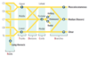
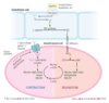
Neurovascular Pairing

Motoneuron Action Potential to Muscle Contraction
T-tubules are extensions of plasma membrane in contact with the sarcoplasmic reticulum, allowing for coordinated contraction of striated muscles.
① Action potential opens presynaptic voltage-gated Ca2+ channels, inducing acetylcholine
(ACh) release.
② Postsynaptic ACh binding leads to muscle cell depolarization at the motor end plate.
③ Depolarization travels over the entire muscle cell and deep into the muscle via the T-tubules.
④ Membrane depolarization induces conformational changes in the voltage-sensitive dihydropyridine receptor (DHPR) and its mechanically coupled ryanodine receptor (RR) → Ca2+ release from the sarcoplasmic reticulum into the cytoplasm.
⑤ Tropomyosin is blocking myosin-binding sites on the actin filament. Released Ca2+ binds to troponin C (TnC), shifting tropomyosin to expose the myosin-binding sites.
⑥ The myosin head binds strongly to actin, forming a crossbridge. Pi is then released, initiating the power stroke.
⑦ During the power stroke, force is produced as myosin pulls on the thin filament. Muscle shortening occurs, with shortening of H and I bands and between Z lines (HIZ shrinkage). The A band remains the same length (A band is Always the same length). ADP is released at the end of the power stroke.
⑧ Binding of new ATP molecule causes detachment of myosin head from actin filament. Ca2+ is resequestered.
⑨ ATP hydrolysis into ADP and Pi results in myosin head returning to high-energy position (cocked). The myosin head can bind to a new site on actin to form a crossbridge if Ca2+ remains available.

Types of Muscle Fibers:
- slow twitch
- red fibers resulting from ↑ mitochondria and myoglobin concentration (↑ oxidative phosphorylation) → sustained contraction
- proportion ↑ after endurance training
Type 1
1 slow red ox
Types of Muscle Fibers:
- fast twitch
- white fibers resulting from ↓ mitochondria and myoglobin concentration (↑ anaerobic glycolysis)
- proportion ↑ after weight/resistance training or sprinting
Type 2
Smooth Muscle Contraction and Relaxation
Bone Formation:
- bones of axial skeleton, appendicular skeleton, and base of skull
- Cartilaginous model of bone is first made by chondrocytes.
- Osteoclasts and osteoblasts later replace with woven bone and then remodel to lamellar bone.
- In adults, woven bone occurs after fractures and in Paget disease.
- defective in achondroplasia
Endochondral Ossification
Bone Formation:
- bones of calvarium, facial bones, and clavicle
- woven bone formed directly without cartilage
- later remodeled to lamellar bone
Membranous Ossification
Bone Histology:
- builds bone by secreting collagen and catalyzing mineralization in alkaline environment via ALP
- differentiates from mesenchymal stem cells in periosteum
- activity is measured by bone ALP, osteocalcin, and propeptides of type I procollagen
Osteoblast
Bone Histology:
- dissolves bone by secreting H+ and collagenases differentiates from a fusion of monocyte/macrophage lineage precursors
- RANK receptors on _____ are stimulated by RANKL (RANK ligand, secreted by osteoblasts)
- RANK receptors blocked by OPG (osteoprotegerin, a RANKL decoy receptor) → ↓ osteoclast activity
Osteoclast
Bone Endocrinology:
- at low, intermittent levels, exerts anabolic effects (building bone) on osteoblasts and osteoclasts (indirect)
- chronically ↑ levels (1° hyperparathyroidism) cause catabolic effects (osteitis fibrosa cystica)
Parathyroid Hormone
Bone Endocrinology:
- inhibits apoptosis in bone-forming osteoblasts and induces apoptosis in bone-resorbing osteoclasts
- causes closure of epiphyseal plate during puberty
- deficiency (surgical or postmenopausal) → ↑ cycles of remodeling and bone resorption → ↑ risk of osteoporosis
Estrogen
Musculoskeletal Pathologies:
- failure of longitudinal bone growth (endochondral ossification) → short limbs
- membranous ossification is affected → large head relative to limbs
- constitutive activation of fibroblast growth factor receptor (FGFR3) actually inhibits chondrocyte proliferation
- > 85% of mutations occur sporadically
- autosomal dominant with full penetrance (homozygosity is lethal)
- associated with ↑ paternal age
- most common cause of dwarfism
Achondroplasia
Musculoskeletal Pathologies:
- Trabecular (spongy) and cortical bone lose mass and interconnections despite normal bone mineralization and lab values (serum Ca2+ and PO43−).
- Most commonly due to ↑ bone resorption related to ↓ estrogen levels and old age
- Can be 2° to drugs (eg. steroids, alcohol, anticonvulsants, anticoagulants, thyroid replacement therapy) or other medical conditions (eg. hyperparathyroidism, hyperthyroidism, multiple myeloma, malabsorption syndromes).
- Diagnosed by bone mineral density measurement by DEXA (dual-energy X-ray absorptiometry) at the lumbar spine, total hip, and femoral neck, with a T-score of ≤ −2.5 or by a fragility fracture (eg. fall from standing height, minimal trauma) at hip or vertebra.
- One time screening recommended in women ≥ 65 years old.

Osteoporosis
Osteoporosis Prophylaxis
- regular weight-bearing exercise
- adequate Ca2+ and vitamin D intake throughout adulthood
Osteoporosis Treatment:
- Bisphosphonates
- Teriparatide
- SERMs
- Calcitonin
- Denosumab (monoclonal antibody against RANKL)
Osteoporosis can lead to _____—acute back pain, loss of height, kyphosis. Also can present with fractures of femoral neck, distal radius (Colles fracture).
Vertebral Compression Fractures
Musculoskeletal Pathologies:
- Failure of normal bone resorption due to defective osteoclasts → thickened, dense bones that are prone to fracture.
- Mutations (eg. carbonic anhydrase II) impair ability of osteoclast to generate acidic environment necessary for bone resorption.
- Overgrowth of cortical bone fills marrow space → pancytopenia, extramedullary hematopoiesis.
- Can result in cranial nerve impingement and palsies due to narrowed foramina.
- X-rays show diffuse symmetric sclerosis (bone-in-bone, “stone bone”).
- Bone marrow transplant is potentially curative as osteoclasts are derived from monocytes.
Osteopetrosis
Musculoskeletal Pathologies:
- Defective mineralization of osteoid (osteomalacia) or cartilaginous growth plates (rickets, only in children).
- Most commonly due to vitamin D deficiency.
- Children have pathologic bow legs (genu varum), bead-like costochondral junctions (rachitic rosary), craniotabes (soft skull).
- ↓ vitamin D → ↓ serum Ca2+ → ↑ PTH secretion → ↓ serum PO43−
- hyperactivity of osteoblasts → ↑ ALP
Osteomalacia/Rickets
Musculoskeletal Pathologies:
xrays show osteopenia and “Looser zones” (pseudofractures)
Osteomalacia
Musculoskeletal Pathologies:
x-rays show epiphyseal widening and metaphyseal cupping/fraying
Rickets
Musculoskeletal Pathologies:
- Common, localized disorder of bone remodeling caused by ↑ osteoclastic activity followed by ↑ osteoblastic activity that forms poor-quality bone
- Serum Ca2+, phosphorus, and PTH levels are normal
- ↑ ALP
- Mosaic pattern of woven and lamellar bone (osteocytes within lacunae in chaotic juxtapositions); long bone chalk-stick fractures.
- ↑ blood flow from ↑ arteriovenous shunts may cause high-output heart failure.
- ↑risk of osteogenic sarcoma.
- Hat size can be increased due to skull thickening.
- Hearing loss is common due to auditory foramen narrowing.
- Treatment: bisphosphonates
Paget Disease of Bone (Osteitis Deformans)
Stages of Paget Disease
- Lytic—osteoclasts
- Mixed—osteoclasts + osteoblasts
- Sclerotic—osteoblasts
- Quiescent—minimal osteoclast/osteoblast activity
Musculoskeletal Pathologies:
- infarction of bone and marrow
- usually very painful
- most common site is femoral head (watershed zone) due to insufficiency of medial circumflex femoral artery

Osteonecrosis (Avascular Necrosis)
Causes of Osteonecrosis (Avascular Necrosis)
CAST Bent LEGS:
- Corticosteroids
- Alcoholism
- Sickle cell disease
- Trauma
- “the Bends” (caisson/decompression disease)
- LEgg-Calvé-Perthes disease (idiopathic)
- Gaucher disease
- Slipped capital femoral epiphysis
Bone Disorders:
- — Serum Ca2+
- — PO43−
- — ALP
- — PTH
- ↓ bone mass
Osteoporosis
Bone Disorders:
- —/↓ Serum Ca2+
- — PO43−
- — ALP
- — PTH
- dense, brittle bones
- Ca2+ ↓ in severe, malignant disease
Osteopetrosis
Bone Disorders:
- — Serum Ca2+
- — PO43−
- ↑ ALP
- — PTH
- abnormal “mosaic” bone architecture
Paget Disease of Bone
Bone Disorders:
- ↑ Serum Ca2+
- ↓ PO43−
- ↑ ALP
- ↑ PTH
- “brown tumors” due to fibrous replacement of bone
- subperiosteal thinning
- idiopathic or parathyroid hyperplasia, adenoma, carcinoma
Osteitis Fibrosa Cystica
- Primary Hyperparathyroidism
Bone Disorders:
- ↓ Serum Ca2+
- ↑ PO43−
- ↑ ALP
- ↑ PTH
- often as compensation for CKD (↓ PO43 excretion and production of activated vitamin D)
Osteitis Fibrosa Cystica
- Secondary Hyperparathyroidism
Bone Disorders:
- ↓ Serum Ca2+
- ↓ PO43−
- ↑ ALP
- ↑ PTH
- soft bones
- vitamin D deficiency also causes 2° hyperparathyroidism
Osteomalacia/Rickets
Bone Disorders:
- ↑ Serum Ca2+
- ↑ PO43−
- — ALP
- ↓ PTH
- caused by oversupplementation or granulomatous disease (eg. sarcoidosis)
Hypervitaminosis D
_____ disease is more common than 1° bone tumors.
Metastatic
Primary Benign Bone Tumors
- Osteochondroma
- Osteoma
- Osteoid Osteoma
- Osteoblastoma
- Chondroma
- Giant Cell Tumor
Primary Benign Bone Tumors:
- most common benign bone tumor
- males < 25 years old
- metaphysis of long bones
- lateral bony projection of growth plate (continuous with marrow space) covered by cartilaginous cap
- rarely transforms to chondrosarcoma
Osteochondroma

Primary Benign Bone Tumors:
- middle age
- surface of facial bones
- associated with Gardner syndrome
Osteoma
Primary Benign Bone Tumors:
- adults < 25 years old
- males > females
- cortex of long bones
- presents as bone pain (worse at night) that is relieved by NSAIDs
- bony mass (< 2 cm) with radiolucent osteoid core
Osteoid Osteoma

Primary Benign Bone Tumors:
- vertebrae
- similar histology to osteoid osteoma
- larger size (> 2 cm)
- pain unresponsive to NSAIDs
Osteoblastoma
Primary Benign Bone Tumors:
- medulla of small bones of hand and feet
- benign tumor of cartilage
Chondroma
Primary Benign Bone Tumors:
- 20–40 years old
- epiphysis of long bones (often in knee region)
- locally aggressive benign tumor
- neoplastic mononuclear cells that express RANKL and reactive multinucleated giant (osteoclast-like) cells
- “Osteoclastoma”
- “soap bubble” appearance on x-ray
Giant Cell Tumor

Primary Malignant Bone Tumors
- Osteosarcoma (Osteogenic Sarcoma)
- Chondrosarcoma
- Ewing sarcoma
Primary Malignant Bone Tumors:
- 20% of 1° bone cancers
- peak incidence of 1° tumor in males < 20 years
- less common in elderly
- usually 2° to predisposing factors, such as Paget disease of bone, bone infarcts, radiation, familial retinoblastoma, and Li-Fraumeni syndrome
- metaphysis of long bones (often in knee region)
- pleomorphic osteoid-producing cells (malignant osteoblasts)
- presents as painful enlarging mass or pathologic fractures
- Codman triangle (from elevation of periosteum) or sunburst pattern on x-ray
- aggressive
- 1° usually responsive to treatment (surgery, chemotherapy)
- poor prognosis for 2°
Osteosarcoma (Osteogenic Sarcoma)

Primary Malignant Bone Tumors:
- medulla of pelvis and central skeleton
- tumor of malignant chondrocytes
Chondrosarcoma
Primary Malignant Bone Tumors:
- most common in Caucasians
- boys < 15 years old
- diaphysis of long bone (especially femur), pelvic flat bones
- anaplastic small blue cells of neuroectodermal origin (resemble lymphocytes)
- differentiate from conditions with similar morphology (eg. lymphoma, chronic osteomyelitis) by testing for t(11;22) (fusion protein EWS-FLI1)
- “onion skin” periosteal reaction in bone.
- aggressive with early metastases, but responsive to chemotherapy
Ewing Sarcoma

Arthritis:
- mechanical
- wear and tear destroys articular cartilage (degenerative joint disorder) → inflammation with inadequate repair
- chondrocytes mediate degradation and inadequate repair
Osteoarthritis
Arthritis:
- age
- female
- obesity
- joint trauma
Osteoarthritis
Arthritis:
- pain in weight-bearing joints after use (eg. at the end of the day)
- improving with rest
- asymmetric joint involvement
- knee cartilage loss begins medially (“bowlegged”)
- no systemic symptoms
Osteoarthritis
Arthritis:
- osteophytes (bone spurs)
- joint space narrowing
- subchondral sclerosis and cysts
- synovial fluid noninflammatory (WBC < 2000/mm3)
- DIP—Heberden nodes
- PIP—Bouchard nodes
- involves 1st CMC; not MCP
Osteoarthritis

Arthritis:
Osteoarthritis Treatment
- Acetaminophen
- NSAIDs
- intra-articular glucocorticoids
Arthritis:
- autoimmune
- inflammation induces formation of pannus (proliferative granulation tissue) which erodes articular cartilage and bone
Rheumatoid Arthritis
Arthritis:
- female
- HLA-DR4
- smoking
- ⊕ rheumatoid factor (IgM antibody that targets IgG Fc region; in 80%)
- anti-cyclic citrullinated peptide antibody (more specific)
Rheumatoid Arthritis
HLA-DR4 (4-walled “rheum”)
Arthritis:
- pain, swelling, and morning stiffness lasting > 1 hour
- improving with use
- symmetric joint involvement
- systemic symptoms (fever, fatigue, weight loss)
- extraarticular manifestations common
Rheumatoid Arthritis
Arthritis:
- rheumatoid nodules (fibrinoid necrosis with palisading histiocytes) in subcutaneous tissue and lung (+ pneumoconiosis → Caplan syndrome)
- interstitial lung disease
- pleuritis
- pericarditis
- anemia of chronic disease
- neutropenia + splenomegaly (Felty syndrome)
- AA amyloidosis
- Sjögren syndrome
- scleritis
- carpal tunnel syndrome
Rheumatoid Arthritis
Arthritis:
- erosions
- juxta-articular osteopenia
- soft tissue swelling
- subchondral cysts
- joint space narrowing
- Deformities:
- cervical subluxation
- ulnar finger deviation
- swan neck
- boutonniere
- involves MCP, PIP, wrist; not DIP or 1st CMC
- synovial fluid inflammatory
Rheumatoid Arthritis

Arthritis:
- NSAIDs
- glucocorticoids
- disease-modifying agents (methotrexate, sulfasalazine, hydroxychloroquine, leflunomide)
- biologic agents (eg. TNF-α inhibitors)
Rheumatoid Arthritis
Musculoskeletal Pathologies:
- acute inflammatory monoarthritis caused by precipitation of monosodium urate crystals in joints
- Risk Factors:
- male sex
- hypertension
- obesity
- diabetes
- dyslipidemia
- strongest risk factor is hyperuricemia
- crystals are needle shaped and ⊝ birefringent under polarized light (yellow under parallel light, blue under perpendicular light B )
Gout
Musculoskeletal Pathologies:
- Strongest risk factor is hyperuricemia, which can be caused by:
-
Underexcretion of Uric Acid (90% of patients)
- largely idiopathic, potentiated by renal failure;can be exacerbated by certain medications (eg. thiazide diuretics)
-
Overproduction of Uric Acid (10% of patients)
- Lesch-Nyhan syndrome, PRPP excess, ↑ cell turnover (eg. tumor lysis syndrome), von Gierke disease
-
Underexcretion of Uric Acid (90% of patients)
Gout
Musculoskeletal Pathologies:
- asymmetric joint distribution
- joint is swollen, red, and painful
- classic manifestation is painful MTP joint of big toe (podagra)
- tophus formation (often on external ear, olecranon bursa, or Achilles tendon)
- acute attack tends to occur after a large meal with foods rich in purines (eg. red meat, seafood), trauma, surgery, dehydration, diuresis, or alcohol consumption (alcohol metabolites compete for same excretion sites in kidney as uric acid → ↓ uric acid secretion and subsequent buildup in blood)
Gout
Treatment for Gout
- Acute:
- NSAIDs (eg. Indomethacin)
- Glucocorticoids
- Colchicine
- Chronic (preventive):
- Xanthine Oxidase Inhibitors (eg allopurinol, febuxostat)
Musculoskeletal Pathologies:
- previously called Pseudogout
- deposition of calcium pyrophosphate crystals within the joint space
- occurs in patients > 50 years old
- both sexes affected equally
- usually idiopathic, sometimes associated with hemochromatosis, hyperparathyroidism and joint trauma
- pain and swelling with acute inflammation (pseudogout) and/or chronic degeneration (pseudo-osteoarthritis)
- knee most commonly affected joint
- chondrocalcinosis (cartilage calcification) on x-ray
- crystals are rhomboid and weakly ⊕ birefringent under polarized light (blue when parallel to light)
- Acute Treatment:
- NSAIDs
- Colchicine
- Glucocorticoids
- Prophylaxis:
- Colchicine
Calcium Pyrophosphate Deposition Disease
The Blue P’s
- blue (when Parallel)
- Positive birefringent
- Calcium Pyrophosphate
- Pseudogout
Musculoskeletal Pathologies:
- childhood arthritis seen in < 12 year olds
- usually presents with daily spiking fevers, salmon-pink macular rash, uveitis, and arthritis (commonly 2+ joints) frequently presents with leukocytosis, thrombocytosis, anemia, ↑ ESR, ↑ CRP
- Treatment:
- NSAIDs
- Steroids
- Methotrexate
- TNF Inhibitors
Systemic Juvenile Idiopathic Arthritis
Musculoskeletal Pathologies:
- autoimmune disorder characterized by destruction of exocrine glands (especially lacrimal and salivary) by lymphocytic infiltrates
- women 40–60 years old
- Findings:
- inflammatory joint pain
- keratoconjunctivitis sicca (↓ tear production and subsequent corneal damage)
- xerostomia (↓ saliva production)
- presence of antinuclear antibodies, rheumatoid factor (can be in the absence of rheumatoid arthritis), antiribonucleoprotein antibodies: SS-A (anti-Ro) and/or SS-B (anti-La)
- bilateral parotid enlargement
Sjögren Syndrome
Musculoskeletal Pathologies:
- a common 1° disorder or a 2° syndrome associated with other autoimmune disorders (eg. rheumatoid arthritis, SLE, systemic sclerosis)
- Anti-SSA and Anti-SSB may also be seen in SLE
- ⊕ Anti-SSA in pregnant women with SLE → ↑ risk of congenital heart block in the newborn
- Complications:
- dental caries
- Mucosa-Associated Lymphoid Tissue (MALT) lymphoma (may present as parotid enlargement)
- focal lymphocytic sialadenitis on labial salivary gland biopsy can confirm diagnosis
Sjögren Syndrome
Musculoskeletal Pathologies:
- S. aureus, Streptococcus, and Neisseria gonorrhoeae are common causes
- affected joint is swollen, red, and painful
- synovial fluid purulent (WBC > 50,000/mm3)
Septic Arthritis
Musculoskeletal Pathologies:
STI that presents as either purulent arthritis (eg. knee) or triad of polyarthralgia, tenosynovitis (eg. hand), dermatitis (eg. pustules)
Gonococcal Arthritis
Musculoskeletal Pathologies:
- arthritis without rheumatoid factor (no anti-IgG antibody)
- strong association with HLA-B27 (MHC class I serotype)
- subtypes share variable occurrence of inflammatory back pain (associated with morning stiffness, improves with exercise), peripheral arthritis, enthesitis (inflamed insertion sites of tendons, eg. Achilles), dactylitis (“sausage fingers”), uveitis
Seronegative Spondyloarthritis
Seronegative Spondyloarthritis Subtypes
PAIR:
- Psoriatic Arthritis
- Ankylosing Spondylitis
- Inflammatory Bowel Disease
- Reactive Arthritis
Seronegative Spondyloarthritis Subtypes:
- associated with skin psoriasis and nail lesions
- asymmetric and patchy involvement
- dactylitis and “pencil-in-cup” deformity of DIP on x-ray
- seen in fewer than 1 ⁄3 of patients with psoriasis
Psoriatic Arthritis
Seronegative Spondyloarthritis Subtypes:
- symmetric involvement of spine and sacroiliac joints → ankylosis (joint fusion), uveitis, aortic regurgitation
- bamboo spine (vertebral fusion)
- can cause restrictive lung disease due to limited chest wall expansion (costovertebral and costosternal ankylosis)
- more common in males
Ankylosing Spondylitis
Seronegative Spondyloarthritis Subtypes:
Crohn disease and ulcerative colitis are often
associated with spondyloarthritis
Inflammatory Bowel Disease
Seronegative Spondyloarthritis Subtypes:
- formerly known as Reiter syndrome
- Classic Triad:
- conjunctivitis
- urethritis
- arthritis
- Shigella, Yersinia, Chlamydia, Campylobacter, Salmonella
Reactive Arthritis
Can’t see, can’t pee, can’t bend my knee.
- conjunctivitis
- urethritis
- arthritis
ShY ChiCS:
- Shigella
- Yersinia
- Chlamydia
- Campylobacter
- Salmonella
Musculoskeletal Pathologies:
- systemic, remitting, and relapsing autoimmune disease
- organ damage primarily due to a type III hypersensitivity reaction and, to a lesser degree, a type II hypersensitivity reaction associated with deficiency of early complement proteins (eg. C1q, C4, C2) → ↓ clearance of of immune complexes
- Classic Presentation:
- rash
- joint pain
- female
- reproductive age
- African-American or Hispanic
Systemic Lupus Erythematosus
Systemic Lupus Erythematosus:
nonbacterial, verrucous thrombi usually on mitral or aortic valve and can be present on either surface of the valve (but usually on undersurface)
Libman-Sacks Endocarditis
LSE in SLE
Systemic Lupus Erythematosus:
- glomerular deposition of DNA-anti-DNA immune complexes
- can be nephritic or nephrotic (causing hematuria or proteinuria)
- most common and severe type is diffuse proliferative
Lupus Nephritis
Common Causes of Death in SLE
Lupus patients die with Redness In their Cheeks.
- Renal disease (most common)
- Infections
- Cardiovascular disease (accelerated CAD)
SLE Criteria
RASH OR PAIN:
- Rash (malar or discoid)
- Arthritis (nonerosive)
- Serositis (eg. pleuritis, pericarditis)
- Hematologic disorders (eg. cytopenias)
- Oral/nasopharyngeal ulcers (usually painless)
- Renal disease
- Photosensitivity
- Antinuclear antibodies
- Immunologic disorder (anti-dsDNA, anti-Sm, antiphospholipid)
- Neurologic disorders (eg. seizures, psychosis)
Musculoskeletal Pathologies:
- 1° or 2° autoimmune disorder (most commonly in SLE)
- diagnose based on clinical criteria including history of thrombosis (arterial or venous) or spontaneous abortion along with laboratory findings of Lupus Anticoagulant, Anticardiolipin, Anti-β2 Glycoprotein Antibodies
- treated with systemic anticoagulation
- Anticardiolipin Antibodies can cause false positive VDRL/RPR
- Lupus Anticoagulant can cause prolonged PTT that is not corrected by the addition of normal platelet-free plasma
Antiphospholipid Syndrome
Musculoskeletal Pathologies:
- features of SLE, systemic sclerosis, and/or polymyositis
- associated with anti-U1 RNP antibodies (speckled ANA)
Mixed Connective Tissue Disease
Musculoskeletal Pathologies:
- pain and stiffness in proximal muscles (eg. shoulders, hips), often with fever, malaise, and weight loss
- does not cause muscular weakness
- more common in women > 50 years old
- associated with giant cell (temporal) arteritis
- ↑ ESR, ↑ CRP, normal CK
- rapid response to low-dose corticosteroids
Polymyalgia Rheumatica
Musculoskeletal Pathologies:
- most common in women 20–50 years old
- chronic, widespread musculoskeletal pain associated with “tender points,” stiffness, paresthesias, poor sleep, fatigue, cognitive disturbance (“fibro fog”)
- Treatment:
- regular exercise
- antidepressants (TCAs, SNRIs)
- neuropathic pain agents (eg. gabapentin)
Fibromyalgia
Musculoskeletal Pathologies:
- ↑ CK, ⊕ ANA (nonspecific), ⊕ anti-Jo-1 (histidyl-tRNA synthetase) (specific), ⊕ anti-SRP (specific), ⊕ anti-Mi-2 (specific) antibodies
- associated with interstitial lung disease
- Treatment:
- steroids followed by long-term immunosuppressant therapy (eg. methotrexate)
Polymyositis/Dermatomyositis
Musculoskeletal Pathologies:
- progressive symmetric proximal muscle weakness, characterized by endomysial inflammation with CD8+ T cells
- most often involves shoulders
Polymyositis
Musculoskeletal Pathologies:
- clinically similar to polymyositis, but also involves malar rash (similar to that in SLE but involves nasolabial folds), Gottron papules, heliotrope (violaceous periorbital) rash, “shawl and face” rash, darkening and thickening of fingertips and sides resulting in irregular, “dirty”-appearing marks
- ↑ risk of occult malignancy
- perimysial inflammation and atrophy with CD4+ T cells
Dermatomyositis
Neuromuscular Junction Diseases:
- most common NMJ disorder
- autoantibodies to postsynaptic ACh receptor
- ptosis, diplopia, weakness (respiratory muscle involvement can lead to dyspnea)
- worsens with muscle use
- improvement after Edrophonium (tensilon) test
- thymoma, thymic hyperplasia
- AChE inhibotor reverses symptoms
- Edrophonium to diagnose
- Pyridostigmine to treat
Myasthenia Gravis
Neuromuscular Junction Diseases:
- uncommon
- autoantibodies to presynaptic Ca2+ channel → ↓ ACh release
- proximal muscle weakness, autonomic symptoms (dry mouth, impotence)
- improves with muscle use
- small cell lung cancer
- AChE inhibitor has minimal effect
Lambert-Eaton Myasthenic Syndrome
Musculoskeletal Pathologies:
- ↓ blood flow to skin due to arteriolar (small vessel) vasospasm in response to cold or stress: white (ischemia) → blue (hypoxia) → red (reperfusion)
- most often in the fingers and toes
- called _____ disease when 1° (idiopathic), _____ syndrome when 2° to a disease process such as mixed connective tissue disease, SLE, or CREST syndrome (limited form of systemic sclerosis)
- digital ulceration (critical ischemia) seen in 2° _____ syndrome
- treat with Ca2+ channel blockers
Raynaud Phenomenon
Musculoskeletal Pathologies:
- triad of autoimmunity, noninflammatory vasculopathy, and collagen deposition with fibrosis
- commonly sclerosis of skin, manifesting as puffy, taut skin without wrinkles, fingertip pitting
- can involve other systems, eg. renal (renal crisis; treat with ACE inhibitors), pulmonary (interstitial fibrosis, pulmonary HTN), GI (esophageal dysmotility and reflux), cardiovascular
- 75% female
Scleroderma (Systemic Sclerosis)
Scleroderma:
- widespread skin involvement, rapid progression, early visceral involvement
- associated with anti-Scl-70 antibody (anti-DNA topoisomerase I antibody)
Diffuse Scleroderma
Scleroderma:
- limited skin involvement confined to fingers and face
- Also with CREST Syndrome:
- Calcinosis Cutis
- Anti-Centromere Antibody
- Raynaud Phenomenon
- Esophageal Dysmotility
- Sclerodactyly
- Telangiectasia
- more benign clinical course
Limited Scleroderma
Skin Layers
Skin has 3 layers:
- epidermis
- dermis
- subcutaneous fat (hypodermis, subcutis)
Epidermis Layers (surface → base):
Californians Like Girls in String Bikinis.
- Stratum Corneum (keratin)
- Stratum Lucidum (most prominent in palms and soles)
- Stratum Granulosum
- Stratum Spinosum (desmosomes)
- Stratum Basale (stem cell site)
Epithelial Cell Junctions

Dermatologic Macroscopic Terms:
- flat lesion with well-circumscribed change in skin color < 1 cm
- freckles
Macule
Dermatologic Macroscopic Terms:
- macule > 1 cm
- large birthmark (congenital nevus)
Patch
Dermatologic Macroscopic Terms:
- elevated solid skin lesion < 1 cm
- mole (nevus)
- acne
Papule
Dermatologic Macroscopic Terms:
- papule > 1 cm
- psoriasis
Plaque
Dermatologic Macroscopic Terms:
- small fluid-containing blister < 1 cm
- chickenpox (varicella)
- shingles (zoster)
Vesicle
Dermatologic Macroscopic Terms:
- large fluid-containing blister > 1 cm
- bullous pemphigoid
Bulla
Dermatologic Macroscopic Terms:
- vesicle containing pus
- pustular psoriasis
Pustule
Dermatologic Macroscopic Terms:
- transient smooth papule or plaque
- hives (urticaria)
Wheal
Dermatologic Macroscopic Terms:
- flaking off of stratum corneum
- eczema
- psoriasis
- SCC
Scale
Dermatologic Macroscopic Terms:
- dry exudate
- impetigo
Crust
Dermatologic Microscopic Terms:
- ↑ thickness of stratum corneum
- psoriasis
- calluses
Hyperkeratosis
Dermatologic Microscopic Terms:
- retention of nuclei in stratum corneum
- psoriasis
Parakeratosis
Dermatologic Microscopic Terms:
- ↑ thickness of stratum granulosum
- lichen planus
Hypergranulosis
Dermatologic Microscopic Terms:
- epidermal accumulation of edematous fluid in intercellular spaces
- eczematous dermatitis
Spongiosis
Dermatologic Microscopic Terms:
- separation of epidermal cells
- pemphigus vulgaris
Acantholysis
Dermatologic Microscopic Terms:
- epidermal hyperplasia (↑ spinosum)
- acanthosis nigricans
Acanthosis
Pigmented Skin Disorders:
- normal melanocyte number with ↓ melanin production due to ↓ tyrosinase activity or defective tyrosine transport
- ↑ risk of skin cancer
Albinism
Pigmented Skin Disorders:
hyperpigmentation associated with pregnancy (“mask of pregnancy”) or OCP use
Melasma (Chloasma)
Pigmented Skin Disorders:
- irregular patches of complete depigmentation
- caused by autoimmune destruction of melanocytes
Vitiligo
Skin Disorders:
- erythematous, well-demarcated plaques with greasy yellow scales in areas rich in sebaceous glands, such as scalp, face, and periocular region
- common in both infants and adults
- associated with Parkinson disease
- sebaceous glands are not inflamed, but play a role in disease development
- possibly associated with Malassezia spp.
- treated with topical antifungals and corticosteroids
Seborrheic Dermatitis
Common Skin Disorders:
- multifactorial etiology
- ↑ sebum/androgen production, abnormal keratinocyte desquamation, Cutibacterium (formerly Propionibacterium) acnes colonization of the pilosebaceous unit (comedones), and inflammation (papules/pustules, nodules, cysts)
- Treatment:
- Retinoids
- Benzoyl Peroxide
- Antibiotics
Acne
Common Skin Disorders:
- pruritic eruption, commonly on skin flexures
- associated with other atopic diseases (asthma, allergic rhinitis, food allergies)
- ↑ serum IgE
- mutations in filaggrin gene ↑ risk (via skin barrier dysfunction)
- often appears on face in infancy and then in antecubital fossa in children and adults
Atopic Dermatitis (Eczema)
Common Skin Disorders:
- type IV hypersensitivity reaction that follows exposure to allergen
- lesions occur at site of contact (eg. nickel, poison ivy, neomycin)
Allergic Contact Dermatitis
Common Skin Disorders:
- common mole
- benign, but melanoma can arise in congenital or atypical moles
- Intradermal Nevi are papular
- Junctional Nevi are flat macules
Melanocytic Nevus
Common Skin Disorders:
- foreign body inflammatory facial skin disorder characterized by firm, hyperpigmented papules and pustules that are painful and pruritic
- located on cheeks, jawline, and neck
- commonly occurs as a result of shaving (“razor bumps”)
- primarily affects African-American males
Pseudofolliculitis Barbae
Common Skin Disorders:
- papules and plaques with silvery scaling, especially on knees and elbows
- acanthosis with parakeratotic scaling (nuclei still in stratum corneum)
- Munro microabscesses
- ↑ stratum spinosum, ↓ stratum granulosum
- Auspitz Sign
- pinpoint bleeding spots from exposure of dermal papillae when scales are scraped off
- associated with nail pitting and _____ arthritis
Psoriasis
Common Skin Disorders:
- inflammatory facial skin disorder characterized by erythematous papules and pustules, but no comedones
- may be associated with facial flushing in response to external stimuli (eg. alcohol, heat)
- phymatous _____ can cause rhinophyma (bulbous deformation of nose)
Rosacea
Common Skin Disorders:
- flat, greasy, pigmented squamous epithelial proliferation with keratin-filled cysts (horn cysts)
- looks “stuck on”
- lesions occur on head, trunk, and extremities
- common benign neoplasm of older persons
- Leser-Trélat Sign
- sudden appearance of multiple lesions, indicating an underlying malignancy (eg. GI, lymphoid)
Seborrheic Keratosis
Common Skin Disorders:
- warts
- caused by low-risk HPV strains
- soft, tan-colored, cauliflower-like papules
- epidermal hyperplasia, hyperkeratosis, and koilocytosis
- condyloma acuminatum on anus or genitals
Verrucae
Common Skin Disorders:
- hives
- pruritic wheals that form after mast cell degranulation
- characterized by superficial dermal edema and lymphatic channel dilation
Urticaria
Vascular Tumors of the Skin:
- rare blood vessel malignancy typically occurring in the head, neck, and breast areas
- usually in elderly, on sun-exposed areas
- associated with radiation therapy and chronic postmastectomy lymphedema
- hepatic angiosarcoma associated with vinyl chloride and arsenic exposures
- very aggressive and difficult to resect due to delay in diagnosis
Angiosarcoma
Vascular Tumors of the Skin:
- benign capillary skin papules found in AIDS patients
- caused by Bartonella infections
- frequently mistaken for Kaposi sarcoma, but has neutrophilic infiltrate
Bacillary Angiomatosis
Vascular Tumors of the Skin:
- benign capillary hemangioma of the elderly
- does not regress
- frequency ↑ with age
Cherry Hemangioma
Vascular Tumors of the Skin:
- cavernous lymphangioma of the neck
- associated with Turner syndrome
Cystic Hygroma
Vascular Tumors of the Skin:
- benign, painful, red-blue tumor, commonly under fingernails
- arises from modified smooth muscle cells of the thermoregulatory glomus body
Glomus Tumor
Vascular Tumors of the Skin:
- endothelial malignancy most commonly of the skin, but also mouth, GI tract, and respiratory tract
- associated with HHV-8 and HIV
- rarely mistaken for bacillary angiomatosis, but has lymphocytic infiltrate
Kaposi Sarcoma
Vascular Tumors of the Skin:
- polypoid lobulated capillary hemangioma that can ulcerate and bleed
- associated with trauma and pregnancy
Pyogenic Granuloma
Vascular Tumors of the Skin:
- benign capillary hemangioma of infancy
- appears in first few weeks of life (1/200 births)
- grows rapidly and regresses spontaneously by 5–8 years old
Strawberry Hemangioma
Bacterial Skin Infections:
- very superficial skin infection
- usually from S. aureus or S. pyogenes
- highly contagious
- honeycolored crusting
- bullous _____ has bullae and is usually caused by S. aureus
Impetigo
Bacterial Skin Infections:
- infection involving upper dermis and superficial lymphatics, usually from S. pyogenes
- presents with well-defined, raised demarcation between infected and normal skin
Erysipelas
Bacterial Skin Infections:
- acute, painful, spreading infection of deeper dermis and subcutaneous tissues
- usually from S. pyogenes or S. aureus
- often starts with a break in skin from trauma or another infection
Cellulitis
Bacterial Skin Infections:
- collection of pus from a walled-off infection within deeper layers of skin
- offending organism is almost always S. aureus
Abscess
Bacterial Skin Infections:
- deeper tissue injury, usually from anaerobic bacteria or S. pyogenes
- pain may be out of proportion to exam findings
- results in crepitus from methane and CO2 production
- “flesh-eating bacteria”
- causes bullae and a purple color to the skin
- surgical emergency
Necrotizing Fasciitis
Bacterial Skin Infections:
- exotoxin destroys keratinocyte attachments in stratum granulosum only (vs. toxic epidermal necrolysis, which destroys epidermal-dermal junction)
- characterized by fever and generalized erythematous rash with sloughing of the upper layers of the epidermis that heals completely
- ⊕ Nikolsky Sign
- separation of epidermis upon manual stroking of skin
- seen in newborns and children, adults with renal insufficiency
Staphylococcal Scalded Skin Syndrome
Viral Skin Infections:
- HSV1 and HSV2 infections of skin can occur anywhere from mucosal surfaces to normal skin
- these include _____ labialis, _____ genitalis, _____ whitlow (finger)
Herpes
Viral Skin Infections:
- umbilicated papules caused by a poxvirus
- while frequently seen in children, it may be sexually transmitted in adults
Molluscum Contagiosum
Viral Skin Infections:
- causes varicella (chickenpox) and zoster (shingles)
- varicella presents with multiple crops of lesions in various stages from vesicles to crusts
- zoster is a reactivation of the virus in dermatomal distribution (unless it is disseminated)
Varicella Zoster Virus
Viral Skin Infections:
- irregular, white, painless plaques on lateral tongue that cannot be scraped off
- EBV mediated
- occurs in HIV-positive patients and organ transplant recipients
- contrast with thrush (scrapable) and leukoplakia (precancerous)
Hairy Leukoplakia
Blistering Skin Disorders:
- potentially fatal autoimmune skin disorder with IgG antibody against desmoglein (component of desmosomes, which connect keratinocytes in the stratum spinosum)
- flaccid intraepidermal bullae caused by acantholysis (separation of keratinocytes, resembling a “row of tombstones”)
- oral mucosa is also involved
- type II hypersensitivity reaction
- immunofluorescence reveals antibodies around epidermal cells in a reticular (net-like) pattern
- Nikolsky sign ⊕
Pemphigus Vulgaris
Blistering Skin Disorders:
- less severe than pemphigus vulgaris
- type II hypersensitivity reaction: involves IgG antibody against hemidesmosomes (epidermal basement membrane)
- tense blisters containing eosinophils affect skin but spare oral mucosa
- immunofluorescence reveals linear pattern at epidermal-dermal junction
- Nikolsky sign ⊝
Bullous Pemphigoid
Blistering Skin Disorders:
- pruritic papules, vesicles, and bullae (often found on elbows)
- deposits of IgA at tips of dermal papillae
- associated with celiac disease
- Treatment:
- Dapsone
- gluten-free diet
Dermatitis Herpetiformis
Blistering Skin Disorders:
- associated with infections (eg. Mycoplasma pneumoniae, HSV), drugs (eg. sulfa drugs, β-lactams, phenytoin), cancers, autoimmune disease
- presents with multiple types of lesions—macules, papules, vesicles, target lesions (look like targets with multiple rings and dusky center showing epithelial disruption)
Erythema Multiforme
Blistering Skin Disorders:
- characterized by fever, bullae formation and necrosis, sloughing of skin at dermal-epidermal junction, high mortality rate
- typically 2 mucous membranes are involved, and targetoid skin lesions may appear, as seen in erythema multiforme
- usually associated with adverse drug reaction.
- a more severe form of with > 30% of the body surface area involved is Toxic Epidermal Necrolysis (TEN), 10–30% involvement denotes SJS-TEN
Stevens-Johnson Syndrome
Skin Disorders:
- epidermal hyperplasia causing symmetric, hyperpigmented thickening of skin, especially in axilla or on neck
- associated with insulin resistance (eg. diabetes, obesity, Cushing syndrome), visceral malignancy (eg. gastric adenocarcinoma)
Acanthosis Nigricans
Skin Disorders:
- premalignant lesions caused by sun exposure
- small, rough, erythematous or brownish papules or plaques
- risk of squamous cell carcinoma is proportional to degree of epithelial dysplasia
Actinic Keratosis
Skin Disorders:
- painful, raised inflammatory lesions of subcutaneous fat (panniculitis), usually on anterior shins
- often idiopathic, but can be associated with sarcoidosis, coccidioidomycosis, histoplasmosis, TB, streptococcal infections, leprosy, inflammatory bowel disease
Erythema Nodosum
Skin Disorders:
- mucosal involvement manifests as Wickham striae (reticular white lines) and hypergranulosis
- sawtooth infiltrate of lymphocytes at dermal-epidermal junctiona
- associated with hepatitis C
Lichen Planus
6 P’s: Pruritic, Purple, Polygonal Planar Papules and Plaques
Skin Disorders:
- “Herald patch” followed days later by other scaly erythematous plaques, often in a “Christmas tree” distribution on trunk
- multiple pink plaques with collarette scale
- self-resolving in 6–8 weeks
Pityriasis Rosea
Skin Disorders:
- acute cutaneous inflammatory reaction due to excessive UV irradiation
- causes DNA mutations, inducing apoptosis of keratinocytes
- exposure to UVA and UVB ↑ risk of skin cancer
- can also lead to impetigo
Sunburn
UVA—tAnning, photoAging
UVB—sunBurn
Burn Classifications:
- superficial
- through epidermis (eg. common sunburn)
- painful, erythematous, blanching
First-Degree Burn
Burn Classifications:
- partial-thickness burn through epidermis and dermis
- skin is blistered and usually heals without scarring
- painful, erythematous, blanching
Second-Degree Burn
Burn Classifications:
- full-thickness burn through epidermis, dermis, and hypodermis
- skin scars with wound healing
- painless, waxy or leathery appearance, nonblanching
Third-Degree Burn
Skin Cancer:
- most common skin cancer
- found in sun-exposed areas of body (eg. face)
- locally invasive, but rarely metastasizes
- waxy, pink, pearly nodules, commonly with telangiectasias, rolled borders, central crusting or ulceration
- also appear as nonhealing ulcers with infiltrating growth or as a scaling plaque (superficial)
- “palisading” nuclei
Basal Cell Carcinoma
Skin Cancer:
- second most common skin cancer
- associated with excessive exposure to sunlight, immunosuppression, chronically draining sinuses, and occasionally arsenic exposure
- commonly appears on face, lower lip, ears, and hands locally invasive, may spread to lymph nodes, and will rarely metastasize
- ulcerative red lesions with frequent scale
- Histopathology: keratin “pearls”
- Actinic Keratosis, a scaly plaque, is a precursor
- Keratoacanthoma is a variant that grows rapidly (4–6 weeks) and may regress spontaneously over month
Squamous Cell Carcinoma
Skin Cancer:
- common tumor with significant risk of metastasis
- S-100 tumor marker
- associated with sunlight exposure and dysplastic nevi
- fair-skinned persons are at ↑ risk
- depth of tumor (Breslow thickness) correlates with risk of metastasis
- At least 4 different types:
- superficial spreading
- nodular
- lentigo maligna
- acral lentiginous (highest prevalence in African-Americans and Asians)
- often driven by activating mutation in BRAF kinase
- primary treatment is excision with appropriately wide margins
- metastatic or unresectable lesions in patients with BRAF V600E mutation may benefit from Vemurafenib, a BRAF kinase inhibitor
Melanoma
ABCDEs:
- Asymmetry
- Border irregularity
- Color variation
- Diameter > 6 mm
- Evolution over time
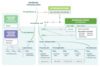
Arachidonic Acid Pathway
- LTB4 is a neutrophil chemotactic agent.
- Neutrophils arrive “B4” others.
-
PGI2 inhibits platelet aggregation and promotes vasodilation.
- Platelet-Gathering Inhibitor
MSK Drugs:
- reversibly inhibits cyclooxygenase, mostly in CNS
- inactivated peripherally
- antipyretic, analgesic, but not anti-inflammatory
- used instead of aspirin to avoid Reye syndrome
- in children with viral infection
- overdose produces hepatic necrosis
- metabolite (NAPQI) depletes glutathione and forms toxic tissue byproducts in liver
- N-acetylcysteine is the antidote—regenerates glutathione
Acetaminophen
MSK Drugs:
- NSAID that irreversibly inhibits cyclooxygenase (both COX-1 and COX-2) by covalent acetylation → ↓ synthesis of TXA2 and prostaglandins
- ↑ bleeding time
- no effect on PT/PTT
- effect lasts until new platelets are produced
- Low Dose (< 300 mg/day): ↓ platelet aggregation
- Intermediate Dose (300–2400 mg/day): antipyretic and analgesic
- High Dose (2400–4000 mg/day): anti-inflammatory
- causes gastric ulceration, tinnitus (CN VII), allergic reactions (especially in patients with asthma or nasal polyps)
- chronic use can lead to acute renal failure, interstitial nephritis, and GI bleeding
- risk of Reye syndrome in children with viral infection
- toxic doses cause respiratory alkalosis early, but transitions to mixed metabolic acidosis-respiratory alkalosis
Aspirin
MSK Drugs:
- reversibly and selectively inhibits the cyclooxygenase (COX) isoform 2, which is found in inflammatory cells and vascular endothelium and mediates inflammation and pain
- spares COX-1, which helps maintain gastric mucosa, thus, does not have the corrosive effects of other NSAIDs on the GI lining
- spares platelet function as TXA2 production is dependent on COX-1
- used for rheumatoid arthritis and osteoarthritis
- ↑ risk of thrombosis, sulfa allergy
Celecoxib
Nonsteroidal Anti-Inflammatory Drugs
- Ibuprofen
- Naproxen
- Indomethacin
- Ketorolac
- Diclofenac
- Meloxicam
- Piroxicam
MSK Drugs:
- reversibly inhibit cyclooxygenase (both COX-1 and COX-2)
- block prostaglandin synthesis
- antipyretic, analgesic, and anti-inflammatory
- Indomethacin is used to close a PDA
- causes interstitial nephritis, gastric ulcer (prostaglandins protect gastric mucosa), renal ischemia (prostaglandins vasodilate afferent arteriole), and aplastic anemia
Nonsteroidal Anti-Inflammatory Drugs
MSK Drugs:
- reversibly inhibits dihydroorotate dehydrogenase, preventing pyrimidine synthesis
- suppresses T-cell proliferation
- used for rheumatoid arthritis and psoriatic arthritis
- causes diarrhea, hypertension, hepatotoxicity, and teratogenicity
Leflunomide
Bisphosphonates
- Alendronate
- Ibandronate
- Risedronate
- Zoledronate
MSK Drugs:
- pyrophosphate analogs
- bind hydroxyapatite in bone, inhibiting osteoclast activity
- used for osteoporosis, hypercalcemia, Paget disease of bone, metastatic bone disease, and osteogenesis imperfecta
- causes esophagitis (if taken orally, patients are advised to take with water and remain upright for 30 minutes), osteonecrosis of jaw, and atypical femoral stress fractures
Bisphosphonates
MSK Drugs:
- recombinant PTH analog
- ↑ osteoblastic activity when administered in pulsatile fashion
- used for osteoporosis
- causes ↑ bone growth compared to antiresorptive therapies (eg. bisphosphonates)
- ↑ risk of osteosarcoma (avoid use in patients with Paget disease of the bone or unexplained elevation of alkaline phosphatase)
- avoid in patients who have had prior cancers or radiation therapy
- causes transient hypercalcemia
Teriparatide
Chronic Gout Drugs (Preventive)
Prevent A Painful Flare.
- Probenecid
- Allopurinol
- Pegloticase
- Febuxostat
Chronic Gout Drugs:
- inhibits reabsorption of uric acid in proximal convoluted tubule (also inhibits secretion of Penicillin)
- can precipitate uric acid calculi
Probenecid

Chronic Gout Drugs:
- competitive inhibitor of xanthine oxidase → ↓ conversion of hypoxanthine and xanthine to urate
- also used in lymphoma and leukemia to prevent tumor lysis–associated urate nephropathy
- ↑ concentrations of azathioprine and 6-MP (both normally metabolized by xanthine oxidase)
Allopurinol

Chronic Gout Drugs:
recombinant uricase catalyzing uric acid to allantoin (a more water-soluble product)
Pegloticase

Chronic Gout Drugs:
inhibits xanthine oxidase
- Allopurinol
- Febuxostat

Acute Gout Drugs
- NSAIDs
- Glucocorticoids
- Colchicine
Acute Gout Drugs:
- antipyretic, analgesic, and anti-inflammatory
- use salicylates with caution (may decrease uric acid excretion, particularly at low doses)
NSAIDs
Acute Gout Drugs:
oral, intra-articular, or parenteral
Glucocorticoids
Acute Gout Drugs:
- binds and stabilizes tubulin to inhibit microtubule polymerization, impairing neutrophil chemotaxis and degranulation
- acute and prophylactic value
- GI and neuromyopathic side effects
Colchicine
TNF-α Inhibitors:
- fusion protein (decoy receptor for TNF-α + IgG1 Fc)
- produced by recombinant DNA
- intercepts TNF
- used for rheumatoid arthritis, psoriasis,and ankylosing spondylitis
- causes predisposition to infection, including reactivation of latent TB, since TNF is important in granuloma formation and stabilization
- can also lead to drug-induced lupus
Etanercept
TNF-α Inhibitors:
- anti-TNF-α monoclonal antibody
- used for inflammatory bowel disease, rheumatoid arthritis, ankylosing spondylitis, and psoriasis
- causes predisposition to infection, including reactivation of latent TB, since TNF is important in granuloma formation and stabilization
- can also lead to drug-induced lupus
- Infliximab
- Adalimumab
- Certolizumab
- Golimumab
