Liver Disease and Peptic Ulcer Flashcards
What is the main blood supply to most of the liver? What is this vessel a conjunction of?
The portal vein. The portal vein is formed by the union of the splenic and superior mesenteric veins.
Blood in the central veins of the ___________ lobules then drain into the _______ ________. This subsequently drains into the _____.
hepatic, hepatic vein, IVC.

Explain the architecture of the hepatic lobules? (Hint: what is in the middle, what is on each corner of the lobule?).

The _____ triad contains what 3 structures?
portal vein, hepatic artery and a bile duct.
Do periportal hepatocytes or centrilobular hepatocytes have an advantage in survival?
Periportal ones have an advantage because they are closest to the blood supply.
Explain bile flow in the liver, where does it eventually end up and how?

The right and left hepatic ducts form the ___________, this joins to the ________ (which is connected to the _______) to form the ___________. This structure then meets with the _________ to form the ________ which enters into the ________
common hepatic duct, cystic hepatic duct, gall bladder, common bile duct, pancreatic duct, Ampulla of Vater which enters into the 2nd portion of the duodenum.

The sinusoid of the liver is lined with _____________ lacking a ____________. The space between the sinusoid and the hepatocytes are called the __________.
fenestrated endothelium lacking a basement membrane. space of Disse.
What cell is important in fibrous tissue deposits in the liver? Where is it normally found? What stimulates them?
Hepatic Stellate cells - found in space of disse.
Kupffer cells in the presence of injurious stimuli produce cytokines to activate them.

What structure are hepatocytes normally supported with? Is this regeneratable after injury
The reticulin frame work

Explain biliruben metabolism and excretion?
What happens if there is a billary block?

Explain these 3 lab evaluations of liver disease: hepatocyte integrity, billary obstruction, hepatocyte synthetic function).

What is the difference between Hep A, B and C diseases? What route of admission do they occur?

What would you see during acute viral hepatitis in a histology? Is regeneration of the lobules possible?

What are the two major clinical findings in acute hepatitis and why?

What are the two hallmark signs of chronic hepatitis? (compared with acute viral hepatitis?)
How does cirrhosis form in the liver (Step by step?).
What is cirrhosis?
Cirrhosis is parenchymal nodules surrounded by fibrosis (and is the end stage of many chronic liver diseases).

How does alcohol cause liver disease? (pathogenesis)

What would you see with fatty liver disease? (under the microscope an macroscopically)

what is seen microscopically with alcohol hepatisis?

what are mallory bodies?
clumped keratin filaments in the cytoplasm of hepatocytes instead of being evenly distributed.
What is alcohol steatofibrosis?

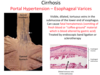
How does portal hypertension occur and what does this cause?
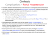
What are porto-systemic anastomoses?
Where do they go?
blood can bypass the vascular obstruction in the liver to reach the IVC.
The possible portosystemic anastomoses are the esophageal varices, in the rectum giving haemorrhoids, around the umbilicus giving caput madusae.