Joints: Upper & Lower Limb Flashcards
the pelvic girdle is the union of the sacrum and 2 hip bones at the (three joints)
the pelvis can be divided into the ____ and _____
sacroilliac joints and pubic symphasis
The pelvis is divided into Greater (false) and Lesser (True) pelvises by terminal line.
the false pelvis is the superior half, the true would be the inferior half due to the contents of the pelvic organs
10

iliofemoral ligament
strongest ligaments in the human body, part of the extracapsular ligaments
13

pubofemoral ligament
part of the extracapsular ligaments
11
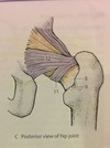
zona orbiculuaris
12

ischiofemoral ligament
part of the extracapsular ligaments
the sacroilliac joint is composed of two parts:
Composed of 2 parts:
- Anterior Synovial joint (Amphiarthrosis) between the auricular surfaces of sacrum and ilium, the joint capsule is very taut and encloses the almost immobile joint and reinforced by ligaments.
- Posterior Syndesmosis between the tuberosities of sacrum and ilium

1 and 2

superior and inferior pubic ligaments
Pubic Symphysis is what kind of joint?
Pubic Symphysis:
non-synovial joint
- Fibrous cartilaginous joint consists of a fibrocartilaginous interpubic disc.
- The disc is generally wider in women, containing a small Non-Synovial cavity
the hip joint is what kind of joint? describe the capsule
- Ball & Socket (Spheroidal) Multiaxial free moving type of joint.
Joint capsule, strong yet loose, has an external fibrous layer and an internal Synovial
membrane. Proximally it attaches around the Acetabular rim and transverse Acetabular
ligament, and distally it attaches to the femoral neck only at the Intertrochanteric line
anteriorly, but posteriorly it crosses the neck to attach to the Intertrochanteric crest.
There are 4 extracapsular ligaments and 1 intercapsular ligament (the femoral ligament)

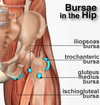
name the 4 hip joint Bursa

- Iliopectineal Bursa: large bursa between the Iliopsoas muscle and the hip joint anteriorly
- Obturator Externus Bursa: a Synovial protrusion beyond the margin of joint capsule into the posterior aspect of the femoral neck. For the tendon of Obturator Externus
- Obturator Internus Bursa
- Gluteus Maximus, Medius and Minimus Bursae: Also called Trochanteric Bursae, found between tendons and greater and lesser trochanters
What are the movements of the hip joint?
- Movements: Free moving, Multiaxial joint, 3 axes:
Anteversion (Flexion) / Retroversion (Extension), at a transverse axis through the head of femur
Abduction / Adduction, at an Anterior-Posterior (Sagittal) axis through the head of femur
Medial / Lateral Rotation, at a vertical axis through the head of femur and the medial femoral condyle
Combined movements of the above produce Circumduction
** Note: the degree of flexion/extension possible at the hip joint depends on the position of the knee. If the knee is flexed (hamstring muscles are relaxed), the
thigh can be actively flexed until it reaches the anterior wall of abdomen. Extension is very limited due to the strong Iliofemoral ligament. Lateral rotation is more powerful than medial rotation.
The hip joint uses what muscles to aid in Flexion? What is the maximum degree of movement?
flexion/Anteversion
Iliopsoas
Rectus Femoris
Pectineus
Sartorius
(With flexed knee Max. 130-140°)
The hip joint uses what muscles to aid in extension? What is the maximum degree of movement?
extension/Retroversion
Gluteus Maximus
Semimembranosus
Semitendinosus
Biceps Femoris- Long Head
(15°, with abduction 45°)
The hip joint uses what muscles to aid in abduction? What is the maximum degree of movement?
Abduction
Gluteus Medius
Gluteus Minimus
Tensor Fascia L
(50°, in flexion 80°)
The hip joint uses what muscles to aid in adduction? What is the maximum degree of movement?
Adduction
Adductor Magnus
Adductor Longus
Adductor Brevis
Pectineus
Gracilis
(10°, in flexion 30°)
The hip joint uses what muscles to aid in lateral rotation? What is the maximum degree of movement?
Lateral Rot.
Gluteus Maximus
Gluteus Medius- Dorsal part
Gluteus Minimus- Dorsal part
Obturator internus
Piriformis
Quadratus Femoris
(15°, in flexion 60°)
The hip joint uses what muscles to aid in medial rotation? What is the maximum degree of movement?
Medial Rot.
Gluteus Medius
Ventral part
Gluteus Minimus
Ventral part
Adductor Longus
Iliopsoas
(30°, in flexion 40°)
The knee joint is what kind of joint and which bones/ surfaces are involved?
mainly a hinge joint combined with gliding
and rolling and with rotation (Torchoid) about a vertical axis, 2 axes joint.
The articular surfaces are large sized, complicated & incongruent shapes. We have to distinguish 3 articulations in the knee joint:
2 Femortibial articulations (lateral & medial) between the lateral and the medial femoral and Tibial condyles
1 Intermediate Femoropatellar articulation between patella and the femur
The fibula is not involved in the joint
2

Patellar ligament:
distal part of quadriceps muscle, thick fibrous band passing from apex of patella to the Tibial tuberosity. On both sides it receives the lateral and medial patellar retinacula (expansions of Vastus muscles and overlying
fascia) which make up the joint capsule on both sides of the patella. Maintain the patella in appropriate articular position with the femur.
5

lateral patellar retinaculum
6

medial patellar retinaculum
9

tibial collateral ligament
strong, flat capsular band
that extends from medial epicondyle of femur to the medial condyle of tibia (&medial superior surface). Its deep fibers are strongly attached and fused to the
medial meniscus and joint capsule. It is weaker than the fibular collateral ligament and more often damaged.
15

Fibular collateral ligament (lateral collateral ligament):
cord-like strong extracapsular ligament from lateral epicondyle of femur to the lateral surface of
head of fibula. It is separated from the lateral meniscus and capsule by the tendon of popliteus and it splits the tendon of Biceps Femoris into 2.
22

suprapatellar bursa
medial view, 9

tibial collateral ligament
14

tendon of semimembranosus
18

Oblique Popliteal ligament:
expansion of the tendon of Semimembranosus that
reinforces the joint capsule posteriorly. Arises posterior to the medial Tibial condyle and passes superolaterally toward the lateral femoral condyle.
19

Arcuate Popliteal ligament:
from posterior aspect of fibular head, passes
superiorly above the tendon of popliteus and spreads on the posterior surface of knee joint. Strengthens the joint Posteriorly
23

medial bursa subtendinous of gastocnemius
24 and 25

medial and lateral head of gastrocnemius
21

popliteus
1

semimembranosus bursa

2

suprapatellar bursa

3

subcutaneous prepatellar bursa

4

subcutaneous infrapatellar bursa

5

deep infrapatellar

6

subsartorial bursa (pes anserinus bursa)

Where is the cruciate ligament pair? How do they restrict movement and allow movement?

Cruciate Ligaments (2) (cross ligaments): crisscross obliquely like X within the
joint capsule AND outside the Synovial cavity.
During medial rotation of knee they wind around each other and limit this movement to 10° only, but during lateral rotation of the knee they unwind and
then it’s available to rotate up to 40° -60° (during flexed knee). Due to their oblique orientation, in every possible movement of knee, at least one of the cruciate ligaments is taut
3 and 7

medial and lateral meniscus
the knee joint menisci are made of what? which bone are they attached to?

crescentic plates of fibrocartilage on the articular surface of the tibia that deepen the surface and play a role in shock absorption. They are attached
firmly to the intercondylar area of the tibia.
1

Anterior Cruciate Ligament:
weaker, from anterior intercondylar area of tibia, extend superiorly posteriorly and laterally and attaches to the medial side of lateral condyle of femur. Prevents hyperextension of knee joint, posterior
displacement of femur on tibia and limits posterior rolling of femoral condyles on the Tibial plateau during flexion.
2

Posterior Cruciate Ligament:
stronger, from posterior intercondylar are of
tibia, passes superiorly and anteriorly and attaches to lateral surface of the medial condyle of femur. Limits anterior Rolling of femoral condyles on the
Tibial plateau during extension, prevents anterior displacement of femur from tibia and Hyperflexion of knee joint. Stabilizes the knee joint in weight bearing
flexed knee (like when walking downhill).
3 and 7

medial and lateral menisci
What are the movements associated with the knee joints?

2 axis
Flexion / Extension of knee joint, at a Transverse axis through the femoral condyles
Lateral / Medial Rotation of knee joint, at a vertical long axis of leg, possible only in the flexed knee position & impossible during extended knee.
what is very special about the knee joint? what is obligatory terminal rotation?
Summary: the knee rotates 5 degrees medially in order to extend fully which allows the leg to be stronger and sturdier. to unlock the knee, the politeus must contract as to rotate the femur the 5 degrees laterally and then can flex the knee joint.
During the extension of knee joint towards maximum, the femoral condyles glide over the Tibial condyles. During the last 10° of extension before completion, the
Obligatory Terminal Rotation of 5° occurs Medially due to the stretching of the Anterior Cruciate Ligament which is permitted by the shape of the medial femoral
condyle (constant, while the Lateral condyle is wider anteriorly) assisted by the Iliotibial tract. This Passive lock or screwing home movement during maximal
extension makes the lower limb a solid column and adapts it for weight bearing and relaxes the thigh muscles partially. (The collateral ligaments are taut, and the cruciate ligaments are relaxed) To unlock the knee, the popliteus muscle must contract to rotate the femur laterally about 5° on the Tibial plateau so the flexion of knee can occur.

When can the knee rotate? what is it called?
Active rotation can only occur in the flexed knee joint
after unlocking the
knee joint. Because the collateral ligaments are loose during flexion and the cruciate
ligaments are taut, the rotation is controlled by the cruciate ligaments. During medial rotation of knee they Wind around each other and limit this movement to 10° only, but during lateral rotation of the knee they unwind and then it’s available to rotate up to 40° -60° (during flexed knee)
what muscles are involved in flexion of the knee joint?
Flexion
Semimembranosus
Semitendinosus
Biceps Femoris
Gastrocnemius
Sartorius
(Max. 130°)
what muscles are involved in extension of the knee joint?
Extension
Quadriceps Femoris
Tensor fasciae Latae
(Max. 180°)
what muscles are involved in lateral rotation of the knee joint?
Lateral Rot.
Biceps Femoris
Tensor fasciae Latae
(40°-60°)
what muscles are involved in medial rotation of the knee joint?
Medial Rot.
Semimembranosus
Semitendinosus
Sartorius
Gracilis
(10°)
12

Superior Tibiofibular joint:
Plane type of Synovial joint that is almost immobile between the head of Fibula and the lateral Tibial condyle.
Tense joint capsule, surrounds margins, Reinforced by:
Anterior / Posterior Fibular Head ligaments
15

Interossious membrane
16

Tibiofibular Syndesmosis:
compound fibrous joint, consisting of the union
between fibula and tibia by the Interossious membrane (shafts) and the Anterior, Interossious & Posterior Tibiofibular ligaments.
The superior tibiofibular joint is known as a compensatory joint due to what?

During Maximal Dorsiflexion of the ankle (talocrural joint), there is an expansion of the Malleolar Mortise as a result of wedging of trochlea of talus, this expansion
results in compensatory movement in the Superior Tibiofibular joint, that’s why
this joint is also known as the Compensatory Joint.
13

acromioclavicular ligament
14

trapazoid ligament
15

conoid ligament
17
coraco-acromial ligament
Acromioclavicular joint is what kind of joint and it has what kind of joint composition? Capsule compostion?
Acromioclavicular joint:
- Plane type of joint that functions as a very limited Ball & socket (spheroidal). 3 axes of movement, Mobility at the AC joint allows the scapula to move in
three dimensions so that it follows the contours of the ribcage.
- The joint contains an incomplete articular disk.
- Articular surfaces covered with fibrocartilage, joint capsule is relatively loose, attached to the margins of articular surfaces. Synovial membrane lines the
fibrous layer

The scapula moves in how many axis of the acromioclavicular joint? What are the movements? practice these movements now.
The scapula moves around each of the three acromioclavicular axes:
- The scapula Protracts (4) & Retracts (3) (Wings) around a vertical axis
- The scapula can be Elevated (1) & Depressed (2) in a Frontal plane
- The scapula tips around a transverse axis.
- The scapula rotates upward (5) or downward (6) around saggital axis through the joint’s capsule.
https://www.youtube.com/watch?v=rRIz6oOA0Vs

What muscles aid in protraction of the scapula?
Protraction: Muscles moving the Acromial end of the clavicle forwards and the scapula ventrolaterally:
Serratus Anterior
Pectoralis Maj.
Pectoralis Minor
(25-30º)

What muscles aid in retraction of the scapula?
Retraction: Muscles moving the Acromial end of the clavicle backward and the scapula dorsomedially:
Trapezius- Transverse part
Rhomboids
Lattisimus Dorsi
(20-25º)

What muscles aid in elevation of the scapula?
Elevation: Muscles elevating the Acromial end of clavicle and scapula: (max. 40º)
Trapezius- Descendig part
Levator Scapulae
Sternocliedomastoid
Rhomboids

what muscles aid in depression of the scapula?
Depression: Muscles depressing the Acromial end of clavicle and scapula: (max. 10º)
Trapezius- Ascending part
Pectoralis minor
Lattisimus Dorsi
Pectoralis Maj.- Abdominal part
Serratus Anterior- Inferior part

what muscles aid in upward rotation of the scapula?
Upward Rotation
Trapezius- Descending part
Trapezius- Ascending part
Serratus Anterior- Inferior part

What are the muscles associated with the Downward Rot. of the scapula?
Downward Rot.
Lattisimus Dorsi
Rhomboids
Pectoralis Major- Abdominal part
Pectoralis Minor

THe shoulder joint, also known as the ____, is what kind of joint and has what sort of joint characteristics?
Glenohumeral Joint (Shoulder joint):
- Ball & socket type of joint (shperoidal). 3 axes of movement. Free moving.
- The large round humeral head articulates with the shallow glenoid cavity of scapula. (3:1 ratio in size)
- Fibrocartilage glenoid labrum (lip): around the margins of glenoid cavity, deepens and enlarges the shallow cavity. Hylaine cartilage covers the articular surfaces.
Can only extend 90 degrees until the scapula needs to move with it

10

coraco-acromial ligaments
7

coracohumeral ligament
9

subscapular muscle tendon
6

Axillary recess:
the articular capsule is slack when the arm is down and the axillary recess hangs down in a pouch like shape

Glenohumeral ligaments:
3 fibrous bands, strengthens the caspule
anteriorly.
1 and 2

1 Transverse humeral ligament:
hold the tendon of long head of biceps
inside the intertubercular sulcus.
2 Enlongation of the senovial membrane for the tendon of biceps brachii

what are the two main bursa of the should joint?
Subscapular bursa:
between the tendon of subscapularis muscle and the neck of scapula. Communicates with joint cavity anteriorly.
Subacromial bursa (subdeltoid):
between the acromion, coracoacromial ligament and deltoid superiorly and the supraspinatus tendon and joint caspule inferiorly. Facilates the movement of the supraspinatus tendon under the coracoacromial arch, and the Deltoid movement over the greater tubercle.

What are the 3 axises of movement in the shoulder joint?
Movements: free moving joint, 3 axes:
Anteversion / Retroversion (extension / flexion) along transverse axis.
Abduction / Adduction along saggital axis.
Outward (lateral) / Intward (medial) Rotation along vertical axis.
The combination of these 3 types of movements produces circumduction.
What muscles are involved in Abduction of the shoulder joint?
Abduction
Max. 90º
Deltoid-Acromial part
Suprapinatus
Biceps Brachii-Long Head
Trapzeius-Descending part
What muscles are involved in Anteversion (flexion) of the shoulder joint?
flexion
To Horizontal plane/ 90
Pectoralis Maj.-Calvicular part
Deltoid- Calvicular part
Biceps Brachii
Coracobrachialis
Pectoralis minor
What muscles are involved in Retroversion (extension) of the shoulder joint?
extension
Max. 40º
Lattisimus Dorsi
Deltoid-Spinal part
Teres Major
Triceps Brachii-Long Head
What muscles are involved in Adduction of the shoulder joint?
Adduction
Pectoralis Maj.-Sternocostal pt.
Lattisimus Dorsi
Teres Major
Coracobrachialis
Pectoralis minor
What muscles are involved in Inward Rot. of the shoulder joint?
Inward Rot.
Max. 70º
Subscapularis
Teres Major
Pectoralis Maj.
Lattisimus Dorsi
Deltoid- Clavicular part
What muscles are involved in Outward Rot. of the shoulder joint?
Outward Rot.
Max. 60º
Infraspinatus
Teres Minor
Deltoid- Spinal part
In order to abduct the arm further than 90 degrees, what needs to move? why?
The abduction is limited anteriouly untill 90º and lateraly untill 75º due the greater and lesser tubercles touching the Coracoacromial arch. Beyond that, we will
need the scapula to move with the humerus to produce Elevation of the arm.
What muscles are involved in elevation of the shoulder joint?
Elevation of Arm
Up to 160º - 180º
Serratus Anterior
Trapezius
Descending part
What kind of joint is the elbow joint? What kind of motion can it actually do? What are the two parts of the joint?
The Elbow joint:
- Hinge type of joint, 1 axis of movement through the condyles of humerus.
* The humeroradial articulation is ball & socket, but restricted functionally into hinge by the collateral ligaments.
- *Humeroulnar and Humeroradial joints**
- The spool shaped Trochlea and Spheroidal Capitulum of the humerus articulate with the trochlear notch of the Ulna and the Superior articular surface of Radius respectively, making the Humeroulnar and Humeroradial joints that compose the Elbow joint.
describe the capsule of the elbow joint
The articular surfaces are covered with Hyaline cartilage. The fibrous joint capsule is
attached to the humerus at the margins of lateral and medial ends of the Capitulum and
trochlea and proximal to the Coronoid process anteriorly, and posteriorly it reaches above
the Olecranon fossa. Synovial membrane lines it from the inside. The capsule is weak
anteriorly and posteriorly but strengthened laterally by the collateral ligaments.

1

radial collateral ligament
Fan like, blends with the annular ligament.

2

Annular ligament of the Radius: encircles and holds the head of radius
in the radial notch of Ulna, forming proximal Radioulnar joint.

3

lateral ulnar collateral ligament

1

anterior ulnar collateral ligaments

2

oblique ulnar collateral ligaments

3

posterior ulnar collateral ligaments

what muscles aid in extension of the elbow joint?
Extension:
Triceps Brachii
Anconeus
what muscles aid in flexion of the elbow joint?
Flexion:
Biceps Brachii
Brachialis
Brachioradialis
Pronator Teres
Proximal Radioulnar Joint is what kind of joint, where, allows movement of what, and what is the joint capsule like?
Proximal Radioulnar Joint:
- Pivot type of joint. 1 rotatory axis.
- The articular circumference of the head of the Radius articulates with the Radial notch of the Ulna. The head of radius is held in position by the annular ligament of the head of Radius. This joint allows the movement of the Head of Radius on the Ulna. pronationa nd supination
- The fibrous capsule is continuous with elbow joint. The Synovial membrane continues distally as a sacciform recess under the annular ligament. (so the head of Radius wont tear or damage the membrane while rotating)

What is the ligament involved in the proximal radioulnar joint?
Ligaments: Annular ligament of Head of Radius.
this ligament holds the radius to the ulna and allows it to rotate/pivot over.

What is the distal radioulnar joint? what is involved? what kind of joint?
Distal Radioulnar Joint:
- Pivot type of joint. 1 rotatory axis.
- Rounded head of Ulna articulates with the ulnar notch on the Radius.
The joint allows the movement of the Head of Ulna on the Radius.

the distal end of the radius rotates around the ulnur head
describe the capsule of the distal radioulnar joint
The joint has a fibrocartilaginous articular disk (triangular ligament), that binds the ulna
and the radius at the distal end.
- The joint cavity is L-Shaped, the joint capsule is deficient superiorly, and the Synovial
membrane extends as a sacciform recess of the distal RU joint.
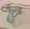
What is the name of the blue area?

triangular articular disk

what muscles are involved in pronation?

Pronation:
Pronator Teres
Pronator Quadratus
Flexor Carpi Radialis
* Brachioradialis muscle: brings arm into mid-position (like the army Salute)
what muscles are involved in supination?

Supination:
Supinator
Biceps Brachii
Extensor Pollicis longus
Extensor Carpi Radialis longus
* Brachioradialis muscle: brings arm into mid-position (like the army Salute)
