Bones: Appendicular Skeleton Flashcards

1
Q

number 2

A

acromial extremity of clavicle

  • flatter end, articulates with acromion of scapula
2
Q

14

A

acromial facet of clavicle

3
Q

12

A

Articular Facet for Sternum

4
Q

1

A

conoid tubercle / coracoid tuberosity

5
Q

inferior view

A

Costal Tuberosity

  • inferomedial side of clavicle
  • it’s an “Impression for (the) Costoclavicular Ligament” and is sometimes known as such, rather than costal tuberosity
6
Q

number 1

A

sternal extremity of clavicle

  • articulates with manubrium
  • pyramidal
7
Q

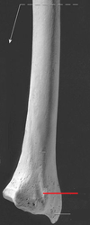
marked by line w blue box

A

Subclavian Groove

  • attachment for subclavius muscle on inferior side
8
Q

17

A

trapezoid line

9
Q
A

clavicle

  • collar bone, 2 paired
  • s-shaped
  • articulates with scapula and sternum

know:

  • sternal extremity
  • acromial extremity
10
Q

which clavicle is this? left or right

A

LEFT

on the superior view looking down, from sternum it arches straight out first and then bends back

11
Q

which clavicle is this? left or right

A

RIGHT

this is a superior view of the right clavicle (since no conoid tubercle is visible)

  • flatter acromial end is concave anteriorly, rounder sternal end is convex anteriorly
12
Q

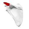
green highlighted posterior, proximal region

A

Intertrochanteric Crest

- posterior ridge from greater to lesser trochanter

13
Q
A

femur

  • proximal bone of leg, longest/largest in body, articulates above w/ acetabulum and below w/ tibia

know:

  • head
  • neck
  • greater trochanter
  • lesser trochanter
  • medial condyle
  • lateral condyle
  • medial epicondyle
  • lateral epicondyle
  • line aspera
14
Q
A

adductor tubercle

  • an elevation on the medial epicondyle of the femur
15
Q

big red part D

A

anterior surface of femur

16
Q

1

A

Fovea for ligament of Head of Femur

17
Q

B

A

Gluteal Tuberosity

- attachment for gluteus maximus

  • often elongated proximally into roughened crest with tubercle known as “third trochanter”
18
Q
A

greater trochanter

  • muscle attachment
19
Q
A

head of femur

20
Q
A

Intercondylar Fossa of Femur

21
Q

green circled structure

A

intertrochanteric line

  • marks transition from neck to shaft
22
Q
A

lateral condyle of femur

23
Q
A

lateral epicondyle of femur

24
Q
A

lesser trochanter

25
Q
A

linea aspera

  • posterior ridge for muscle attachment
  • splits proximally into its medial and lateral lips
  • extends distally into the medial and lateral supracondylar lines
26
Q
A

medial condyle of femur

  • on medial side of articular notch at distal end
27
Q
A

medial epicondyle of femur

28
Q
A

neck of femur

29
Q
A

patellar surface

30
Q

feature marked by red box

A

Pectineal Line

- distal coninuation of lesser trochanter

  • attachment for pectineus muscle
31
Q

from where (what bone marking) does this tendon originate?

A

popliteal groove of femur

  • groove that separates lateral condyle and epicondyle
32
Q
A

Popliteal Surface of Femur

  • the “kneepit”
33
Q
A

Quadrate Tubercle of Femur

- between trochanters on posterior side

  • attachment for quadratus femoris
34
Q
A

shaft of femur

35
Q

A

A

Supracondylar Lines of Femur

  • one medial, one lateral
  • join proximally to form linea aspera
36
Q
A

trochanteric fossa

37
Q

What is the bone?

What are its surfaces and borders to know?

A

fibula

  • lateral bone of lower leg
  • thinnest bone proportional to length

Lateral and Medial Surfaces, separated by anterior border** (somewhat more lateral and flatter) and **interosseous border (more medial ridge-like border for connection of interosseous membrane).

38
Q

tiny blue highlighted portion at very superior tip of the lateral bone

A

apex of fibula

39
Q

3

A

Articular Facet of Lateral Malleolus

40
Q
A

head of fibula

  • proximal end of fibula
41
Q

number 5

A

lateral malleolus

  • lateral prominence of ankle
42
Q

blue portion

A

neck of fibula

43
Q

A, M and P

A

Anterior, Middle and Posterior Talar Articular Surfaces of the Calcaneus

  • all face superiorly and articulate with talus
44
Q
A

Calcaneal Tuberosity

  • faces posteriorly and is the bony portion of the heel
45
Q

25

A

cuboid bone

  • articulates w/ “pinky and ring” metatarsals IV and V
46
Q

9

A

position of cuneonavicular joint

47
Q

the anterior portion of the bone indicated by the arrow, which contains articular surfaces for the navicular tarsal

A

head of talus

48
Q

8

A

intermediate cuniform bone

articulates with metatarsal II

49
Q

23

A

lateral cuniform bone

articulates w/ metatarsal III

50
Q

29

A

lateral malleolar surface of the talus

51
Q

7

A

medial cuneiform bone

- articulates with metatarsal I

52
Q

10

A

navicular bone

53
Q

green area

A

neck of talus

54
Q
A

phalanges of foot

  • 14 each foot (2 for big toe, 3 for others), 28 total
  • distal, middle, and proximal
55
Q

15

its overall name and its parts

A

posterior talar process

  • has medial and lateral tubercles and a groove for the tendon of flexor hallucis longus
56
Q

C

A

Sustentaculum Tali of Calcaneus

  • anterosuperior eminence on medial side of calcaneus
57
Q

What is the space between these bones, circled here in red?

A

tarsal sinus

58
Q

What are all these bones individually? And what do they make up as a whole?

A

tarsals (7 each ankle, 14 total)

  • make up the tarsus
59
Q
A

calcaneus

  • largest tarsal, contains the “heel”
60
Q
A

metatarsals

  • numbered I through V, most medial is I
61
Q
A

talus

  • smaller than calcaneus
62
Q

What tarsal articular surface do these arrows indicate the complex nature of?

A

Trochlea of Talus

63
Q

24

A

tuberosity of 5th metatarsal

64
Q
A

Capitate carpal

  • articulates w/ metacarpal III (middle finger)
  • largest carpal
65
Q
A

Hamate Carpal

  • articulates w/ metacarpals IV and V (ring and pinky finger)
  • has “hook of hamate” or hamulus process
66
Q
A

Lunate Carpal

  • articulates directly w/ radius
67
Q

D

A

pisiform carpal

Sits atop triquetrum, more easily seen in palmar view

68
Q
A

Scaphoid carpal

  • articulates directly w/ radius
  • has tubercle
69
Q
A

Trapezium carpal

  • articulates w/ metacarpal I (thumb)
  • has tubercle
70
Q

F

A

Trapezoid Carpal

  • articulates w/ metacarpal II (index finger)
71
Q
A

triquetrum carpal

  • articulates w/ ulna via an articular disc
72
Q
A

Carpals

  • 8 on each wrist, 16 total
73
Q

B

A

Tubercle of Scaphoid

74
Q

D

A

tubercle of trapezium

75
Q
A

metacarpals

  • five in each hand, make up palm
  • numbered I-V, thumb is I
76
Q
A

phalanges

  • fourteen in each hand, 2 in thumbs, 3 in fingers
  • named Proximal, Distal, Middle
  • numbered I through V, thumb is I
77
Q

2

A

Hamulus of Hamate carpal

  • from ‘hamus’ meaning hook
78
Q
A

humerus

  • bone of brachial region of arm, articulates with scapula proximally and radius/ulna distally
  • in addition to all markings know: anteromedial/lateral surfaces and medial/lateral borders
79
Q
A

anatomical neck of humerus

  • former epiphyseal plate
80
Q

15

what surface?

A

anteromedial surface

81
Q
A

body of humerus

82
Q
A

capitulum

  • located laterally in anatomical position
  • articulates with radius
83
Q
A

coronoid fossa of humerus

  • anteromedial accomodation for coronoid process of ulna
84
Q
A

Crest of Greater Tubercle

85
Q

humerus right anterior superior

3

A

crest of lesser tubercle

86
Q
A

deltoid tuberosity of humerus

  • roughened, lateral area where deltoid muscle attaches
87
Q
A

greater tubercle of humerus

  • lateral and large
88
Q

22

midshaft posterior humerus

type of indentation

A

groove for radial nerve

89
Q

21

inferior view posterior humerus

look for indentation

A

groove for ulnar nerve

90
Q
A

head of humerus

  • articulates with glenoid fossa of scapula
91
Q

Not where the red arrow points to the crest of the lesser tubercle, but just lateral to that where #5’s pointer ends

A

intertubercular groove (or sulcus)

92
Q
A

lateral epicondyle of humerus

93
Q

9

inferior view

A

lateral supracondylar ridge

94
Q
A

lesser tubercle of humerus

  • smaller, medial
95
Q
A

medial epicondyle of humerus

  • bony distal projection, muscle attachment
96
Q

Part D

A

Medial Supracondylar Ridge

97
Q
A

olecranon fossa

  • posterior accomodation for olecranon of ulna
98
Q
A

radial fossa of humerus

  • anterolateral accomodation for radius
99
Q
A

surgical neck of humerus

  • named because it’s a common break site
100
Q

part D

A

trochlea of humerus

  • like pulley, articulates with trochlear notch of ulna
101
Q
A

patella

  • thick flat triangular “kneecap”
  • increases quadriceps leverage

know:

  • base
  • apex
  • articular facets
102
Q

1

A

apex of patella

(view is POSTERIOR and INVERTED, RIGHT patella. base is proximal and apex is distal)

103
Q

4

A

base of patella

(view is POSTERIOR and INVERTED, RIGHT patella. base is proximal and apex is distal)

104
Q

3

A

facet for articulation with lateral femoral condyle

(view is POSTERIOR and INVERTED, RIGHT patella. base is proximal and apex is distal)

105
Q

2

A

facet for articulation with medial femoral condyle

(view is POSTERIOR and INVERTED, RIGHT patella. base is proximal and apex is distal)

106
Q

18

A

Acetabular Fossa

107
Q

19/Red Arrow

Right Hip Bone

A

Acetabular Notch

108
Q
A

acetabulum

  • socket for articulation with head of femur
  • formed by meeting of lateral faces of ilium, ischium and pubis
109
Q

red arrow

A

anterior superior iliac spine

110
Q

11

lateral aspect right hip bone

A

anterior gluteal line

111
Q
A

anterior inferior iliac spine

112
Q

24

medial aspect right hip bone

A

arcuate line of pelvis

113
Q
A

auricular surface

114
Q

13

lateral aspect right hip bone

A

external lip of iliac crest

115
Q

general reference inferior portion

right hip bone lateral aspect

A

B = ischium
C = pubis
1 Posterior superior iliac spine
2 Posterior gluteal line
3 Posterior inferior iliac spine
4 Greater sciatic notch
5 Ischial spine
6 Lesser sciatic notch
7 Body of ischium
8 Ischial tuberosity
9 Obturator foramen

15 Inferior gluteal line
16 Anterior inferior iliac spine
17 Lunate surface of acetabulum
18 Acetabular fossa
19 Acetabular notch
20 Pecten pubis
21 Pubic tubercle
22 Body of pubis

116
Q

general reference superior portion

lateral view right hip bone

A

A = ilium
1 Posterior superior iliac spine
2 Posterior gluteal line
3 Posterior inferior iliac spine
4 Greater sciatic notch
10 Iliac crest
11 Anterior gluteal line
12 Internal lip of iliac crest
13 External lip of iliac crest
14 Anterior superior iliac spine
15 Inferior gluteal line
16 Anterior inferior iliac spine
17 Lunate surface of acetabulum
18 Acetabular fossa

117
Q
A

greater sciatic notch

118
Q

the whole ridge along the top of the pelvis

A

iliac crest

119
Q
A

iliac fossa

120
Q

25

medial aspect right pelvis

A

iliopubic eminence

121
Q
A

Ilium

  • dorsal superior section of os coxa
122
Q
A

inferior ramus of pubis

  • forms medial, inferior portion of obturator foramen
123
Q

15

lateral aspect right hip bone

A

inferior gluteal line

124
Q

12

medial view right hipbone

A

internal lip of iliac crest

(lateral view)

125
Q
A

ischial spine

  • bony process just below greater sciatic notch
126
Q
A

ischial tuberosity

127
Q

grey part #3

A

ischium

  • dorsal posterior portion of os coxa
128
Q
A

lesser sciatic notch

129
Q

17

A

Lunate Surface (of acetabulum)

130
Q

opening indicated by small pointer at bottom of picture

A

obturator foramen

  • large foramen btwn ischium and pubis
131
Q

34

A

obturator groove

132
Q

20

A

pecten pubis

AKA pectinate line

133
Q
A

posterior inferior iliac spine

134
Q
A

posterior superior iliac spine

135
Q

part E

A

pubic symphysis

  • fibrocartilagenous joint between left and right superior pubic rami
136
Q

21

A

pubic tubercle

137
Q
A

pubis

  • ventral, inferior portion of os coxa
138
Q
A

ramus of ischium

139
Q
A

superior ramus of pubis

  • forms anterosuperior aspect of obturator foramen
140
Q

26

A

symphysial surface of pubis

141
Q

what should the angle of the pelvis be, between plane of pelvic inlet and the horizontal plane?

also called PELVIC INCLINATION

A

should be ~60 degrees

(horizontal plane begins where plane of pelvic inlet ends at junction between Lumbar and Sacral Spine)

142
Q

10

A

Body of the Ilium

143
Q

14

A

Body of Ischium

144
Q

14

What is the area?

A

Body of Pubis

145
Q

What is this area of the ilium?

A

Gluteal Surface

146
Q

male vs. female pelvis

A

Male

  • < 90 degree pubic angle
  • large oval obturator foramen
  • vertical coccyx
  • larger, ateriorly directed acetabulum

Female

  • usually >100 degree pubic angle
  • small triangular obturator foramen
  • posteriorly tilted coccyx
  • smaller, laterally directed acetabulum
146
Q

green area

A

Limbus (or Margin) of Acetabulum

148
Q

3

A

Posterior Gluteal Line

148
Q

this is the area above the pelvis’ articular feature for the head of the femur

what is it called?

A

supra acetabular fossa

149
Q

What is this circular delineation? What does it separate?

And what features make it up?

A

Terminal Line of Pelvis

  • separates the Greater Pelvis from the Lesser Pelvis
  • in a clockwise motion, it starts posteriorly at the Sacral Promontory, then Arcuate Line of left pelvic bone, Iliopubic Eminence, Pectinate Line, and Pubic Symphysis. It continues up the same features on the right pelvic bone and back to the sacrum.
150
Q
A

Wing (or Ala) of Ilium

151
Q
A

radius (2, paired)

  • lateral (on thumb side) when in anatomical position

in addition to markings, know: anterior, posterior and lateral surfaces as well as anterior and interosseous borders

152
Q

17

A

first groove of radius

152
Q

18

A

second groove of radius

153
Q

19

A

third groove of radius

153
Q

20

A

fourth groove of radius

154
Q

2

posterior view

A

articular circumference of the radius

  • circumferential portion of radius’ head which articulates with radial notch of ulna
155
Q

8 … a distal surface of a long bone

A

articular surface of radius

divided into articulation with scaphoid carpal (lateral by styloid process) and lunate carpal (medial by ulnar notch)

156
Q

distal view of radius

indicated by red line

A

Dorsal (or Lister’s) Tubercle

157
Q
A

Head of Radius

  • articulates with capitulum of humerus
158
Q

highlighted blue portion

A

Neck of Radius

159
Q

green part

A

radial tuberosity

  • attachment for biceps brachii
160
Q
A

styloid process of radius

162
Q

which view of which bones is this?

A

radius and ulna anterior view

163
Q

which view of which bones is this?

A

posterior view of radius (right) and ulna (left)

164
Q
A

ulnar notch of radius

  • articulates with distal end of ulna
165
Q
A

acromion of scapula

  • bony tip of shoulder
166
Q
A

coracoid process of scapula

  • looks like bent finger
167
Q
A

glenoid cavity

  • articulates with head of humerus
168
Q

Left scapula posterior view

A

inferior angle

169
Q
A

infraspinous fossa of scapula

  • broad area below spine, posterior aspect
170
Q
A

lateral border of scapula

171
Q
A

medial border of scapula

172
Q
A

spine of scapula

173
Q
A

subscapular fossa

  • smooth, broad anterior surface of scapula
174
Q

letter D (angle)

right anterior costal scapula

A

superior angle

175
Q
A

supraspinous fossa of scapula

  • above spine, posterosuperior aspect
176
Q
A

Scapula (2 paired)

  • shoulder blade, flat/triangular articualtes with acromion of scapula

know:

  • spine
  • acromion
  • coracoid process
  • glenoid cavity
  • medial border
  • lateral border
  • supraspinous fossa
  • infraspinous fossa
  • subscapular fossa
177
Q
A

Acromial Angle of Scapula

178
Q

9

right scapula, anterior

A

articular facet for acromion

179
Q

part H

A

Infraglenoid Tubercle

180
Q
A

Neck of Scapula

  • slightly narrower portion which connects glenoid cavity/tubercle complex to rest of scapula
181
Q

A

A

Superior Margin of Scapula

182
Q

right scapula lateral

11

A

supraglenoid tubercle

183
Q

3

Right scapula posterior superior view

A

Scapular Notch

  • AKA suprascapular notch
184
Q

5

A

Soleal Line of Tibea

- diagonal line on posterior, proximal shaft of tibia

  • attachment for soleus (and others)
185
Q
A

tibia

  • larger of two distal lower limb bones
  • articulates above with femur, below with talus

know:

  • medial condyle
  • lateral condyle
  • tibial tuberosity
  • medial malleolus
186
Q

superior view of tibia

what is part A?

A

Anterior Intercondylar Area of Tibia

  • know it’s anterior because tibial tuberosity (bump seen on top of bone here) is anterior
187
Q

8

A

Anterior Border of Tibia

- prominent anterior ridge that separates lateral/medial surfaces

188
Q

right branch of pointer #8

A

Articular Facet of Medial Malleolus

189
Q

What other borders/surfaces of the tibia should be known? How can you identify them?

A

**Medial Surface - **lies on anterior side of bone, medial to anterior border

**Medial Border - **edge of bone distal to medial epicondyle, separating anterior and posterior sides

**Interosseous Border - **edge of bone distal to lateral epicondyle, facing towards fibula, separating anterior and posterior sides

190
Q

Area marked light blue

A

Fibular Notch of Tibia

191
Q

area marked by green dot

A

Gerdy’s Tubercle

- lateral insertion of IT tract

192
Q

this is the bone which contains the medial malleolus

what is the red marked area?

A

Groove for Tibialis Posterior

(and Flexor Digitorum Longus)

…so it’s the tibia!

193
Q

left branch of pointer #8

A

Inferior Articular Surface of Tibia

194
Q

What is the structure and what are its two parts?

A

Intercondylar Eminence of Tibia

  • made up of medial and lateral intercondylar tubercle
195
Q
A

lateral condyle of tibia

196
Q

9

A

lateral surface of tibia

197
Q
A

medial condyle of tibia

198
Q

number 1

A

medial malleolus of tibia

  • bony prominence on inside of ankle
200
Q

diagonal ridge between 1 and 2

A

oblique line of tibia

202
Q

superior view of tibia, note location of tibial tuberosity

what is part C?

A

Posterior Intercondylar Area of Tibia

- determine it’s posterior based on location of tuberosity (anterior)

203
Q

Area marked light blue

Overall name and separate parts

A

Superior Articular Surfaces of Tibia

Medial and Lateral Facets

(determine which is which via medial malleolus, or fibula if present)

204
Q
A

tibial tuberosity

205
Q
A

ulna

  • medial bone of forearm in anatomical position

know:

  • olecranon process
  • coronoid process
  • trochlear notch
  • radial notch
  • head
  • styloid process
206
Q

16

A

articular circumference of the ulna

207
Q
A

coronoid process

  • articulates with coronoid fossa of humerus
208
Q

distal structure pointed out by thick black line

A

head of ulna

  • note that it’s on the distal end! not all heads are proximal
209
Q

4

A

interosseous border of ulna

210
Q
A

olecranon process

  • fits into olecranon fossa of humerus
211
Q
A

radial notch of ulna

  • lateral to coronoid process
  • accomodates radial head
212
Q
A

styloid process of ulna

213
Q
A

trochlear notch of ulna

  • formed by olecranon and cornoid processes
  • articulates with trochlea of humerus
214
Q

14

anterior superior forearm

A

ulnar tuberosity

215
Q

10

A

Fossa of Lateral Malleolus

- talo- and tibiofibular ligaments attach here

216
Q

What line do the two small arrows indicate?

A

Chopart’s Line

  • a common amputation line between the talus/calcaneus and the rest of the tarsals
217
Q

What line does the red indicate?

A

Lisfranc’s Line

  • an important amputation line between the tarsals and meta-tarsals
218
Q

Posterior view of distal femur

A

Intercondylar Line

219
Q

13

A

Pronator Tuberosity of Radius

220
Q

What is the small blue highlighted feature at the anterior of these two bones?

A

Pubic Crest

  • extends from pubic tubercle to medial border of pelvic bone
221
Q

2

A

Supinator Crest of Ulna

  • extends distally from the radial notch