Bones: Neurocranium Flashcards
16

Arterial Grooves
11, 12, 13

External Table
Diplöe - spongy inner portion analogous to the trabecula in long bones
Internal Table

Superior sagittal sulcus
the groove for the superior sagittal sinus is a depression formed on the internal surface of the frontal, parietal, and occipital bones in the midline.
It courses from the frontoethmoid junction to the internal occipital protuberance.
32

petro-occipital fissure
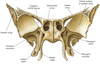
Frontal Bone
- know: frontal sinus, frontal orbits, supraorbital margin

Frontal crest
the union of the superior sagittal sinus sulcus margins
the entire ridge!
1

Fossa for lacrimal gland
houses lacrimal gland

glabella
slightly depressed area of frontal bone that joins the two superciliary ridges.

Frontal or Metopic suture
only the part of the highlighted bone within the eye socket

Frontal Orbits
What is this area (not anterior cranial fossa, but part of a single bone)?
What are the special markings to note on it?

Orbital Plate of the Frontal Bone
- could consider it the “roof” of the orbits
- its impressiones digitataes (“finger impressions”) correspond to the convolutions of the brain
1, not the bone but the portion of it

Squamous Portion of Frontal Bone
2

Frontal Sinus
area defined in red

supraorbital margin of frontal bone
tiny blue-marked area at top of eye socket

supraorbital notch, or foramen if it is more hole-like
2

Trochlear pit or fossa

Supraciliary arch
2

Frontal tuber or eminence
Tiny green area in anterior of skull here:
What bones make it up?

Foramen Caecum
- made up of alae of crista galli (of ethmoid) and frontal bone
- transmits emissary vein from nose to sup. sagittal sinus but is frequently closed
What is this hole and what does it connect?

Jugular Foramen
- connects external base of skull to posterior cranial fossa

blue
piri form aperature
“pear shaped”

Occipital Bone
know: Foramen Magnum, Occipital Condyles, Superior/ Inferior Nuchal Lines

Cerebellar fossa

Cerebral fossa

Clivus of Occipital Bone
- slopes downward posteriorly from the sella turcica of the sphenoid

Condylar canal

Condylar fossa

Occipital Condyles
articulate with atlas vertebra

Cruciform eminence

External occipital protuberance


External occipital crest

Foramen Magnum
passageway for spinal cord
2

Groove for superior sagittal sinus

Groove for transverse sinus

Highest nuchal line
blue arrows

Hypoglossal canal
connects the posterior cranial fossa with the
external base of skull
E

inferior nuchal line

Internal occipital crest

Internal occipital protuberance

Jugular notch of Occipital Bone
the notch in the occipital bone that forms one
boundary of the jugular foramen

Superior Nuchal Line
(Inferior is just below)

jugular process

area marked by blue circle
Nuchal plane

Squamous Portion of Occipital Bone
- superior and posterior portion of occipital in relation to foramen magnum
area marked by red circle
Occipital plane
Cranial Bones
Paired
- Temporal
- Parietal
Unpaired
- Frontal
- Occipital
- Sphenoid
- Ethmoid
Name all 4 Structures

A - Posterior/Occipital Fontanel
B - Mastoid Fontanel
C - Anterior/Frontal Fontanel
D - Sphenoidal Fontanel

parietal bone (paired)
16

Parietal Foramina
- bilateral foramina towards back of parietal bones at their sagittal border
- transmit parietal emissary vein

Parietal eminence
18

Frontal Angle of Parietal Bone
- where one parietal meets the frontal bone and other parietal at the bregma
(marked as 3 below)

the little green dotted features

granular foveolae
several small,
irregular fossae may also be seen on either side of the superior sagittal sulcus, for the
reception of the arachnoid granulations.
14
parietal notch

the lines represent what?
Grooves for the meningeal vessels
19

Occipital Angle of Parietal Bone
- angle where one parietal meets the occipital and other parietal at the lambda
(marked as 2 below)

What are the two divisions of the skull and what do they form?
How many bones are there in the skull?
- Neurocranium encloses brain and attach to some head and neck muscles, divided into cranial vault (calvary) and base
- Viscerocranium form anterior of skull/face
22 bones and 2 x 3 bony ossicles of inner ear
what are all three?
Superior and inferior temporal line noted by the left lines, frontal temporal line on the right


Sphenoid Bone
- key bone of skull, all others connect to it, forms part of cranium and orbits
know:
- Sella Turcica
- Greater Wings
- Lesser Wings
- Sphenoidal Sinuses
- Optic Foramen
- Superior Orbital Fissure
- Foramen Ovale
- Pterygoid Process
What are the 5 facets of the sphenoid bone?
- **Cerebral Facet **(superior, makes up middle/anterior scalae)
- **Temporal Facet **(lateral, exterior of skull along sphenosquamous suture)
- **Infratemporal Facet **(lateral, exterior of skull below zygomatic arch)
- **Orbital Facet **(anterior, on posterior wall of orbit)
- **Pterygomaxillar Facet **(anterior, unseen unless bone is removed from skull, btwn pterygoid processes and maxilla)

Anterior clinoid process

Carotid groove

Dorsum sellae

Foramen lacerum
- between sphenoid, temporal and occipital
-provides passage for a branch internal carotid artery called lacerum segment as well as other nerves and vessels


Foramen Ovale
- blood vessels to meninges, nerves to orbit, face, jaw
inferior view below is marked with red line
Part A

Greater Wing of Sphenoid
- project laterally, form parts of middle cranial fossa/orbits
not the region but the specific functional location

Hypophyseal or pituitary fossa
- fossa hypophysealis
holds the pituitary gland within the region of the sella turcica
1

infratemporal crest
crest dividing the lateral exterior surface of the sphenoid
area below/inferior to the ridge marked as 1

Infratemporal surface
forms the “roof” of the infratemporal fossa

Lateral plate of pterygoid process

B

Lesser Wings of Sphenoid
- horn-shaped, form part of ant. cranial fossa/orbit


Medial plate of pterygoid process


Optic Foramen
- AKA optic canal, optic nerve pases through


posterior clinoid process
area denoted by black bar and arrows
Prechiasmatic Groove
1
pterygomaxillary fissure

2
pterygopalatine fossa


pterygoid process
(terry-goid)
- inferior projections from greater wings, attaches pterygoid muscles (chewing)

posterior view of sphenoid
Pterygoid canal
an opening through the base of the
pterygoid process of the sphenoid bone


Pterygoid fossa

3

Pterygoid hook or hamulus
little hook at the bottom of the medial pterygoid plate

circled red feature
Round foramen or Foramen Rotundum
- connects middle cranial fossa to pterygopalatine fossa
seen marked with blue from the posterior view
Scaphoid Fossa
- small, shallow oval depression at base of medial plate of pterygoid process
- gives origin to tensor veli palatini


Sella Turcica
(Turkish saddle)
- saddle-shaped process containing a depression for the pituitary gland

3
sphenopalatine foramen

circled in red
foramen spinosum

Superior Orbital Fissure
- connects orbit to middle cranial fossa
- posterior of orbit btwn sphenoid wings
- nerve/vein passage

Tubercle of saddle (tuberculum sellae)
2
vaginal process of sphenoid bone
a thin lamina of bone that extends medially under the bodyof the sphenoid bone from the medial lamina of the pterygoid process;
it articulates with
the vomer and the palatine bones

Sphenoidal Crest
- vertical crest on anterior surface of body
- articulates with perpendicular plate of ethmoid to form part of nasal septum
Sphenoidal Lingula
a slender process projecting posteriorly
between the body and greater wing of the sphenoid bone, on either side, forming the
lateral margin of the carotid groove. In the dry skull, it projects into the foramen lacerum

1
Sphenoidal Rostrum
articulates with the vomer


Sphenoid Sinus
contained within the body of the sphenoid, the “eyes” of the moth if looking at the shenoid process from the frontal position like below

sphenoidal yoke or jugum
a plane surface connecting the two
lesser wings
31

Groove for the Inferior Petrosal Sinus
- formed by junction of petrous part of temporal and basilar part of occipital
What is this red point?

Bregma
- the point at which the coronal and sagittal sutures meet
red line

Coronal suture
- btwn parietals and frontal
What’s this circled area?

Lambda
- the point at which the lambdoid and sagittal sutures meet
red line

Lambdoidal Suture
- btwn parietals and occipital, looks like greek letter lambda

occipitomastoid suture
- between occipital bone and mastoid region of temporal bone
2

Sagittal Suture
-btwn the 2 parietals
9

Sphenoethmoidal Suture
27

Sphenosquamous Suture

squamous suture
-btwn temporal and parietal

External Acoustic Meatus
- forms much of the tympanic part of the temporal bone

internal acoustic meatus
- start of the facial canal
- where blood vessels + nerves enter inner ear

Mastoid Process
- attachment for head/neck muscles that rotate/flex head
which part of base of skull, not of mandible

Mandibular Fossa
- articulates with condylar process of mandible


Petrous Region of Temporal Bone
- thick region that houses sensory structures of inner ear
note the apex


Styloid Process of Temporal Bone
- sharp projection, attachement for tongue/larynx muscles
- think “stylus” like the pen of a PDA


Zygomatic Process of Temporal Bone
- part of cheekbone, articulates with zygomatic bone
13
petrous apex

1
Arcuate Eminence
- bony eminence containing anterior semicircular canal
(#26 in pic below)

Articular tubercle of temporal bone
2
articular tubercle
- just anterior to mandibular fossa
noted below as #3

8
mastoid foramen
- transmits mastoid emissary vein to sigmoid sinus

17
What does it connect?
carotid canal
- external base of skull to scala media
What opening is the pipecleaner coming through?
carotid canal
- internal carotid artery runs superiorly from external opening of canal, then anteromedially past apex of petrous region, by the foramen lacerum and then along the carotid grooves of the sphenoid
1
groove for middle temporal artery
35
What two divisions does this cause in its neighboring foramen?

intrajugular process of temporal bone
- partially divides jugular foramen into smaller anteromedial pars nervosa and larger posterolateral pars vascularis (containing jugular vein)
3
mandibular fossa
13
jugular fossa
jugular fossa is the depression where the jugular vein lies. the jugular foramen is the passage between the extenral base of the skull and the internal cranial fossa

7
mastoid foramen
7
mastoid notch

9
mastoid notch
6
mastoid process
20
musculotubal canal
- begins at petrous portion of the temporal bone and passes to the tympanic cavity
- divided into one canal for the auditory tube and into another canal for the tensor muscle of the tympanic membrane.
9
occipital border
15
external opening of cochlear canal
external orifice of the cochlear aqueduct on the temporal bone medial to the jugular fossa.

4
parietal notch
the angle posteriorly between the squamous and petrous parts of the temporal bone.

What are this bone’s 5 parts?

Temporal Bone
The temporal bones are situated at the sides and base of the skull, each consists of 5 main parts:
- Squamous part (pars squamosa, squama temporalis)
- Petrous part (pars petrosa), pyramid
- Tympanic part (pars tympanica)
- Mastoid part
- Hyoid part (Styloid process)
16
Fossula Petrosa
A small indentation on the inferior surface of the petrous part of the temporal bone, located between the jugular fossa and the external aperture of the carotid canal. It houses the petrous (inferior) ganglion of the glossopharyngeal nerve and is located near the tympanic canalicus, which carries the tympanic branch of the glossopharyngeal nerve.
What is the posterior portion of the line indicated by #4?
From where to where does it lead?
petrotympanic and petrosquamous fissure of temporal bone
- carries chorda tympani from infratemporal fossa to tympanic cavity (eventually connecting with facial canal)
(posterior side borders tympanic region, as opposed to anterior side which is petrosquamous)
21
sphenoid border
What does this hole connect?

stylomastoid foramen
- external base of skull to facial canal (out the internal acoustic meatus to the posterior scala)
(seen as #10 below)
3
supramastoid crest
5
Suprameatal spine

2
suprameatal triangle
between the posterior wall of the external acoustic meatus and the posterior root of the zygomatic process

vaginal process or sheath of the styloid process

5
external aperature for vestibular aqueduct
- posterior to internal acoustic meatus, just inferior to arcuate eminence and somewhat hidden by overhanging bone
- transmits small vein and endolyphatic duct

1
zygomatic process

Carotid Canal
- medial to styloid process, internal carotid artery entrance
Interior View:
11
cerebral side of squamous face
5
(region)

Temporal fossa
24
What leads up to it? What is it? Where does it lead?

Groove for Lesser Petrosal Nerve leads up to it
it’s the Hiatus for the LP Nerve
and it leads to the tympanic cavity
(the tympanic nerve changes into the lesser petrosal nerve in the petrous portion of the temporal bone)
20

Groove for Middle Meningeal Artery
- runs laterally from foramen spinosum
- starts on edge of sphenoid but is mostly on squamous part of temporal

Groove for sigmoid sinus
22 and #23
Where does #22 lead?

Groove and Hiatus for the Greater Petrosal Nerve
- hiatus leads to the facial canal

infratemporal fossa

6
intrajugular process
Area indicated by arrow

Mastoid Region of Temporal Bone

blue area, note spanning two areas

Temporal line of frontal bone
2
mastoid foramen

petrosquamous suture
(not fissure)
What are the features indicated by the blue line?
petrotympanic (Glaserian) fissure AND petrosquamous fissure
separated by (invisible) tegmental crest
(tympanic fissure on posterior tympanic side, squamous on anteror squamous side)
posterior side of the petrous region
the posterior border is at the base of the petrous region of the temporal bone
10, 11, and 13
tympanic region, external acoustic meatus, and mandibular fossa

Squamous Region of Temporal Bone

7
styloid process of the temporal bone
9
Subarcuate fossa

superior border/ridge of the petrous part

Tympanic Region of Temporal Bone

red line indicates
tympanomastoid fissure
between tympanic and mastoid regions

4

Mastoid Air Cells
- reduce weight of skull
- connected to middle ear
21

Trigeminal Impression of Petrous Part of Temporal Bone
23
(just above carotid canal on right side of picture)

Groove for the Auditory Tube
- accommodates the cartilaginous part of the auditory tube before it runs into the musculotubal canal
What is indicated by the pink arrow?

Pharyngeal Tubercle
- on basilar part of occipital bone anterior to foramen magnum

red arrow

Spine of Sphenoid
- sharp inferiorly oriented process off greater wing
- just medial to mandibular fossa in articulated skull