Histology Lecture 3-Connective Tissue Flashcards
Types of Connective Tissue:
1.) Embryonic Connective Tissue 2.) Adult Connective Tissue
Types of Embryonic Connective Tissue:
1.) Mesenchyme 2.) Mucous
Types of Adult Connective Tissue:
1.) Connective Tissue Proper 2.) Specialized Connective Tissue
Types of Connective Tissue Proper:
1.) Loose (areolar) 2.) Reticular 3.) Dense Regular 4.) Dense Irregular 5.) Adipose
Types of Specialized Connective Tissue:
1.) Blood 2.) Supporting Connective Tissue (bone and cartilage)
Functions of Connective Tissue:
1.) Binds, Supports, and Strengthens other body tissues 2.) Protection- surrounds organs and bones 3.) Insulation of internal organs 4.) Compartmentalization of organs 5.) Transport nutrients and oxygen 6.) Storage of Energy Reserves (adipose) 7.) Immune Responses (blood)
All Connective Tissue has what common characteristics?
1.) cells in an extracellular matrix (protein fibers and ground substance) 2.) common origin (all CT comes from mesenchyme) 3.) degrees of vascularity (can be avascular, poorly vascularized, or highly vascularized)
What is mesenchyme?
Embryonic connective tissue that is: -loosely organized CT -has elongated, undifferentiated cells with cytoplasmic processes -has viscous ground substance Mesenchyme can differntiate into other types of cells other than CT cells
What is the general composition of connective tissue?
Cells with a large amount of extracellular matrix between them.
The ECM is made up of:
1.) Protein fibers 2.) Ground substance
The protein fibers found in the ECM of connective tissue are:
1.) Collagen 2.) Reticular fibers 3.) Elastic fibers
Describe Ground Substance:
amorphous material between fibers, provides medium through which diffusion can occur, can be fluid, semifluid, gelatinous, or calcified (no diffusion), GS is hydrophilic
What are the components of the Ground Substance in the ECM?
1.) Glycosaminoglycans (GAGs) (ex. hyaluronic acid and chondroitin sulfate) 2.) Proteoglycans 3.) Multiadhesive Glycoproteins (ex. laminin and fibronectin)
What is the major product or activity of fibroblasts, chondroblasts, osteoblasts, and odontoblasts?
production of fibers and ground substance
What is the major product or activity of plasma cells?
productions of antibodies
What is the major product or activity of lymphocytes?
production of immunocompetent cells
What is the major product or activity of eosinophils?
participation in allergic and vasoactive reactions modulation of mast cell activities and the inflammatory process.
What is the major product or activity of Neutrophils?
phagocytosis of foreign substances, bacteria
What is the major product or activity of macrophages?
secretion of cytokines and other molecules, phagocytosis of foreign substances and bacteria, antigen processing an presentation to other cells.
What is the major product or activity of mast cells and basophils?
liberation of pharmacologically active molecules (histamine)
What is the major product or activity of adipose cells?
storage of neutral fats and thermogenesis
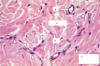
What type of cell is indicated by the black arrows? What does this type of cell do?

Type of cell: fibroblast
Activity: produces ECM fibers
What is the most common cell type in connective tissue?
fibroblast cells
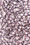
What type of cells can be seen in this image?

Active fibroblasts (seen coming away from ECM which indicates that they are active)
What type of cells are indicated in the images?

fibroblasts
What type of cell is indicated by the black arrows?

Myofibroblasts (Fibroblasts that also have some characteristics of muscle. Elongated cell similar to muscle, and is associated with actin filaments)
What type of cell is shown in the image?

Mast cells (filled with basophilic granules contianing chemicals like heprin).
What type of cell is shown in the image?

Mast cell
What type of cells can be seen in the images?

Plasma cells-
Short life span (10-20) days,
Derived from B lymphocytes,
Produce antibodies
What type of cells is indicated by the letter “A”?

Macrophages-Nucleus is kidney shaped, cellular borders are irregular
Label A-D

A.) Fibroblast nuclei
B.) Plasma Cells
C.) Basophils
D.) Eosinophils
Three types of connective tissue fibers:
- ) Collagen-collagen protein
- ) Reticular- type 3 collagen protein
- ) Elastic- elastin protein
Label A-C

A.) Collagen
B.) blood vessel
C.) Fibroblast
Label B and C

B.) Collagen
C.) Elastin
What type of tissue is pictured and where might you find it?

Reticular Connective Tissue, seen in organs that support other organs like the lymphatic organs
Describe the structure of collagen fibers.
3 alpha chains intertwine to form a triple helix collagen molecule. A bundle of triple helix collagen molecules forms a collagen fibril. A bundle of collagen fibrils forms a collagen fiber

Label a-g

a. ) collagen fibrils
b. ) mitochondria
c. ) rough ER
d. ) collagen fibrils
e. ) fibroblast
f. ) ECM
g. ) rough ER
Label A and B

A.) Elastic fibers
B.) Collagen fibers
Label A and B

A.) Elastic fiber
B.) Collagen fiber
Ground Substance consists of:
A.Glycosaminoglycans (GAGs) (ex. hyaluronic acid and chondroitin sulfate)
B.Proteoglycans
C.Multiadhesive Glycoproteins (ex. laminin and fibronectin)
Describe GAGs
Glycosaminoglycans are unstructured material between cells and fibers, provides medium for diffusion. Hyaluronic acid and chondroitin sulfate are examples. GAGs bind water (can bind a lot of water, can withstand pressure).
What type of tissue is pictured? How do you know?

Mesenchyme. Contains elongated, undifferentiated cells, usually a prominent nucleus, viscous ground substance.
What type of tissue is pictured?

Mesenchyme
What type of tissue is pictured?

Wharton’s Jelly.
Mucous tissue, (Embryonic CT),vsimilar to what is found in pulp cavity in young teeth
Typically collagen fibers
Label A-D

A.) Umbilical Cord
B.) Umbilical arteries
C.) Umbilical vein
D.) Wharton’s Jelly (mesenchyme)
What type of tissue is pictured? Where is it found? What is it’s function? Label A-C

Type: Loose Areolar Connective Tissue
Location: Under Epithelia, forms lamina propria of mucous membranes, packages organs in urinary, reproductive, and respiratory tract
Function: cushions organs, strength, elasticity, support
A.) Elastic Fibers
B.) Collagen Fibers
C.) Fibrobalsts
What type of tissue is pictured? Label A-E

Type: Loose Areolar Connective Tissue
A.) Elastic Fiber
B.) Blood vessel
C.) Mast Cell
D.) Macrophage/lymphocyte
E.) Collagen fiber
What type of tissue is pictured? Where is it found? What is it’s function? Label A-C

Type: Reticular Connective Tissue
Location: Lymphoid organs (lymph nodes, bone marrow, spleen)
Function: Fibers form a soft internal skeleton that supports other cell types including white blood cells, mast cells, and macrophages.
A.) Lymphocyte
B.) Reticular fibers
C.) Mast Cells
What type of tissue is pictured? Where is it found? What is it’s function? Label A and B

Type: Dense Regular Connective Tissue
Location: tendons, most ligaments, aponeuroses.
Function: attaches muscles to bones or to muscles, attaches bones to bones, withstands great tensile stress when pulling force is applied in one direction.
A.) collagen fibers
B.) nuclei of fibroblasts
What type of tissue is pictured? Where is it found? What is it’s function? Label A and B

Type: Dense Irregular Connective Tissue
Location: dermis of skin, submucosa of digestive tract, fibrous capsules of organs and joints
Function: Able to withstand tension exerted in many directions; provides structural strength
A.) fibroblasts
B.) collagen fibers
Label A-F

A.) Dense Connective Tissue
B.) Loose Connective Tissue
C.) Epithelial Tissue
D.) Adipose Tissue
E.) Epithelial Tissue
F.) Dense Connective Tissue
What type of tissue/cells is pictured? Where is it located? What is its function?

Type: White Adipose Tissue/ White adipocytes
Location: subcutaneous layer, mammary gland, greater omentum, mesenteries, orbits, bone marrow cavity
Function: energy storage, insulation, cushioning of vital organs, and secretion of hormones
What type of tissue/cells is pictured? Where is it located? What is its function?

Type: Brown Adipose Tissue
Location:
in Newborns: 5% of total body mass; back, along upper half of spine and toward shoulders
in Adults: retroperitoneal space, around aorta, neck, back, mediastinum
Function: heat production (thermogenesis)