Exam 2 (Pt. 9) Flashcards
Free-Running Rhythms of Three Animals of Different Species - Onset/Offset
The time of activity onset (and offset) occurred earlier (A, B) or later (C) each day. Thus each individual had a free-running period either shorter (A, B) or longer (C) than 24.0 hours.

The Circadian System Model

Suprachiasmatic Nucleus (SNC) & Circadian Rhythms
Double-plotted drinking record from a squirrel monkey before (A) and after (B, C) receiving a histologically verified total SCN lesion. The approximately 25-h drinking rhythm prelesion (A) persisted with a reduced amplitude for over 90 days postlesion (B) before finally decaying into arrhythmia (C).

Projections of the SCN/Subparaventricular Zone Complex and Their Likely Functions

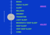
Continuum of States of Arousal
Suprachiasmatic Nucleus (SNC) & Circadian Rhythms - Relationship
Suprachiasmatic Nucleus (SCN) ablation results in loss of circadian rhythms.
The Arousal Continuum from Highest to Lowest Levels

Electrophysiological Correlates of Waking and Sleep Stages - Vertex Spike
Characteristic EEG patterns seen during different stages of sleep in humans are shown here. The sharp wave called a vertex spike appears during stage 1 sleep. Brief periods of sleep spindles are characteristic of stage 2 sleep

Electrophysiological Correlates of Waking and Sleep Stages - Deeper Sleep
Deeper stages of slow-wave sleep show progressively more large, slow delta waves.

Electrophysiological Correlates of Waking and Sleep Stages - Similarity
Note the similarity of activity during waking, stage 1 sleep, and rapid eye movement (REM) sleep.

Properties of Slow-Wave and REM Sleep

Sleep Stages & Sleep Architecture
Note the progressive lengthening of REM episodes (blue) and the loss of stages 3 and 4 sleep as the night goes on.

Evidence for Bremer’s 1937 Passive Sensory Theory of Sleep - Procedure
Transecting the neuraxis at the level between the superior and inferior colliculi removed much of the ascending sensory input to the forebrain.

Evidence for Bremer’s 1937 Passive Sensory Theory of Sleep - Conclusion
This produced a cat that appeared to be sleeping; hence, Bremer concluded that depriving the rostral brain produced sleep.

Evidence for Moruzzi and Magoun’s Active Reticular Activating-System Theory of Sleep - Procedure
In the late 1940’s Moruzzi and Magoun made small lesions in the core of the brain stem so that most of the classic ascending sensory pathways were left intact.

Evidence for Moruzzi and Magoun’s Active Reticular Activating-System Theory of Sleep - Conclusion
Nevertheless, these animals displayed a sleep-like EEG pattern. Hence they concluded that the reticular formation produced sleep through an active process.

The Nature & Extent of the Reticular Formation - Diagram
MBRF=midbrain reticular formation; MRF=medullary reticular formation; PRF=pontine reticular formation; TRF=thalamic reticular formation; HYP=hypothalamus.

Brain Stem Reticular Formation
The reticular formation is thought to activate the rest of the brain.

Ascending Arousal Systems - Projections

The ascending arousal system sends projections from the brainstem and posterior hypothalamus throughout the forebrain.
Ascending Arousal Systems - Cholinergic Fibers

Neurons of the laterodorsal tegmental nuclei and pedunculopontine tegmental nuclei (LDT and PPT) (blue circles) send cholinergic fibers (ACh) to many forebrain targets, including the thalamus, which then regulate cortical activity.
Ascending Arousal Systems - Aminergic Nuclei

Aminergic nuclei (green circles) diffusely project throughout much of the forebrain, regulating the activity of cortical and hypothalamic targets directly.
Ascending Arousal Systems - Neurotransmitter

Neurons of the tuberomammillary nucleus (TMN) contain histamine (HIST), neurons of the raphé nuclei contain 5-HT and neurons of the locus coeruleus (LC) contain noradrenaline (NA).
Ascending Arousal Systems - Sleep-Promoting

Sleep-promoting neurons of the ventrolateral preoptic nucleus (VLPO, red circle) contain GABA and galanin (Gal).
Descending Projections from the Ventrolateral Preoptic Area (VLPO) that Terminate on Major Brain Stem “Arousal Nuclei” - VLPO Axon

Axons from the VLPO directly innervate the cell bodies and proximal dendrites of neurons in the major monoamine arousal groups.


