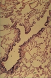
Department Images 98 - 151 (Dustin) Flashcards
What tissue is this slide from?
(really difficult without seeing more of the lumen)

Esophagus
contains inner circular and outer tongitudinal muscles layers with skeletal muscle interspersed throughout
epithelium is stratified squamous non-keratinized

What tissue is this slide from?

Stomach Cardia
Near the esophageal-gastric junction, the bottom part of the image has strat squam non-keratinizing epithelium

Which tissue is this slide taken from?

Fundus of Stomach
Has many different kinds of cells in the gastric pits: know mucus neck cells, parietal cells, chief cells
One protruding fold of cells is called a plica villosa, surrounded on either side by gastric pits

What tissue is this from?
What stain? What is the purpose of this stain?

Fundus of Stomach
PAS-Congo-Hematoxylin stain
Makes mucus (both surface and neck) cells purple as they are PAS positive
Parietal cells turn orange
Chief cells become basophilic / grey

What cells here are orange?
Which ones are red?

Orange = Parietal Cells
Red = Mucus Neck Cells

What are the orange cells?
What are the greyer/ basophilic cells more at the top of the image?
What are the redder cells at the bottom of the image?

Orange = Parietal Cells
Greyer/basophilic = Chief Cells
Redder = Mucus Neck Cells
All in the gastric glands of stomach fundus

What tissue is this slide from?

Pylorus of Stomach
has branched tubular glands, almost entirely mucus secreting cells, stained poorly

What tissue is this slide from?
What major structures do you see?

Pylorus of Stomach
See Gastric Pits, and below are Pyloric Glands - not to be confused with Brunner’s glands of the duodenum which are in the tela submucosa

What is the large fold you see curving rightward in the lower half of the image?
What tissue is this slide from?

Plica Circularis aka Fold of Kerckring
Slide is from Jejunum
(no Peyer’s Patches or Brunner’s Glands)

What do you call the protruding fold of cells that makes up most of the image?

Intestinal Villi

What tissue is this slide from?
What stain?
(not a typical slide)

Jejunum
Indian Ink
(book says it demonstrates the blood vessels of the villus)
What are the cells with reddish granules at the base of the crypt?
Where do these exist in the GI tract?

Paneth Cells
Exist only in small intestine crypts of Lieberkuhn

What do you call the lighter thing sandwiched between the two darker eosinophilic structures?

Myenteric Plexus (of Auerbach)
Takes place between the inner circular and outer longitudinal smooth muscle layers of the GI tract

What tissue is this slide from?

Duodenum
Have typical small intestine structures + Brunner’s Glands in the tela submucosa

What type of glands are at the bottom of the image?
What tissue is this slide from?

Brunner’s Submucosal Glands
From the Duodenum

What tissue is this slide from?
What are the large purple areas in the middle?

Ileum
Purple areas = Peyer’s Patches

What tissue is this slide from?

Colon
Crypts of Lieberkuhn with a bunch of goblet cells and no villi

What tissue is this slide from?

Appendix
Has secondary follicles, NOT Peyer’s Patches
Many goblet cells, few/short crypts, no teniae

What tissue is this image from?
What stain?
What is the majority of the blue stuff in the image?

Pig Liver (Azan)
Blue = interlobular connective tissue that clearly separates the hepatic lobules, and in some corners between lobules you see the (Glisson’s) portal triad of interlobular bile duct, vein, and artery

What 3 important vessels do you see here?
What is the combination of them called?
What stain?

Interlobular Bile Duct: Simple cuboidal-columnar epithelium
Interlobular Artery/arteriole: thick-walled, small diameter.
Interlobular Vein/venule: largest diameter vessel here, typical thin wall
All 3 together = portal triad
Azan Stain

What organ is this from?
What stain?
What does this stain show?

Liver, India Ink Injection
Shows vascular pattern / Kupffer cells

What liver structure is this image focused on?

Portal Triad again
Really only the interlobular veins/venules are clear

What 3 important vessels do you see here?

Portal Triad again
This time all 3 are very clearly here

What type of cells are here?

Hepatocytes
Make up liver sinusoids, lined with Kupffer Cells which are generally not visible with an H-E stain like this

What tissue is this?
What stain? What does that stain show?

Liver
Best’s Carmine Staining
Stains glycogen granules as red
You know liver got that glycogen

What organ is this?
What stain?
What animal is it from?
What does the stain show?

Liver
Golgi silver impregnation stain
Pigeon (that was stressed for some reason)
Stain shows the bile capillaries between hepatocytes. Note that there are Hering ducts at the periphery of hepatic lobules, but I don’t think you can see them

What organ is this from?
What kind of epithelium?
What is unique about its layers?

Gall Bladder
Simple Columnar with Cuticle
Has no lamina muscularis mucosa or submucosa

What organ is this from?
What are most of the cells you see?
What is the lighter cluster at the top-right of the image?

Pancreas
Most cells are serous acini (exocrine)
Lighter area = Islets of Langerhans (endocrine function)
Note there are also NO salivary ducts or adipose tissue, how you can differentiate from Lacrimal and Parotid Glands

What type of cells are these?
What organ are they from?

Serous acini of Pancreas
Pretty bitchy question without more context, but pancreatic serous acini are supposed to be more acidophilic and have a narrower lumen

What organ is this from?
Be able to identify many regions, I’m not gonna label it here
Note also the epithelium transitioning

Larynx - Frontal Section
Epithelium everywhere except the vocal folds should be Respiratory (pseudostratified columnar ciliated)
In vocal folds it is stratified squamous non-keratinizing

Where in the body is this slide from?
What type of epithelium?
What type of tissue makes up the left half of the image?

Trachea
Pseudostratified Columnar Ciliated (Respiratory Epithelium)
Left half = Hyaline (Tracheal) cartilage

What organ is this image from?
1, 2, and 3?
What is the mostly white space between 3 and 3?

Lung
1: Bronchus because it has respiratory epithelium and cartilage
2: Bronchiolus because of smooth muscle
3: A pulmonary artery
Between 2 and 3: Alveoli

What type of airway is the most prominent in this image?
(the big one with the irregular dark lumen)
Terminal Bronchioles
Epithelium becomes simple cuboidal (mostly Club/Clara cells) non-ciliated with no goblet cells
Should probably say Club cells and not Clara cells because Clara was a nazi, and you don’t like nazis, do you?

What type of airway is this?

Alveolar Sac with Alveoli
Look for the Type I and Type II Pneumocytes

What tissue is this?
What stain? What does it show?

Lung
India Ink - shows pulmonary capillary network
What organ is this?
What stain? What does it show?

Lung
Resorcin Fuchsin - shows elastic fibers of the pulmonary connective tissue
What organ is this from? And what part of that organ?
What are the round things you see? What are the 2 layers of those things?

Kidney - Cortex
Round stuff = Renal / Malpighian Corpuscles, which contain Bowman’s capsule and glomerulus
Bowman’s Capsule has 2 layers:
Parietal (simp squam epith)
Visceral (podocytes)

What organ is this from?
What stain?

Kidney
India Ink: shows the vascular pattern, staining the interlobular arteries and glomerulus

What organ is this?
There are some labeled things on the answer card, be able to know where all of them are on this image

Kidney (Cortex)
Note: macula densa is a modified part of distal convoluted tubule (DCT) situated at the vascular pole of the renal corpuscle. Has darker cells, more nuclei
The DCT have simple cuboidal epithelium with a wider, regular, rounder lumen than the PCT, and DCT lumen has no brush border

From the top, what is the first labeled duct?
Second? Third?
Where is this sample from?

1st = Thick Limb of Henle Loop
2nd = Thin Limb of Henle Loop
3rd = Collecting Duct
(I think, anyway..)
This is from outer medulla of kidney

What organ is this from?
What type of epithelium?
Be able to name layers

Urinary Bladder
Transitional Epithelium
(Esophagus can look similar, the umbrella cells are clear in this one though)

What organ?
1 - 5?

Testis
- Convoluted Seminiferous Tubule (make sure you say convoluted!)
- Leydig Cells - large polygonal cells with acidophilic cytoplasm, pale nucleus, visible nuceolus
- Sertoli Cell - pale conical nucleus, prominent nucleolus
- Spermatogonium (closer to the wall of the tubule) and Primary Spermatocyte
- Secondary Spermatocyte

What organ?
What stain?
Be able to identify the types of cells

Testis in a Convoluted Seminiferous Tubule
Iron Hematoxylin
Gotta know different types of cells in spermiogenesis
(Outer layer of squamous-looking cells around a seminiferous tubule = Lamina Limitans)

What organ?
What is 1?
What’s the tubule above #1?
What stain?

Mediastinum Testis
1. Rete Testis of Haller
The tubule above #1 is probably a straight seminiferous tubule (tubuli seminiferi recti)
Iron Hematoxylin Stain

What tissue is this image from?
What type of vessels make up the top half of the image?
and the bottom half?

Epididymis
Top half = Ductus Epididymis - note the pseudostratified columnar epithelium, with stereocilia
Bottom half = Ductus Efferentes Testis with characteristic alternating cell height, simple cuboidal and simple/psuedostratified columnar epithelium, with microvilli, and kinocilia

What type of vessel is this?

Ductus efferentes testis

What vessel is this?
What type of epithelium?

Ductus Epididymis
Pseudostratified Columnar Ciliated

What tissue is this from?
What’s the huge vessel in the middle? (you also should be aware of its layers) & What kind of Epithelium?
What’s the smaller (but still pretty big) vessel in the top-right of the image?

Spermatic Cord
Big thing - Ductus (or Vas) Deferens, with pseudostratified columnar ciliated epithelium and 3 muscular layers
Top-right vessel - Deferential Artery, should always be near the ductus deferens

Which tissue is this image from?

Seminal Vesicle
Looks similar to the ampulla of the oviduct, especially without more in the frame

What type of epithelium is here?
What organ is this from?

Simple Cuboidal to Columnar Epithelium
(it’s variable in this organ, depending on testosterone level. Our green histo book p. 162 says simple cuboidal to simple columnar, but other sources say it’s more between pseudostratified columnar and simple columnar, and I believe that is probably more correct than our book because just look at it)
In the Seminal Vesicle

Which organ is this image from?
What type of gland/secretion is this?

Prostate Gland
Apocrine Gland (although people have recategorized it to “pseudo-apocrine” because it’s really more merocrine. Fucking scientists.)
Has low cuboidal to columnar epithelium, widely dilated lumen, corpora amylacea/ prostatic stones, smooth muscle bundles within the connective tissue septa

What tissue/region is this from?
What type of epithelium in that lumen at the top?
What are the blue arrows pointing at?
What are the red stars showing?

Penis - Corpus Spongiosum
Stratified Columnar Epithelium of Urethra
Blue arrows = Intimal Cushions of Ebner inside the Helicine Arteries
Red Stars = Littre’s Paraurethral Glands

What tissue is this from?
What kind of glands do you see?

Glans Penis (foreskin)
Probably the only place with one external and two internal layers stratified squamous keratinizing epithelium
Has some (holocrine) sebaceous glands
Notable features: prepuce, navicular fossa, strat squam non-ker epithelium in urethra
(Having trouble finding any decent images for this; either it’s shitty histology sections or it’s mutilated dicks)
