Department Images 152 - 209 (Dustin) Flashcards
(58 cards)
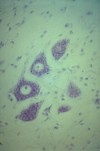
What organ is this from?
What does the arrow point to?
Star?
1, 2, 3?

Ovary
Arrow: Peritoneal Epithelium (simple cuboidal)
Star: Graafian Follicle (although it’s labeled as a Tertiary Follicle, but looks much like a deeper Graafian. Gonna ask about this one)
1: Primary Follicles
2: Primordial Follicles
3: Secondary Follicle (probably)
(lemme know if you think one of these is wrong! if I remember correctly, the Hungarian way to classify these is slightly different than in English books, maybe more similar to German books)
Where is this?
What type of epithelium do you see?
What’s the layer beneath it?
What are the round things?

Ovary
Peritoneal Epithelium (simple cuboidal)
Layer beneath it = Tunica Albuginea
Round things = Primordial Follicles

What type of follicles are the 3 large ones in the bottom half of the image?
And the medium-sized one right in the middle?
And all the little ones at the top right?

3 large ones: Secondary Follicles
Medium sized in middle: Primary Follicles
Top ones: Primordial Follicles

What kind of follicle?

Graafian
But the image is labeled as Tertiary Follicle, going to ask about this later
What is this from?
What are the paler cells that make up most of the image?
What are the darker cells at the top?

Corpus Luteum
Paler cells = Granulosa Lutein Cells
Darker cells = Theca Lutein Cells

What tissue is this from?

Uterine Tube / Oviduct, Ampullary Part
Simple Columnar, some ciliated and some non-ciliated
In some slides, the mesosalpynx might be seen: double layer of peritoneum containing blood vessels
Don’t mix it up with seminal vesicle

What tissue is this?
What type of cells do you see in the epithelium?

Oviduct, Ampulla
See Simple Columnar Epithelium with:
Ciliated Cells
Non-ciliated (secretory) Cells
“Peg” or “Pin” Cells - dark, elongated nuclei protruding into the lumen, degenerating cells

What tissue is this from?

Uterus - Proliferative Phase (early in phase)
Has relatively less glands than secretory phase, thinner endometrium

What tissue is this?

Uterus - Secretory Phase
Has many “tortuous” glands, thick endometrium

What tissue is this from?
What layers do you see?

Fetal portion of the Placenta
At the lowest part of the image is the simple cuboidal amnion epithelium
Above that is the chorionic plate: thick connective tissue
At the top you see chorionic villi
What tissue is this from?
1? 2?
What’s the space between the structures?

Placenta
1: Chorionic Villi (free)
2: Anchoring Villus
Intervillous Space - fills with maternal blood
Note also the syncitiotrophoblasts, cytotrophoblasts and other important placental structures

What tissue is this?
What does the image show?

Placenta
Shows the anchoring villus connecting with the basal plate and decidual cells, with eosinophilic fibrinoid
(I think, anyway)

What’s the big pink blob?

Placental Septum with mainly decidual cells
What tissue is this?
What does this show?

Placenta
Shows the anchoring villus connecting with the basal plate and decidual cells, with eosinophilic fibrinoid
(another one)
What tissue is this?

Vagina
Note:
- Wide lamina propria mucosae has lots of CT with elastic fibers
- Vaginal wall has neither glands nor submucosa
- Esophagus easily distinguished from vagina bc esophagus has muscularis mucosae, tela submucosa, and glands
- Ureter has transitional epithelial lining and regular smooth muscle in its tunica muscularis

What tissue is this from?

Non-Lactating Breast
(Mamma Nonlactans)
What tissue is this?

Lactating Breast
Mamma Lactans
What tissue is this?
What glands are here?

Non-Lactating Breast
There are Areolar Glands of Montgomery here: apocrine secretion
Areolar glands or Glands of Montgomery are sebaceous glands in the areola surrounding the nipple. The glands make oily secretions (lipoid fluid) to keep the areola and the nipple lubricated and protected. Volatile compounds in these secretions may also serve as an olfactory stimulus for newborn appetite.
What tissue is this from?
What stain?
Be able to name the parts

Hypophysis
Chrome-Hematoxylin-Phloxin stain: helps differentiate between acidophils and basophils of the adenohypophysis (the bright red tissue to the right)

What tissue is this from?
What cells do you see?

Adenohypophysis - H-E Stain
Acidophils: bright red
Basophils: larger, basophilic cells
Chromophobes: poorly stained

What tissue is this?
What cells do you see?

Adenohypophysis
(with Chrome-Hematoxylin-Phloxin stain to show more of a difference between cells)
Acidophils: bright red
Basophils: larger blue cells
Chromophobes: poorly stained

What tissue is this?

Adenohypophysis
(Says “Kresazan” for stain, which either google translates as “road traffic,” or it’s some sort of altered Azan stain)
What tissue is this?

Adenohypophysis
(Says “Kresazan” for stain, which either google translates as “road traffic,” or it’s some sort of altered Azan stain)
What 2 parts of the pituitary gland do you see here?

Intermediate Part to the left
Neurohypophysis takes up the rest of the image