Chronic cell injury and adaptations Flashcards

1
Q

If the injury is sublethal and chronic the cells may:

A
  • adapt
  • accumulate normal or abnormal substances
2
Q

What is autophagy?

A
  • self eating
  • survival mechanism
  • consume damaged organelles
  • consume own proteins and carbs as a source of nutrition
  • protects cell from death
  • limits inflammation if cell dies
3
Q

What is heterophagy?

A
  • where a cell phagocytosis another cell or part of another cell
4
Q

What epithelium is this showing?

A
  • normal ep
  • simple columnar of mammary gland
  • purple shows nuclei
5
Q

What is shown here?

A
  • atrophy = decrease in tissue mass due to decreased size and/ or number of cells after it has reached normal size
6
Q

What are the causes of atrophy?

A
  • nutrient deprivation
  • loss of hormonal stimulation
  • decreased workload
  • loss of innervation
  • compression
7
Q

What is hypoplasia?

A
  • tissues decreased in size because they never developed completely
8
Q

What is this showing?

A
  • Hypertrophy = increase in tissue mass due to increased size of cells (parenchymal cells, not stroma or leukocytes)
  • increased size and number of organelles within cells (not water)
9
Q

Causes of hypertrophy?

A
  • increased workload
  • increased hormonal stimulation
10
Q

What is this showing?

A
  • Hyperplasia = increase tissue mass due to increased number of cells
  • subsides if stimulus removed
11
Q

Causes of hyperplasia?

A
  • increased workload
  • increased hormonal stimulation
  • inflammation
  • physical trauma

(can be precursor to neoplastic transformation)

12
Q

What is this showing?

A
  • Metaplasia = change from 1 differentiated cell type to another
  • e.g. squamous metaplasia (replacement of glandular with stratified squamous ep)
  • can be seen in healing after mastitis
13
Q

Purpose of metaplasia?

A
  • protective mechanism
  • can have negative consequences
14
Q

What is this showing?

A
  • Dysplasia = abnormality in formation of a tissue
  • in ep it implies:
    • increase in number of poorly differentiated cells
    • disorganised arrangment
    • variable appearance
    • can be precursor to neoplasia
15
Q

Why do injured cells accumulate exogenous and endogenous substances?

A
  • altered metabolism
  • genetic mutations
  • exposure to indigestable exogenous substances
16
Q

What is Lipidosis?

A
  • accumulation of lipid within parenchymal cells e..g. hepatocytes
17
Q

Causes of lipidosis?

A
  • increased fatty acid metabolism
  • abnormal cell metabolism
  • impaired release of lipoproteins
18
Q

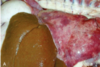
What is the appearance of lipidosis?

A
  • swollen, yellowed liver
  • greasy texture
  • may float in water
19
Q

What is the micro appearance of lipidosis?

A
  • sharply defined large lipid vacuoles
  • distend the cytoplasm
  • displace the nucleus peripherally
  • can see spaces where fat would be (washes out)
20
Q

Where is glycogen normally stored?

A
  • hepatocytes
  • skeletal muscle cells
21
Q

In what situations is glycogen depleted?

A
  • starvation
  • sick animals
22
Q

When does glycogen accumulated intracellularly?

A
  • excessive in glycogen storage disease
  • in the liver with diabetes mellitus and canine hyperadrenocorticism
23
Q

What is the gross appearance of glycogen hepatopathy?

A
  • mottled
  • brown
  • swollen liver
  • if cut with a knife would not be greasy - not lipidosis
24
Q

What is the micro appearance of glycogen hepatopathy?

A
  • poorly defined
  • small
  • irreguarly shaped vacuoles (feathery)
25
What colour does protein stain with H&E?
* pink
26
What colour do nucleic acid stain?
* blue
27
What would you observe with the accumulation of protein in cells?
* Hylaine = glassy appearance
28
When can protein accumulation be normal?
* Russell bodies of plasma cells
29
What is this showing?
* kidney * purple is nucleus * pink is cytoplasm * granular and dark pink cytoplasm = protein that shouldnt be there
30
What are viral inclusion bodies?
* feature of some viruses * can be intranuclear, intracytoplasmic or both * can be eosinophilic, basophilic or amphophilic
31
What is this showing?
* virus (pink blobs in the nucleus and cytoplasm)
32
Name the examples of extracellular accumulations
* hyaline (protein) substances * pathological calcification
33
What is this showing?
* hyaline substance accumulation (extracellular) * stain homogenously eosinophilic on H&E stains * not indicative of a single protein * (kidney)
34
What is this showing?
* hyaline protein accumulation in kidney * protein casts in the lumen of renal tubules (albumin, haemoglobin, myoglobin) * serum or plasma in blood vessels * serum proteins in vessel walls * collagen fibres in some scare * collegaen encusted with proteins from eosinophils * thickened basement membranes * fibrin thrombi * amyloid
35
What is this showing?
* Pathological calcification * deposition of calcium salts in soft tissues
36
What is Metastatic calcification?
* calcification of soft tissues due to elevated serum calcium e.g. * renal failure * PTHrp in some neoplasms * bone invasion and lysis in some neoplasms * Vit D toxicity
37
What is dystrophic calcification?
* calcification of dead tissue (soft tissue necrosis) * body is trying to make the tissue more inert * calcium does not react with anything
38
What is Calcinosis cutis?
* calcification of the ep and collagen of the skin * canine hyperglucocortioidism (Cushings)
39
What is Calcinosis circumscripta?
* localised calcification of the dermis or subcutis * tends to happen on pressure points/ often bony prominences * a form of dystrophic calcification
40
Name the exogenous pigmented substances
* carbon and dust * anthracosis (black carbon pigment) * pneumoconiosis (lung disease due to dust) * tattoos * pigmentation of lymph nodes * carotenoid pigments * yellow colouration to plasam and lipid laden cells * tetracycline * yellow to brown discolouration of teeth if present during tooth development + bone
41
What are the nonhaematogenous pigments?
1. melanin 2. lipofuscin 3. ceroid
42
What is this showing?
* Melanosis = localised deposits of melanin (not a lesion)
43
What does copper deficiency cause?
* fading coat colour (copper required in production of melanin)
44
What is vitiligo?
* autoimmune attack of melanocytes
45
What is lipofuscin?
* yellow-brown lipoprotein that accumulates as residual bodies in secondary lysosomes
46
What is ceroid?
= lipofuscin-like but accumulates in disease states (particularly oxidative stress)
47
What are the haematogenous pigments?
* haemoglobin * haematin * haemosiderin * haematoidin * bilirubin * porhyria
48