Basic principles of cytological examination Flashcards

1
Q

What does the outcome of a cytological examination depend on?

A
  • cellularity
  • cell preservation
  • representative sample
  • (quality of submitted smears)
2
Q

What different processes may appear indistinguishable on cytology?

A
  • fibroplasia and low grade spindle cell neoplasia
  • well-differentiated neoplasms from normal tissue
3
Q

What are the 4 headings on a cytology report?

A
4
Q

What are immunohistochemistry (IHC) and immunocytochemistry (ICC)?

A
  • the use of colour-labelled antibodies to identify certain cell markers on histopathology slides (IHC), or cytology smears (ICC)
5
Q

Why can antibodies be helpful in ICH and ICC?

A
  • antibodies can help us:
    • identify the exact cell type where this is not clear on routine staining (e.g. round cell tumours)
    • recognise a sub-group within a cell type
    • identify certain properties of tissues (e.g. cell proliferation markers such as Ki-67 in mast cell tumours)
6
Q

What is this showing?

A

IHC

7
Q

What is this showing?

A

ICC

8
Q

What question should you always ask yourself?

A
  • am i looking at inflammation, neoplasia, inflam?
9
Q

What questions should be asked if inflammation is seen?

A
  • relevant history - vaccines etc.
  • is there only 1 type of inflam cell present? chronicity? concurrent haemorrhage?
  • are there any tissue-derived, and what is their appearance?
  • any infectious organisms? are they relevant to the inflammation, or contaminants/ normal flora?
  • is there indication for special stains?
  • evidence of foreign body material/ non- biological material presence
  • is there concurrent necrosis?
  • can we rule out underlying neoplasia?
10
Q

When can neutrophilic inflammation be defined?

A
  • when >85% of nucleated cells are neutrophils
11
Q

Causes of neutrophilic inflam?

A
  • bacterial infection
  • trauma
  • tissue necrosis
12
Q

What does this show?

A
  • non-degenerate neutrophil (blood)
13
Q

What are these?

A
  • degenerate neutrophils
    • nuclei more swollen
    • colour is paler
    • cell borders of the neutrophil are not clearly visible
14
Q

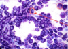
This is from a dogs synovial fluid. What cells can be identified and what arrangement of cells can be seen?

A
  • no infectious organisms seen
  • arrangment: ‘windrowing’ of cells (form lines) - associated with high viscosity
  • synovial fluid - not very cellular (so increased number of cells shows infection)
15
Q

What has happened to these cells, and what is it showing?

A
  • pyknosis/ karyorrhexis of neutrophils
    • nucleus has fragmented (apoptosis)

NEUTROPHILIC INFLAM

16
Q

What is this cell, and what does it mean?

A
  • neutrophil (a lot bigger than normal)
  • so neutrophilic inflammation
  • septic peritonitis
17
Q

Identify the circled cell

A
  • degenerate nuclei containing bacterial cocci
  • in synovial fluid
  • neutrophilic inflam
18
Q

What is this cell?

A
  • very degenerate nuclei of neutrophil
  • contains bacterial rods
19
Q

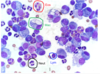
This is from a cats pleural fluid. What can you identify in this image

A
  • neutrophilic inflam
  • short arrow = protein content of fluid
  • long arrow = large group of degenerate neutrophils
  • filamentous bacteria seen
20
Q

What is macrophagic inflam and what are the causes?

A
  • where macrophages are predominating
  • causes:
    • foreign body reaction
    • mycobacterial infection
    • fungal infection
21
Q

When would you classify inflam as chronic inflam?

A
  • multinucleate/ giant macrophages seen
22
Q

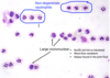
This is a skin nodule from a dog. What is labelled here, and what could be the diagnosis?

A

(from top)

1 - high number of red cells, clumped

2- macrophage

3- abnormal macrophages

Diagnosis = macrophagic inflam and fungal infection

23
Q
A
24
Q

This is a cat skin mass. What is labelled and what is the diagnosis?

A
  • circled = macrophagic inflam and fungal infection
  • arrow = long structures found within and outside macrophages - fungus
25
What cell is this and what does the staining indicate?
* macrophage * has non staining areas - indicates mycobacteria
26
What stain should be used to identify mycobacteria?
* Zlehl-Neelsen stain
27
What can be seen, and what does it suggest?
* giant macrophages * shows chronic inflammatory process * necrosis * mixed/ eosionphilic inflam
28
What is eosinophilic inflam and what are the causes?
* when \>10% of total nucleated cells are eosinophils (among other inflam cells) * causes: * hypersensitivity/ allergic reactions * parasitic infections * feline asthma, eosinophilic bronchpneumopathy in dogs * paraneoplastic eosinophilic inflam (T cell lymphoma, mast cell tumour)
29
Identify the eosinophil species
30
This shows skin from a dog. What is happening?
* eosinophilic inflam * all cells are eosinophils (slighlty lobulated nuclei shown by arrow)
31
What inflammation is occuring, and what is causing it?
* mild eosinophilic inflam * increased mucus * presence of large parasitic larva
32
What is lymphocytic/ plasmacytic inflam and what are the causes?
* when lymphocytes are predominating accmpanied by variable numbers of plasma cells * causes * injection site reactions * other forms of antigenic stimulation
33
What is this showing?
* lymphocytic/ plasmacytic inflam * arrows pointing to lymphocytes
34
What cell is this and what is the arrow pointing to?
* plasma cell * golgi zone
35
What are the features of plasma cells?
* round nuclei * increased amount of blue cytoplasm * golgi zone
36
WHat are the cells shown?
* plasma cells
37
What is mixed inflammation?
* when a mixture of inflam cells are present * one cell type may be predominating
38
What is this an example of?
* mixed inflam
39
What may not be visible with true haemorrhage and why?
* platelets * as they disintegrate quickly
40
What haemoglobin breakdown products form within macrophages over time?
* haemosiderin (dark blue/black) * haemotoidin (golgen rhomoid crystals)
41
What has happened in this slide? What is circled?
* macrophages circled * this slide shows a haemorrhage in abdominal fluid * erythrophagocytosis
42
What is this?
* haemosiderin
43
What are the 2 arrows pointing to?
* top arrow = haemsoderin (blue/black) * bottom arrow = haematoidin (golden rhomoid crystals) * within a macrophage
44
What is inside this macrophage?
* haematoidin (golden rhomoid crystals)
45
How do you classify neoplasms in cytology?
* mesenchymal * cells forming tissues that are considered 'soft tissues' or connective tissues * derived from mesoderm * epithelial * cells derived from external mucus membranes, parenchymal organs and organ surfaces, glandular tissues * round cell * composed of discretely occuring cells e.g. lymphocutes, plasma cell etc
46
Fill in the labels showing the cytological features of mesenchymal cells
47
Fill in
48
What different arrangments of ep cells can you get?
49
What is the origin of the cells seen?
* EPITHELIAL * cells very cohesive - big lump * see the cell borders
50
This shows the cytological features of round cells - fill in gaps
51
What are the 5 types of round cell tumour?
1. mast cell tumour 2. lymphoid tumours 3. plasmacytic tumours 4. histiocytic tumours 5. transmissable venereal tumour (not in UK)
52
What tumour type is shown here? label the cells
* round cell (lymphoma) * green = round cells (neoplastic) * black = lymphocytes
53
What cell type is shown here?
* round cells * large number of them = tumour
54
What type of tumour is this? label
* mast cell tumour (common in dogs)
55
What type of tumour is this? label
* mast cell tumour
56
What are the cytological criteria of malignancy?
* anisocytosis - variable size of cells * anisokaryosis - varible size of nuclei * mulinucleated cells * anisokaryosis within the same cell * cell crowding/ disorganisation * nuclear moulding * enlarged nuclei (macrokaryosis) * micronulcei/ satellite nuclei/ lag chromatin * increased numbers of mitotic figues * abnormal cell divisions * uneven nucleoli within the same cell/ different cells (anisonucleoliosis) * increased numbers of nucleoli within nucleus * variable numbers of nucleoli amongst cells * enlarged nucleoli (\>2/3 RBC in dogs) * irregular nucleolar shapes, angular * hyperchromatic cytoplasm * signet ring
57
What is this arrow pointing to?
* micronuclei
58
What cytological criteria of malignancy is this showing?
* cell crowding * nuclear moulding
59
What is this and how is it causes?
* signet ring * significant dilation of cytoplasm and displacement of nucleus