5.2 EKG Flashcards
What is an ECG?
ECG & EKG = same
- composite of all APs generated by nodal and contracile cells at given time
- waves: deflections above or below baseline
- Segments: sections of baseline between 2 waves
intervals: combination of waves & segments
*ECG is NOT an action potential -> looking at all electrical activity from all heart parts @ same time
what are the different deflections in an ECG


What does the P-R interval represent?
- 16s
- start of atrial excitation to start of ventricular excitation
*sometimes called P-Q interval, but Q wave is often hard to find so finding P-R interval is easier

what is the S-T segment
- 08s
- AP of ventricular myocytes are in plateaus phase
(ventricles depolarize)
*all muscles are contracting here

describe teh QT interval
- start of ventricular depolarization to end of ventricular repolarization


P wave
- atrial depolarization, initated by SA node
*stops at atria -> doesnt spread to ventricles
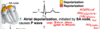

With atrial depolarization complete, impulse is
delayed at AV node


ventricular depolarization
- begins at apex, causes QRS complex
- atrial repolarizaiton occurs


ventricular depolarization is complete
*moment where all ventricular muscles are contracting


ventricular repolarization begins at apex, causing the T wave


ventricular repolarization is complete
- after T wave the ventricles are fully relaxed


junctional rhythm
- Sa node is non functional so P waves are absent
- av node paces heart at 90-60 bpm
* P wave caused by Sa node that causes atrial depolarization

- second degree heart block
- AV node fails to conduct some SA node impulses
- As result there are more P waves than QRS
what is systole
what is diastole
systole = contraciton (1/3 of time)
diastole = relaxation (2/3 of time)
what is the first phase of the cardiac cycle
ventricular filing
(mid to late diastole)
- AV valves open; semilunar vales are closed
- 80% of blood passively flows into ventricles
- Atrial systole occurs, delivering remaining 20%
- end diastolive volume: volume fo blood in each ventircle at end of ventricular diastole
what is end diastolic volume
happens at end of venricular filling
volume of blood in each ventricle at end of ventricular diastole
describe the second phase of the cardiac cycle
- ventricular systole
- atria relax (atrialdiastole); venticles begin to contract
- INC venticular pressure results in closing of AV valves (dec in vol so inc P)
- Isovolumetirc contraction phase: all valves are closed
- Ejection phase: ventricular pressure > pressure in large arteries; semilunar values open
*end systolic volume
describe end systolic volume
volume of blood remaining in each ventricle at end of ventricular systole
describe the 3rd pahse of the caridac cycle
isovolumetirc relaxation
- occurs in early diastole
- > ventricles relax
- > backflow of blood in aorta & pulmonary trunk closes SL valves & causes dicrotic notch
*breif rise in aortic pressure
describe the sounds of the heart
- two sounds associated with closing of heart vlaves
- 1st:
- Av valves closing
- beginning of ventricualr systole
- “lub”
- 2nd
- occurs when SL vlaves close
- beginning of ventricular diastole
- “dub”
what is a heart murmer
abnormal heart sounds, most often indicative of value probelms


