3.1 Oral Cavity & stomach Flashcards
what are the functions of saliva
Cleanses mouth
Moistens & dissolves food chemicals
Aids in bolus formation
Contains enzymes to begin digestion
Buffers pH (bicarbonate)
Lubrication
what are the 2 types of salivary glands
intrinsic (buccal): sacttered in oral mucosa
Extrinsic: parotid, submadnibular and sublingual


what cells are found in salivary galnds
serous cells: procue watery secretion of enzymes, ions and some mucin
*stin pink
mucous cells: dont stain well

what are the 4 salivary galnds, describe theri cell composition
- Submandibular: serous and mucous cells
*make 60-70% of saliva volume
- Parotid glands: mostly serous cells
- Sublingual glands: mucous cells
- Intrinsic (buccal) glands: serous & mucous cells
what is saliva compsed of
* rpoduce 1-1.5L a day
-97-99.5% water -> almsot neutral
contains:
electrolyes: Na, K, Cl, PO43-, HCO3-
- enzymes: salivary amylase (starch-> maltose) & lingual lipase (fat breakdown)
- proteins: mucin , lysozyme, IgA, albumin
- metabolic wastes: urea and uric acid
- lysozyme, IgA, defensins & cyandide comp to protect aginst microorganisms
how is salivation controlled
- intrinsic gland continuously keep mouth moist
- extrinsic glands produce secretions due to smell, taste, sound or sight or pressure in mouth
- > higher centers of brain -> salivary nucleus of medulla -> PSNS -> CN IX and CN VII
* sleep, fatigue and fear inhibtis it

describe the structure of the tooth
external neatures
- Crown: exposed part above gingiva, covered by enamel (hardest substance in body: calcum salts and hydrozyapatite crystals)
Neck: conencts crown and root
Root: portion embedded in mandible amd maxilla bone

descibre the strcuture of a tooth: internal features
dentin: bonelife materail maintained by odontoblasts of pulp cavity
^ extends trhu all 3 regions
Pulp: connective tissue, blood vessels and nerves
^contains pulp cavity (crown cavity w/ pulp) and root canal (pulp in root)
Cementum: calcified connective tissue: holds teeth in bone, covers root, anchord by peridontal ligament
describe eruption times of human dentition
around 6, 12 and 24
deciduous (milk) teeth:
- central incisor, lateral, first molar, canine, second molar
Permanet teeth: Central incisor + first molar, lateral incisor, 1st and 2nd premolars/bicuspid + second molar, 3rd molar/wisdom tooth

pharynx in the digestive system
- oropharynx and laryngopharynx (naso not part of digestive)
- allows passage of food, fluids and air
describet he esophagus
- inner lumen -> mucosa (stratified squamous epithelium) -> submucosa (aerolat conenctive tissue) -> muscularis externa (circular layer and longitudinal layer) -> Adventita (fibrous connective tissue)
- flat muscule tube (25cm long)
- > pierces diaphagm at esophageal hiatus
- > collased when not involved in food propulsion
- glands in submucosa secrete mucus (lubircation)

desrcibe the buccal phase of deglutition
*chewing- mechanically breaking down
- upper esophageal sphincter is contracted
- tongue presses against hard palate (voluntary)
- bolus forced into oropharyx (involuntary phase begins)

describe the pharyngeal esophageal phase of of deglutition
*Esophageal Phase - involuntary no control
- Uvula & larynx rise -> epiglottis closes larynx (blocks larynx)
- tongue blocks off anterior mouth
- upper esophageal sphincter relaxes
- constrictor muscles of pharynx contract (food forced into esophagus)
- Upper esophageal sphincter contracts
- uvula drops back
- food mvoes through esophagus by peristalsis
- gastroesophageal sphincter opens (food enters stomach)
descibr pharynxgeal esophgeal phase (getting to stomach)
- food mvoes through esophagus by peristalsis
- gastroesophageal sphincter opens (food enters stomach)
anatomy of infant vs adult
(swallowing)
- infant: small oral cavity, tongue palate is flatter, epiglottis almost attached to soft palate
*airway and foodway are separated except when swallowing
- in adult, larynx is lower in neck, food and airway cros in pharynx
*infants can drink and breathe at the same time

descrube the vomiting reflex
- start w/ salivation and nausea
- reverse peristalsis from upper SI & stomach
- glottis closes (prevents aspiration), breath held mid inspiration
- diaphragm & abdominal wall muscles contract, inc intra0abdominal pressure
- esophagus and sphincters relax
- gastric contents ejected

what are the digestive processes of the stomach
- Mechanical breakdown
- Denaturation of proteins
- Enzymatic digestion of proteins by pepsin (+ rennin in infants)
- Secretes intrinsic factor
- Absorption of lipid-soluble substances
- Delivers chyme to small intestine
how does the body respond to the stomach filling
- can hold 50mL to 4L
- stomach pressure remains constant until about 1.5L of food is ingested
- relatice uncahnging pressure results from
- > receptive relaxation: as food travels in esophagus, stomach muscles relax
- > Gastric accommodation: intrinsic ability of smooth muscle to exhibit stress- relaxation response
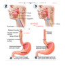
gross anaomy of the stoamch


decsibr the mucsa epithelial cells in the stomach
*Gastric mucosa produced 3L of gastric jucie/day
- it surface have mucus surface cells that secrete alkaline mucus
- then into mucuous neck cells: secrete acidic mucus (less protective, more activating)
- as go further get 3 pronged parietal cells (secrete HCL and intrinsic factor -> inc stomach acidity)
- further into gland get cheif cells: secretes pepsinogen to activate into pepsin
- bottom have some enteroendocrine cells: hormone producitng G cells: secrete gastrin

describe mucosal proection
- layer of bicarbonate rich mucus (protects epithelial cells so juice doesnt eat away at you
- in tight junctions between epithelial cells
- damaged epithelial cells are qucily replaced by dividion of stem cells (surface wells lifespan is 3-6 days)

describe the secretions from parietal cells
- intrinsic factor:: Glycoprotein requred for absorption of vitamin B12 in SI (w/o intrinsic factor cant abs B12)
- HCl: stomach pH 1.5-3.5, deatures protein in food, activates pepsin and kills many bacteria
how do parietal cells produce HCl
- CO2 — from interstitial fluid to parietal cell —> CO2 + H2O
- CO2 + H2O — carbonic anhydrase —> H2CO3
3a. H2CO3 -> H+ can be pumped into stoach lumen while K+ pumped in (H+ K+ ATPase) or
3b. H2CO3 -> leaves cell goes itno interstitial fluid and Cl- pumped in

