3.3 Gastric enteroendocrine Flashcards
what does secretion of gastrin do
- Inc HCl secretion care0 8 3 activators
- Stimulates gastric emptying (minor effect)
- Stimulates parietal cell maturation
- Stimulates chief cells to secrete pepsinogen
- INC intestine muscle contraction
- Relaxes ileocecal valy and stimulates mass movements

what does secretion of histamine do
- secreted from enterochromaffin cells
- inc parietal cells HCl release (one of the 3 thigns needed to activates secretion of HCl)
what does secretion of serotonin do
- from enterochromaffin like cells
- inc contraction of stomach muscle
(makes 90% of total body serotonin, SSRI)
what does release of Somatostatin do
- inhibits secretion from stomach and pancreas
- inhibts small intestine absorption
- inhibits gall bladder and liver release of bile

describe the regulation of gastric secretion
- neural & hormonal mechansims that alter secretions
- Stimulatory and Inhibitory events occur in phases
- Cephalic (reflex) phase: few min prior to food entry (short)
- Gastric phase: food entering stomach t0 3-4 hours later
- intestinal phase: brief sitmulatory effect as partially digested food enters duodenum, followed by inhibitory effects
describe the Cephalic Phase of gastric digestion
- starts with sight, smell, taste or thgouht of food
- > CNS sends impulse via CNX
- Stim mucous cells -> muscus, Chief cells -> pepsinogen, Parietal cells -> HCl, and G cells to make Gastrin
describe the Hastric phase of gastric secretion
- can be triggered by
- psrtially digested proteins, caffeine, inc in pH
- acts on G cells to secrete gastrin which encourages all the stomach actions
- Chemo and stretch receptors
- trigger mucous, chief, parietal and G cells
- psrtially digested proteins, caffeine, inc in pH

describe the intestinal phase of gastric secretion
- decreased pH (more acidic) or prescence of lipids and carbs caues release of secretin, GIP and CCK -> goes in blood stream and inhibits chief and parietal cells, and peristalsis
- duodenal stretch & chemoreceptors act via enterogastic reflax and inhibt myenteric plexus

describe the gross anatomy of the small intestine
* major organ of digestion and absorption
- diameter: 2.5-4cm
- Duodenum: 25cm, jejunum (upper left) 2.5m, ileum (lower right) 3.5 m

what surface modifcation are made to the SI to increase SA
- need inc SA for nutrient abs
- circular fold (plicae circulares) ~1 cm deep (ones you can see)
- villi 1mm high (on folds make it look fuzzy)
- microvilli ~100-2000nm high
- total SA of SI = 200m2
describe the structure of the intestinal mucosa
- Absorptive cells (simple columnar epithelium)
goblet cells and enteroendocrine cells in epithelium
- lacteal running thi middle
- Paneth cells: found deep in crypts that release antimicrobial agents
- villus epithelium replaced every 2-4 days

what are the modifcations in the diff regions of the SI
- Duodenum: in submucosa -> secretes bicar
- Duodenal glands
- Serosa
- muscularis externa
- Ileum
- aggregated lymphoid modules (big colelction of lymphocytes
- Jejunum
* call have plicucircularis
describe the secretions of your SI
- secreted in response to distension or iritation of mucosa by hypertonic or acidic chyme
- slightly alkaline pH (7.4-7.8) & isotonic with blood plasma
- largely water, enzyme-poor, but contains mucus
- facilitates transport and absorption of nutrients
describe the process of digestion in the small intestine
- Chyme from stomach contains
- > patially digested carbohydrates & proteins
- > undigested fats (lipase so far has been minmal
- spends 3-6 h in SI: most water is absorbed, all nutriends are absorbed
what are the requirements for digestion and absorptoin in the small intestine
- Slow delivery of acidic, hypertonic chyme
- Delivert of bile, enzymes & bicarbonate ions from liver and pancreas
- mixing
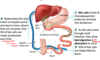
how are things released into SI
- Propfulsion: peristaltic waves move from fundus -> pylorus (starts @ cardiac region)
- Grinding: Most vigorous peristalsis and mixing occur close to pylorus
- Retropulsion: pyloric end of stomach act as pump to deliver 3mL of chyme to duodenum, when it closes material that doesnt go throuhg splashes back (helps with mixing)
describe the ileocecla sphincter/valve
- closed when chyme exerts backward pressure (prevents regurgitation into ileum
- relaxes to admit chyme into LI when
- > gastroileal reflex enhances force of segmentation in ileum
- > gastrin increase motility of ileum
describe the liver
- largest gland in body (3 lbs)
- 4 lobes based on surface features (8 based on vascular & biliary suply)
- right, left, caudate and quadrate lobe
- has a falciform ligament (actually a mesentary)

what does the calciform ligament do
Suspends liver from diaphragm & anterior abdominal wall (a mesentery)
Separates right & leftl obes
Round ligament (ligamentum teres): Remnant of fetal umbilical vein
How does blood get to the liver
blood enters liver at Porta hepatis via
- Hepatic arteries
- Hepatic portal veins
- blood exits liver through hepatic veins (into IVC)
*have two capillary networks in row: artery -> cap -> vein -> vap

locations of hepatic veins and arteries

Describe the microscopic anatomy of Liver
shape of lovules
- liver lobules: hexagonal structure
- filter & process nutrient rich blood
- copmosed of plates of hepatocytes radiating from longitudinal central vein

liver microscopic anatomy
describe the innervation
- middle central vein
- Portal triad: at each corner of lobule (bile duct, branch of hepatic portal bein and branch of hepatic artery)
- liver sinusoids (leaky capillaries between hepatic plates)
- kupffer cells (stellate macrophages)