(14.1) Pulmonary Pathology (Singh) Flashcards
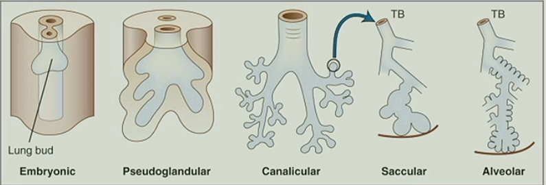
Describe the progression of normal fetal lung development
Embryonic (lung bud) –> Pseudoglandular –> Canalicular –> Saccular –> Alveolar

What are the clinical uses for knowing the stage of fetal lung development?
Knowing the stage can help the clinician determine the age of the fetus
What are the requirements for normal fetal lung development?
- Space in the thoracic cavity
- Ability to inhale (chest call must be able to move, and there must be enough material (amniotic fluid) present to inhale
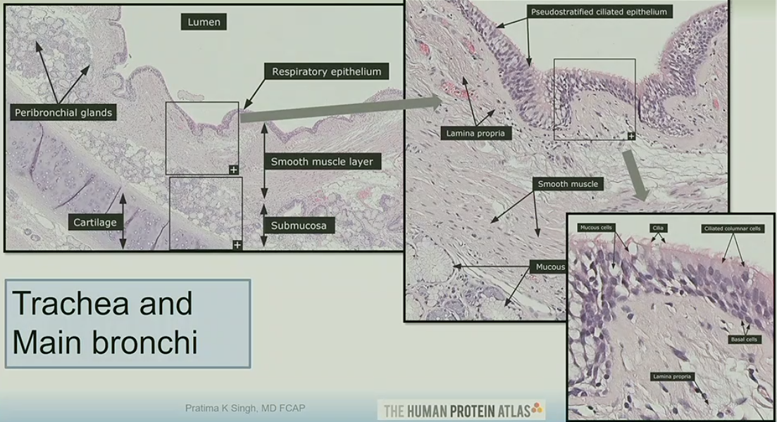
What are the main structures you will see histologically of the trachea and main bronchi?
Cartilage
Glandular tissue
Respiratory epithelium
Smooth muscle layer
Lamina propria

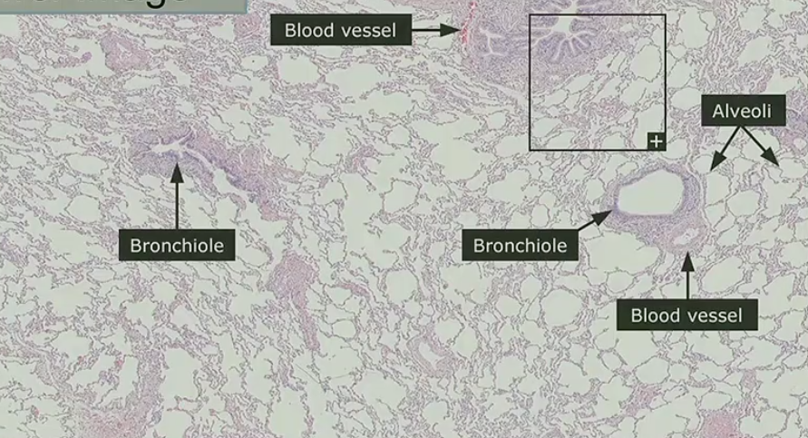
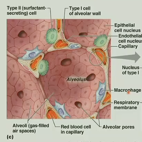
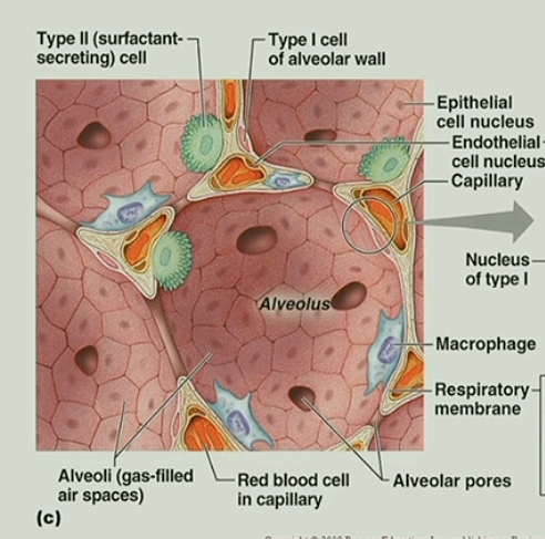
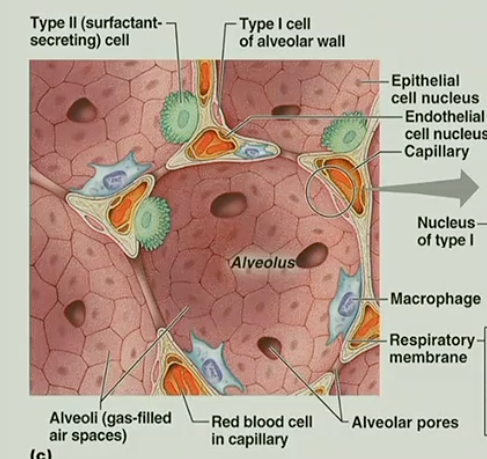
Describe what you would encounter with a low-power image of lung parenchyma
Alveoli
Blood vessels
Bronchioles

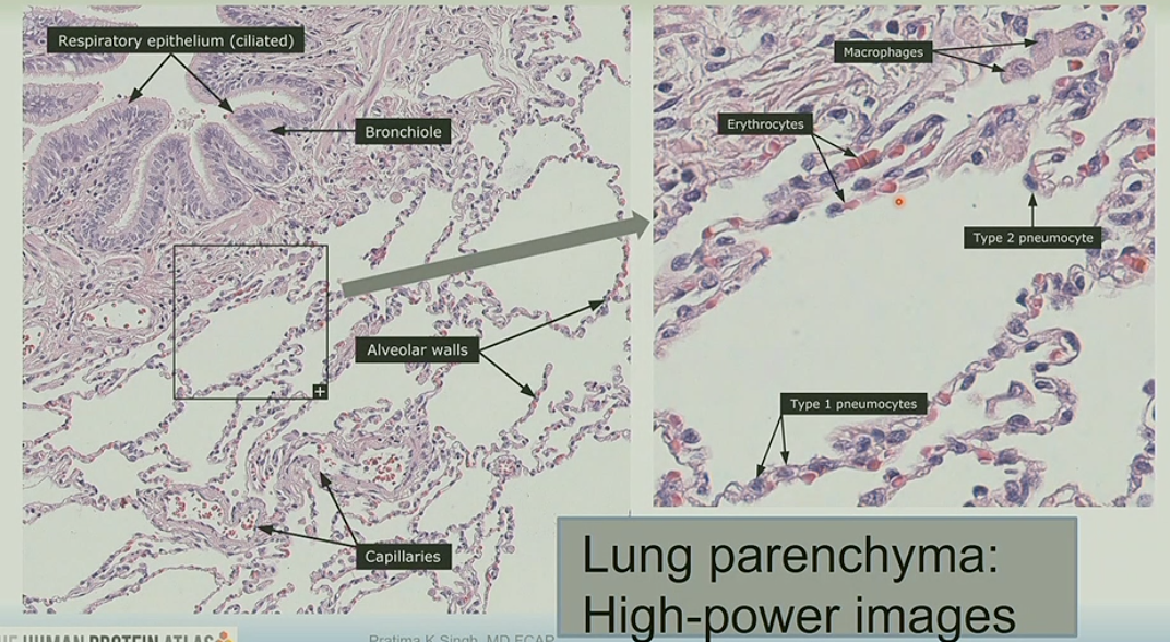
Describe what you would encounter with a high-power image of lung parenchyma
Erythrocytes
Macrophages
Type 1/2 pneumocytes

What is CRUCIAL to the sucessful oxygen exchange occuring in the lungs?
A very THIN layer between the circulating erythrocytes and alveolar lumen

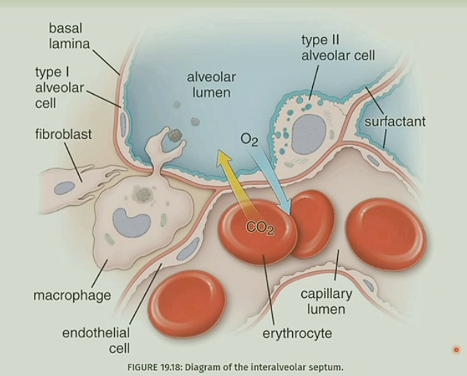
Describe the function of Type I pneumocytes
Facilitate gas exchange
*Support and line the alveoli

Describe the function of Type II pneumocytes
Product surfactant
Replace type 1 pneumocytes
***THEY ARE MODIFIED STEM CELLS HOW COOL IS THAT WOWOWOW

What is the function of alveolar pores?
Allow aeration but also bacteria/cells/exudate to travel between alveoli

What is this pathology?

Pulmonary hypoplasia

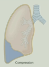
What causes pulmonary hypoplasia?
Reduced space in the thoracic cavity (eg. diaphragmatic hernia)
Impaired ability to inhale (eg. oligohydraminos, airway malformation, chest wall motion disorders)
What is the mortality rate of pulmonary hypoplasia?
HIGH! (up to 95%)
If lung weight is <40%, immediate death occurs in neonatal period
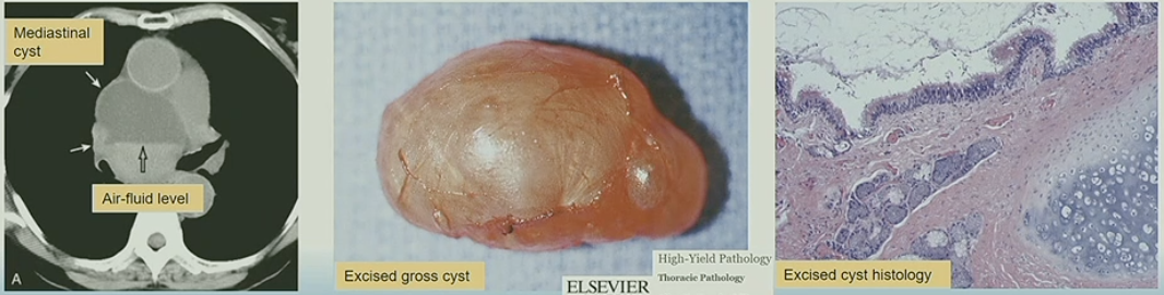
What are foregut cysts?

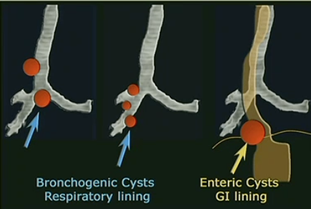
Detached outpouchings of foregut
Seen along hilum and mediastinum
Can be respiratory, esophageal or gastroenteric
Often seen incidentally; complications include rupture, infection, or airway compression
Tx: Excision is curative

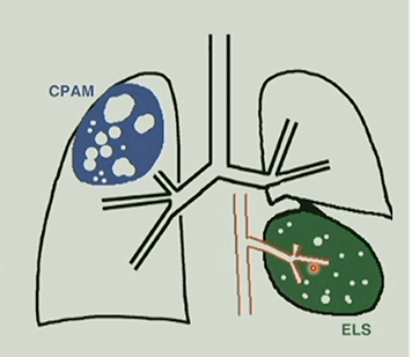
What is congenital pulmonary adenomatoid malformation (CPAM)?
“Arrested development” of pulmonary tissue with formation of intrapulmonary cystic masses
aka, a specific type of pulmonary tissue decides to only make a certain type of tissue for the rest of it’s life
What are pulmonary sequestrations?
Nonfunctioning lung tissue that forms as an aberrant accessory“lung bud”
*Typically found in the region of the left lower lobe
How are pulmonary sequestrations characterized?
Lack of connection to the tracheobronchial tree
Independent (systemic) arterial supply
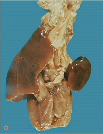
What is this image an example of?

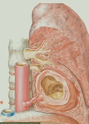
Intralobar pulmonary sequestration (ILS)
What are intralobar pulmonary sequestration (ILS)s susceptible to?
Lack of airway perfusion makes ILS’s susceptible to infection and absess formation

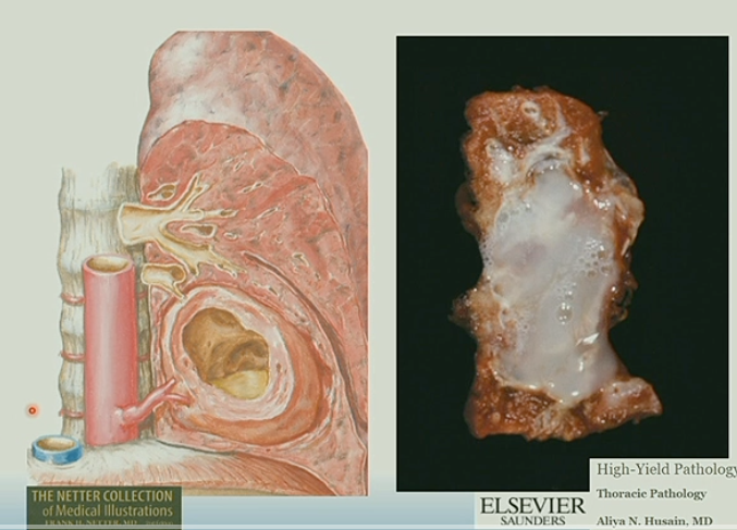
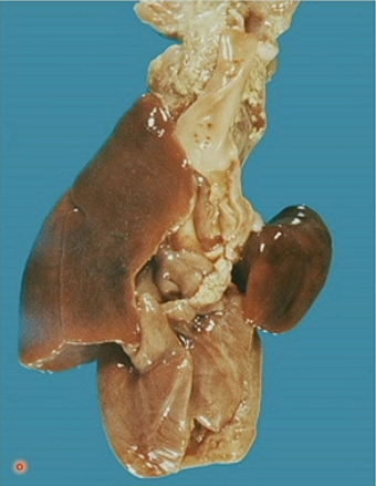
What is this an example of?

Extralobar pulmonary sequestrations (ELS)

How are extralobar pulmonary sequestrations (ELS) different from ILS?
ELS have their own PLEURA
What comes to attention sooner…
ILS or ELS?
ELS
Describe the differences between these congenital anomalies:
CPAM/CCAM
vs
Pulmonary sequestraion
CPAM/CCAM = INTRApulmonary cystic malformation w/ CONNECTION to tracheobronchial airways and pulmonary vasculature
Pulmonary sequestration = INTRA or EXTRA pulmonary lung tissue with NO CONNECTION to pulmonary vasculature or tracheobronchial tree

What is atelectasis?
Partial or complete collapse of the lung
What are the three major types of atelectasis?
- Resorption
- Compression
- Contraction

What causes resorption atelectasis?
Airway obstruction with gradual resorption of air reduces lung expansion

What causes compression atelectasis?
Accumulated material in pleural cavity compresses the lung parenchyma

What causes contraction atelectasis?
Fibrotic or other innate restrictive process in the pleura or peripheral lung restricts lung expansion

What is the LEAST common form of atelectasis?
Contraction atelectasis

What pathologic process is occuring here?

Pulmonary edema

What are the major causes of pulmonary edema?
“Pushing out” –> left sided heart failure
“Leaking out” –> liver disease
Injury to alveolar wall
What are the clinical criteria for acute lung injury (ALI)?
Acute onset
Hypoxemia
Bilateral infiltrates
No evidence of cardiac failure
What is the clinical criteria for acute respiratory distress syndrome (ARDS)?
Worsening hypoxemia
What is diffuse alveolar damage (DAD)?
Histologic manifestation of ARDS
What is the pathology?

Acute respiratory distress syndrome (ARDS)
What are the clinical indications of acute respiratory distress syndrome (ARDS)?
- Abrupt obsent of symptoms
- Hypoxemia (PaO2/FiO2) <200
- Bilateral infiltrates
- Non-cardiac in nature

What is the pathology?

Diffuse alveolar damage (DAD)
Edema + Fibrin + Cell debris = HYALINE MEMBRANES

Describe the histologic differences b/w a normal lung and a lung w/ hyaline membranes
Lung w/ hyaline membranes has THICKENED walls that make oxygen exchange nearly impossible
This is what accounts for the decreased PaO2/FiO2 values observed

What are the three stages of ARDS?
- Exudate
- Proliferative
- Fibrotic
What occurs during the exudative stage of ARDS?
Edema
Hyaline membranes
Neutrophils
What occurs during the proliferative stage of ARDS?
Fibroblast proliferation
Organizing pneumonia
Early fibrosis
What occurs during the fibrotic stage of ARDS?
Extensive fibrosis
Loss of normal alveolar architecture
ARDS : resolution
After the fibroproliferative phase, 2 pathways may ensue… what are they?
Resolution OR Fibrosis (irreversible)

What is acute interstitial pneumonia (AIP)?
Same clinical presentation as ARDS
Same histology as ARDS/DAD
***CANNOT BE ATTRIBUTED TO A SPECIFIC ETIOLOGY
What is a restrictive lung disease?
Volume restriction
FEV1/FVC is normal
FVC is reduced
(FEV=force expiratory volume, FVC=forced vital capacity)

What is obstructive lung disease?
Decreased flow
LOW FEV1/FVC ratio
***(FEV=force expiratory volume, FVC=forced vital capacity)

What are examples of obstructive pulmonary diseases?
Emphysema
Chronic bronchitis
Asthma

What is the most common cause of COPD/Chronic bronchitis?
Smoking
Describe what is occuring during a chronic bronchitis state
- Mucus hypersecretion
- Inflammation
- Infection
*Typically is a response to cigarrette smoke

Define:
Chronic bronchitis
Persistent cough w/ sputum production for 3 months out of 2 consecutive years
What is the predominant pathophysiologic mechanism of chronic bronchitis?
Mucous gland hyperplasia

What is the pathology?

Emphysema
*Notice the extremly dilated spaces within the lung parynchema

Why is emphysema considered an obstructive process?
There is a compressed duct proximal to the alveolar sac

What is the clinical presentation of emphysema?
Enlarged lungs on CXR
“Barrel chest”
FEV1/FVC ratio is reduced

What does “blue bloaters” refer to?
Chronic bronchitis

What does “pink puffers” refer to?
Emphysema

What is alpha1-antitrypsin?
Alpha1-antitrypsin coats the lungs protecting them from neutrophil elastase
Neutrophil elastase is produced by white blood cells to break down harmful bacteria

What is an alpha1-antitrypsin deficiency?
Lungs LACK alpha1-antitrypsin coating
This leaves lungs vulnerable to damage by neutrophil elastase

Describe the type of emphysema associated with each image


Alpha1 antitrypsin deficiency demonstrates a ___________ emphysema pattern
BASILAR panacinar

What are the basic mechanisms of asthma?

What are the classifications of asthma?
Atopic (extrinsic)
Non-atopic (intrinsic)
Describe atopic asthma
2/3 of all patients
May be any age, usually children
FH of asthma
Elevated IgE levels (type I hypersensitivity)
Triggers may include a variety of allergens
Describe non-atopic asthma
1/3 of all patients
Often older pts
NORMAL IgE levels
Triggers include cold, exercise, infection
Why is it SO important to make sure your asthmatic patients’ have “controlled” asthma?
High potential for irreversible airway remodeling

What are some of the results of airway remodeling?
Fibrosis
Smooth muscle hyperplasia
Increased goblet cells and submucosal glands

Pharmacologically, what is a huge problem in patients that have undergone airway remodeling?
DECREASED response to therapeutic agents:
Bronchodilators
Corticosteroids
What is status asthmaticus?
Unremitting, potentially fatal asthma attack
Characterized by bronchial occlusion by thick mucus
*Hallmark = curschmann spirals (coiled mucus plugs)

What is aspirin-sensitive asthma?
Unique sensitivity to aspirin that induces asthma
Highly associated with: nasal polyps and recurring rhinitis

What is bronchiectasis?
Necrotizing inflammatory response that is an end stage process of multiple processes that can include infection or obstruction

Describe the path of cystic fibrosis
CFTR gene is affected
Chloride transport is affected
W/out it, cells absorb extra Na+ and H20 –> Leads to dehydrated mucus

What is kartagener’s syndrome?
Aka Primary ciliary dyskinesia
–> Dysfunction of dynein arm of microtubules
Triad of : Sinusitis, bronchiectasis, situs inversus
***Often male infertility

What is this pathology?

Allergic Bronchopulmonary Aspergillosis (ABPA)

What is allergic bronchopulmonary aspergillosis (ABPA)?
Exaggerated hypersensitivity response to Aspergillus infection overlying chronic lung disease
–> Background of asthma or CF
-Increased IgE on serum testing, positive skin test
***Thick mucus in bronchi

What is pneumoconiosis?
Reaction by the lungs to inhaled mineral or organic dust
*occupational exposure, air pollution
Name the pathology

Coal worker’s pneumoconiosis

What is silicosis?
Disease resulting from inhaled silicon dioxide
*mining/quarry work/concrete repair/demolition
Describe the histologic findings for silicosis
Dense collagenous nodules

Describe the radiographic findings of silicosis
Eggshell calcifications
(calcified hilar lymph nodes)

What populations are at risk for asbestosis exposure?
Insulation workers
Shipyard workers
Paper mill workers
Oil or chemical refinery workers
What are histologic hallmarks of asbestos inhalation?
Asbestos bodies

What is this pathology?

“Candlewax drippings” on pleura w/ asbestosis infiltration

What cancer is highly associated with asbestos exposure?
Mesothelioma