Study guide 3-4 Flashcards
(24 cards)
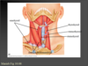
What is the function of the suprahyoid muscles in relation to laryngeal function?
Elevate the hyoid bone and larynx. These muscles help connect the hyoid bone and larynx to the mandible and skull – being located above these structures, when the muscles contract, they draw the hyoid bone and larynx upwards. This mainly occurs when you increase the pitch of your voice – test this out by placing your fingers on your larynx (around the laryngeal prominence/Adam’s apple), then say “aaaaa” at a low, followed by a very high pitch. You should be able to feel your whole larynx moving upwards (elevation) as you increase pitch – this is due to the actions of the suprahyoid muscles.
What is the function of the infrahyoid muscles in relation to laryngeal function?
Directly the opposite of the suprahyoids – they depress the larynx and hyoid bone. So, with the same exercise as above, decrease your vocal pitch from very high to very low, you should feel your whole larynx move downwards.
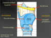
A wedge-shaped rima glottidis would indicate what type of function?
Normal respiration
What is the function of movements at the cricothyroid joint in relation to speech?
The thyroid cartilage rocks anteriorly and downwards at the cricothyroid joint, this has the effect of stretching out the vocal folds (which connect from the arytenoid cartilages posteriorly to the internal anterior surface of the thyroid cartilage-see Fig. 16, page 39 of Module 1). Just like if you tighten a guitar string, this will cause increased vocal pitch. To decrease pitch, the thyroid cartilage rocks posteriorly, decreasing the tension in the vocal folds. You can test this by placing your fingers on your laryngeal prominence and try a few pitch changes – you should feel the laryngeal prominence moving forwards (anteriorly) during increased pitch, and backwards during decreased pitch.
What is the purpose of movements at the cricoarytenoid joints in relation to speech?
These movements alter the position of the vocal folds. The arytenoid cartilages can slide, rock and rotate on the cricoid cartilage. Lateral rotation of the arytenoid cartilages causes adduction of the vocal folds, bringing them towards the midline and into the air stream. This occurs during speech production. Medial rotation has the opposite effect, abducting the vocal folds, drawing them out of the air stream (we do this when we stop speaking). During adduction and abduction the arytenoid cartilages also slide medially or laterally. Rocking movements can occur during speech to fine tune the tension in the vocal folds, leading to fine changes in vocal pitch.
What muscles are responsible for the following movements at the cricoarytenoid joints? a. Sliding the arytenoid cartilages closer together b. Rocking the arytenoid cartilages forwards and medially ) c. Rocking the arytenoid cartilages backwards and laterally
a. Sliding the arytenoid cartilages closer together TRANSVERSE and OBLIQUE ARYTENOIDS b. Rocking the arytenoid cartilages forwards and medially ) LATERAL CRICOARYTENOID c. Rocking the arytenoid cartilages backwards and laterally POSTERIOR CRICOARYTENOID
name the cartlidges of the larynx


Name the “happy “ and the “sad”,muscles
Happy muscles__Sad muscles
Zygomatic major (F) Depressor anguli oris (L)
Zygomatic minor (G) Depressor labii inferioris (M)
Levator labii superioris (D) Orbicularis oris (K)
Levator labii superioris alaeque nasi (C) Mentalis (N)
Risorius (H) Platysma (O)
Note: orbicularis oris may be involved in both smiling and frowning, although it’s role is more dominant in frowning as its main action is to pucker the lips.

Describe the bony components and ligaments that make up the TMJ
Bones: the condyle of the mandible articulates with the mandibular fossa of the temporal bone.
Ligaments: three ligaments support and stabilise the TMJ – the stylomandibular ligament which runs from the styloid process to the angle of the mandible, the sphenomandibular ligament which runs from the sphenoid bone to the inner surface of the mandible, and the lateral (or temporomandibular) ligament, which acts as part of the joint capsule and surrounds most of the TMJ.
Where does the Temporomandibular joint derive its sensory and motor innervation from?
Both sensory and motor (to the 4 TMJ muscles-masseter, temporalis, lateral and medial pterygoids) innervation are supplied by the mandibular branch of the trigeminal nerve (cranial nerve V, or fifth cranial nerve). This is all the detail you need to know about this for now, until we cover the cranial nerves in detail in a later topic.
Describe the pathway of the facial artery, taking into account its location in relation to the muscles of facial expression
The facial artery branches off the external carotid artery. It crosses the lower border of the mandible just anterior to the angle of the mandible, and courses in a medial and superior direction across the face, before terminating at the medial corner of the eye (this occurs on both sides, for the left and right facial arteries). It lies deep to the zygomatic major and levator labii superioris.
If you have an itchy forehead, what nerve is transmitting the information to your brain?
Itching is information carried by SENSORY nerves (try not to get confused between sensory (something you feel) and motor (movement using muscles). So, sensory information is carried TO the brain from the face by the trigeminal nerve (cranial nerve V).
Just for interest, motor information TO the muscles of facial expression, is via the facial nerve (cranial nerve VII).
Describe the detailed motor innervation of the muscles of facial expression
The facial nerve, cranial nerve VII, supplies all of the muscles of facial expression with motor nerve fibres (this is what makes them move!). It emerges from inside the skull at the stylomastoid foramen and forms 5 branches – the temporal, zygomatic, buccal, marginal mandibular, and cervical branches. These innervate muscles as follows:
Temporal – orbicularis oculi (upper part)
Zygomatic – upper lip muscles (zygomatic major and minor, levator labii superiors, levator labii superioris alaeque nasi, nasalis)
Buccal – muscles in the cheek region – buccinator, risorius, upper part of orbicularis oris
Marginal mandibular – lower lip muscles (lower part of orbicularis oris, depressor anguli oris, mentalis, depressor labii inferioris)
Cervical – platysma
Paralysis of the depressor anguli oris muscle would result from damage to which nerve and branch?
This type of question tests whether you can relate structure to function and dysfunction – is a muscle is paralysed, it cannot move, hence its motor innervation (not sensory) is likely to be affected. If there is a loss of sensation then we’re talking about sensory nerves.
The answer to this question then is the facial nerve, marginal mandibular branch (see the question above where we established that this is the nerve and branch that supplies motor innervation to the depressor anguli oris).
What is the purpose of the sublingual and submandibular glands?
These are salivary glands, they produce saliva which lubricates the oral cavity, and helps to lubricate food during mastication.
What’s the dangly bit at the back of the oral cavity called? What does it represent?
The uvula. It represents the cone-shaped ending of the soft palate.
