Pelvis 1 Flashcards
Where does the true pelvis exist? What is the false pelvis and what cavity is it apart of?

What is the shape of the pelvic inlet (males vs females)?
What is the shape of the pelvic outlet?
Inlet males: Heart shaped.
Females: Oval (for giving birth).
Pelvic outlet: Diamond shape.


Explain the tilt of the pelvis
Pelvis actually lies in a tilted position.
In the coronal plane is the anterior supeior iliac spine and the pubic tubercle.

Female vs male sex differences in pelvis

Female subpubic angle is obtuse.
In males it is V shaped (angled).
Plus pelvic inlet differences.
The sacrum in female is straighter as well as coccyx, in males it can be more curved (more concave).
From supeior aspect in the females it is more cylcindrical shape and doesn’t taper (the pelvic space).
Male bony structures are bigger bc of heavier loads.

What are the 3 parts of the hip bone? How do they meet? When do they ossify?
Ileum (big one), Ischium and pubic bone.

Join in a Y shape suture in ascetabulum.
Completes ossification 22 years old (ascetabulum).

Piriformis goes out via the greater trochanter to the greater trochanter of the femur.

The two pelvid floor muscles: 1: _______ and 2: _________.
Muscle 1 can be split into 3: _________ , and 4: ________.
4: has different parts called _________ and ___________ / ____________.
1: Levator ani, 2: coccygeus (ischiococcygeus).
3: Illiococygeus, 4: pubococygeus.
called the puborectalis (not connected to the median raphe) and the pubovaginalis (which forms the sphincter of the vagina) /puboprostatis (wraps around the prostate).

Pubococcygeus is separated further. Purple is the puborectalis, it does a turn around motion (doesn’t attach to median raphe), this helps with angulation between anus and rectum.
The part of the pubococcygeus part forms the pubovaginalis, this forms sphincter of vagina. In the males it goes around the prostate and is called the puboprostatis.

What are the pelvic floor muscles innervated by?
Branches of pudendal nerve (S2-S4) - external anal sphincter - inferior rectal branch of pudendal.


What are the two sections of the perineum (anterior and posterior)?
Urogenital triangle and anal triangle.

What are the contents of the pelvic cavity? (males vs females)


Rectovesical pouch in males.
Females: Vesicouterine and rectouterine pouch (pouch of Douglas).
Vesicle = relating to bladder.

Contents in the pelvis are ____peritoneal.
Infraperitoneal


This is the ligamentous structures which the ovary is suspended, name the individual bits.


The bladder is _____ peritoneal.
The bladder is covered by the ____ muscle.
The smooth area of the bladder is called the ________. It has _ openings they are….
Retro.
Detrusor.
Trigone: 3 opennings, one for the internal urethral orifice, 2 for the ureteric orifaces.

Explain the nerve supply to the muscle of the bladder.
Nerve supply motor by the parasympathetic nerves via pelvic splancnic nerves.
Sympathetic supplies the internal urethral sphincter and trigone from L1 and L2, this is more important in males because before ejaculation these will be closed off so that there is no retrograde ejaculation.
What is the blood supply to the bladder?
Lymphatics?
Blood supply comes from superior and inferior vesical artereis - branches of internal iliac.
Lymphatics follow artereis (internal and external iliac nodes).


The bladder ______ is where the urethra starts.
The ________ in males, or the _______ in females supports the neck of the bladder.
neck.
pubovesicul ligament, puboprostatic.

The ureter is crossed near the base of the bladder by the ________ _________ in men, and the _______ _______ in women. This can be remembered by the saying “_______ ______ ______ _______”.
ductus deferens, uterine artery. “Water under the bridge”.

The male urethra is __ cm long with __ bends.
Explain the 3 parts of the male urethra.
30cm with 2 bends.
Prostatic (as it travels through the prostate), membranous and spongy (which is the longest as it travels through the corpus spongiosum and

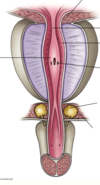
What is the function of the yellow bulbs?

In the external sphincter area there are bulbourethral glands which open in spongy urethra and lubricates urethra and neutralises acidity.

How long is the female urethra?
Where does it open?

Is the rectum covered by peritoneum?
Upper 1/3 covered anteriorly and sides, middle 1/3 only anteriorly, lower 1/3 no peritoneum.
How does the rectum differ from the rest of the colon?
No appendices epiploicae, no tinea coli, no mesentary.
The __________ muscle must relax before defication to straighten out the _________ angle.
puborectalis, puborectal.

How long is the rectum?
How many curves does it have?
What are rectal folds?
12 cm.
3 curves: upper and lower to the right, middle to the left.
Transverse rectal folds formed by circular smooth muscles.

What is the arterial supply to the rectum and the arteries origins?
Continuation of inferior mesenteric artery called the superior rectal artery.

Explain the venous drainage of rectum?
What are the clinical implications?
Same as the arteries.

Venous drainage goes toward the abdomen (superior rectal vein or inferior mesenteric vein - this joins splanchnic to go to portal vein - this means it is to the portal system).
Middle rectal means that the internal iliac vein goes to IVC - same as the inferior rectal.
There are lot of anastomosis between all of these - a mixing of the portal and systemic veins. This is where shunts can occur.
What is this fat?

In the perineum there is the ischioanal fossa with lots of fat.

Continuation of the ___________ __________ into the ________ triangle deep to the perineal membrane.

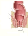
The upper _/_ of the anal canal has _______ which turn into ______.
The _______ ______ delineates the superior part to the inferior part. What is the physiological significance of this deliniation?
2/3, columns which turn into valves.
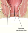
Pectinate line. Above = autonomic innervation and derived from endoderm, below = somatic innervation and derived from ectoderm.


Explain the difference between the internal anal sphincter and external anal sphincter.
Internal = smooth muscle, circular continuation, involuntary control.
External = voluntary, attached anteriorly to perineal body, blends with puborectalis muscle.


