PATHO - Term Test I (Hematologic System) Flashcards

1
Q

What are the 4 chief functions of blood?

A

1) delivery of substances needed for cellular metabolism
2) removal of wastes
3) defence against microorganisms and injury

maintenance of acid-base balance

2
Q

Blood volume is approximately ___ L in adults.

A

5.5L (~6 quarts)

3
Q

Plasma is made up of ___% water and ___% solutes. What is it and what % of blood volume is plasma?

A
  • 91% water, 9% solutes
  • component of blood that is an aqueous liquid containing variety of organic and inorganic elements
  • accounts for 50-55% of blood volume
4
Q

How is serum different than plasma?

A

serum is free of clotting proteins (which may be advantageous as some clotting proteins may interfere with diagnostic tests)

5
Q

What gases exist in arterial plasma?

A

CO2

O2

N2

6
Q

The following are waste products found in the arterial blood. They are the end products of what?

1) urea
2) creatinine
3) Uric acid
4) bilirubin
5) individual hormones

A

1) Urea: from protein catabolism
2) Creatinine: from energy metabolism
3) Uric acid: from protein metabolism
4) Bilirubin: from heme, from RBC destruction
5) Individual hormones: functions specific to target tissue

7
Q

All of the following functions of proteins are true EXCEPT:

a) acting as buffers
b) providing colloid osmotic pressure of plasma
c) clotting factors, antibodies, hormones, transporters
d) binding other plasma constituents
e) all the above are functions of proteins

A

e) all the above are functions of proteins

8
Q

What is contained in the Buffy coat?

A

platelets, WBCs

9
Q

The 2 major plasma protein groups are ________. They are mostly produced by the _______.

A

albumins and globulins

mostly produced in the liver

10
Q

What is the essential role of albumin? What happens when there is a decreased production/excessive loss of albumin?

A
  • 57% of plasma protein (majority)
  • essential role: regulation of passage of water and solutes through capillaries
  • Too large so cannot diffuse freely through blood vessels therefore maintains colloid osmotic pressure instead
  • if decreased/lost: fluid is retained in tissues and does not return to vascular system
11
Q

What are globulins and how are they classified in the blood?

A
  • accounts for ~38% of plasma protein
  • classified by their movement relative to albumin:
    • Alpha globulins: moving most closely to albumin (HDLs, prothrombin, hormone transporters)
    • Beta globulins: LDLs
    • Gamma globulins: least movement, primarily antibodies
12
Q

Plasma proteins are classified by the following functions:

a) clotting
b) Defense/protection
c) transport
d) regulation

Please list an example or how these proteins specifically accomplish each function.

A

a) Clotting: fibrinogen ⇒ promotes clotting and stops bleeding from damaged blood vessels
b) Defense/protection: antibodies and complement proteins
c) Transport: binding and carrying inorganic/organic materials (ex. transferrin carrying iron, lipids, lipoproteins carrying steroid hormones)
d) Regulation: enzymatic inhibitors (protect from tissue damage); precursor molecules that become active when needed; protein hormones (cytokines) that communicate between cells

13
Q

Describe the following of an erythrocytes:

a) structure
b) function
c) life span

A

A) no nucleus, biconcave disk containing Hb

b) gas transport to and from tissue cells and lungs
c) 80-120 days

14
Q

Describe the following of a leukocyte:

a) structure
b) function
c) life span

A

a) nucleated (varied sizes, depends on the leukocyte)
b) body defence mechanisms
c) varied (depends on the leukocyte)

15
Q

Describe the following re: a neutrophil.

a) structure
b) function
c) life span

A

a) granulocyte; several-lobed nucleus (polymorphonuclear)
b) phagocytosis (especially during early stage of inflammation)
c) 4 days

16
Q

Describe the following re: eosinophil.

a) structure
b) function
c) life span

A

a) granulocyte, several lobed nucleus
b) control inflammation, phagocytosis, defense against parasites, allergic reactions
c) unknown life span

17
Q

Describe the following re: basophils.

a) structure
b) function
c) life span

A

A) granulocyte, several lobed nucleus

b) mast cell-like functions, associated with allergic reactions (release histamine); also release heparin
c) unknown life span

18
Q

Describe the following re: monocytes and macrophages.

a) structure
b) function
c) life span

A

a) large phagocyte, one nucleus (mononuclear)
b) phagocytosis
c) months to years

19
Q

Describe the following re: lymphocyte.

a) structure
b) function
c) life span

A

a) mononuclear immunocyte
b) humoral and cell-mediated immunity
c) days to years, depending on type

20
Q

Describe the following re: NK cells.

a) structure
b) function
c) life span

A

a) large granular lymphocyte
b) defense against some tumors/viruses
c) unknown

21
Q

Describe the following re: platelets.

a) structure
b) function
c) life span

A

a) irregularly shaped cytoplasmic fragment
b) hemostasis after vascular injury; coagulation/clot formation and retraction; release of growth factors
c) 8-11 days

22
Q

Erythrocytes account for ~___% of blood volume in men, and _____% in women. The range of RBCs is between ________ RBCs/mm3 of blood.

A

48% in men

42% in women

4.2-6.2 million

23
Q

Describe the properties and function of an erythrocyte.

A
  • Primary function: tissue oxygenation (Hb carries the gas, electroyles regulate gas diffusion through cell membranes)
  • No organelles so no other cell functions or mitotic divisions
  • Biconcave shape and reversibly deformable
    • biconcave shape increases SA/V ratio optimal for gas diffusion
    • reversible deformity into a torpedo shape to undergo diapedesis and squeeze through microcirculation
24
Q

Describe the function and properties of leukocytes.

A
  • Function: defence against organisms that cause infections, removes debris
  • Avg: 5000 - 10 000 WBCs/mm3 blood
  • Classified by structure:
    • Granulocytes: neutrophils, basophils, eosinophils
    • Agranulocytes: monocytes, macrophages, lymphocytes
  • Classified by function:
    • Phagocytes: neutrophils, basophils, eosinophils, monocytes, macrophages
    • Immunocytes: lymphocytes
25
Q

True or False: Granulocytes contain enzymes for killing microorganisms and catabolizing debris ingested via phagocytosis, and are capable of amoeboid movement.

A

True

26
Q

What is the most numerous of the granulocytes? Describe its properties.

A
  • neutrophil (65-75% of total WBC count)
  • chief phagocytes of early inflammation but quickly dies in 1-2 days due to how sensitive it is to the environment of damaged tissue
  • neutrophils breakdown and release digestive enzymes that dissolve cell debris and prepare site for healing
27
Q

Describe the properties and function of eosinophils.

A
  • have large, coarse granules and account for 2-5% of WBC count
  • uses pattern recognition receptors (PRRs) to allow for diapedesis and phagocytosis
  • induced by IgE-mediated hypersensitivity reactions to attack parasites
  • control inflammation processes (release proinflammatory molecules)
  • high levels in Type I hypersensitivity allergic reactions and asthma
28
Q

Describe the properties and function of basophils.

A
  • <1% of WBCs, structurally similar to mast cells
  • contain histamine, chemotactic factors, preoteolytic enzymes, and heparin
  • when stimulated, induces synthesis of leukotrienes and cytokines that affect Th1 and Th2 cell differentiation
  • rich in IL-4 that guides B cell differentiation into plasma cells to secrete IgE
29
Q

_______ are immature macrophages and are formed and released by bone marrow.

A

Monocytes

30
Q

Mononuclear Phagocyte System (MPS)

A

consists of monocytes that differentiate without dividing and reside in tissues and ingests/destroys unwanted materials

31
Q

Primary cells for immune response are ________.

A

lymphocytes

32
Q

Platelets are formed in bone marrow by fragmentation of __________.

1/3 of the body’s available platelets are in a reserve pool in the _________.

Platelets are removed by _______ after circulating for 8-11 days.

A

megakaryocytes

spleen

macrophages primarily in the spleen

33
Q

Primary and secondary lymphoid organs include:

A

Primary lymphoid organs:

  • thymus
  • bone marrow

Secondary lymphoid organs:

  • spleen
  • lymph nodes
  • tonsils
  • Peyer patchs in ileum of small intestine
34
Q

The largest lymphoid organ is the _________. What are its functions?

A
  • Spleen

Functions:

  • site of fetal hematopoiesis
  • filters blood-borne antigens and cleanses blood through MPS
  • initimates immune response to blood-borne microorganisms
  • destroys old RBCs
  • blood reservoir
35
Q

The spleen is enclosed in a capsule that is divided into compartments by strands of connective tissue called ________. These compartments contain masses of lymphoid tissue called ___________.

A

trabeculae; splenic pulp

36
Q

Splenic pulp (white vs. red pulp)

A

Splenic pulp: masses of lymphoid tissue containing macrophages, lymphocytes, and lymphoid follicles; divided into white and red pulp

  • White pulp: contains lymphocytes and macrophages; where immune function occurs because this is where antigens encounter lymphocytes causing B cells to proliferate and differentiate (humoral immune response)
  • Red pulp: contains RBCs and venous sinuses; also has some macrophages for splenic filtration (this is the principal site)
37
Q

All of the following are true regarding venous sinuses except

a) highly distensible storage areas in spleen
b) have lots of gaps between endothelial lining to allow blood cells to exit circulation
c) can store more than 300mL of blood but cannot expel this as this blood is needed for critical function of the spleen
d) has slow circulation

A

c) it can store more than 300mL of blood BUT up to 200mL can be expeled into venous circulation when BP drops; increases Hct up to 4%

Other notes:

  • it has slow circulation to allow for macrophages to easily eat up old, damaged, dead blood cells, debris, microorganisms etc.
  • iron from catabolized RBCs are released back into the blood which the blood then goes to the liver
38
Q

True or False. The spleen is not absolutely necessary for life or for adequate hematologic function.

A

True.

39
Q

What effects would there be if the spleen was removed?

A
  • leukocytosis
  • decreased circulating iron levels
  • severely diminished immune response to encapsulated bacteria (decreased IgM levels) = increased risk of infections
  • increase in structurally defective RBCs in circulation
40
Q

Describe the pathway of lymph entering and exiting a lymph node.

A

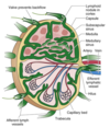
Lymph enters through small afferent lymphatic vessels ⇒ subcapsular sinus ⇒ drains into cortical sinuses ⇒ medullary sinuses ⇒ lymph collected and leaves via efferent lymphatic vessel

41
Q

Describe the pathway of blood flow through lymph nodes.

A

Blood flows in via lymphatic artery ⇒ post-capillary venules in outer cortex ⇒ drained through lymphatic vein

42
Q

The lymph node is the primary site for first encounters between antigens and lymphocytes (which is why when lymph nodes are enlarged you can feel them). Briefly describe how this works (i.e. what is happening in the lymph node at this time).

A
  • lymphocytes enter the lymph node via post-capillary venules and diapedesis
  • macrophages already reside inside the lymph node to phagocytize things
  • once an antigen is present, B cells are sitmulated to proliferate and differentiate into memory cells and plasma cells ⇒ causes lymph node enlargement
43
Q

Red bone marrow vs. yellow marrow

A

Red marrow: aka myeloid tissue; active marrow where hematopoietic stem cells (HSCs) are created and reside

Yellow marrow: inactive marrow

44
Q

Bone cavities at birth only contain _____ marrow.

A

red

45
Q

Active red bone marrow is found primarily where in adults?

A
  • flat bones of pelvis (34%)
  • vertebrate (28%)
  • cranium and mandible (13%)
  • sternum and ribs (10%)
  • humerus and femur (4-8%)
46
Q

Bone marrow niches contain two populations of stem cells. These are:

A

1) hematopoietic stem cells (HSCs) - progenitors of all hematologic cells

2) mesenchymal stem cells (MSCs) - stomal cells (connective tissue cells) that differentiate into osteoblasts, adipocytes, chondrocytes, sinusoidal endothelial cells, fibroblasts

47
Q

Osteoblasts vs osteoclasts

A

Osteoblasts: derived from fibroblasts, responsible for bone construction

Osteoblasts: multinucleate cells from monocytes that remodel bone by resorption

48
Q

Hematopoietic stem cells differentiate into two lineages. What are they and what are the end cell products of each lineage?

A

1) Lymphoid lineage: T and B lymphocytes, NK cells

2) Myeloid lineage: monocytes, macrophages, neutrophils, basophils, eosinophils, platelets, RBCs

49
Q

True or False. Yellow marrow cannot be converted to red marrow.

A

False. Conversion of yellow marrow ⇒ red marrow can be accomplished via erythropoietin (EPO - produced in kidney to stimulate RBC production) which will increase circulating levels of hematologic cells

50
Q

Hematopoiesis

A
  • production of blood cells
  • constantly ongoing, occurs in liver and spleen (in fetus) and ONLY in bone marrow (after birth)
  • continues throughout life and increases in response to need for replenishment or response to infection
51
Q

What is extremedullary hematopoiesis?

A
  • Blood cell production in tissues other than bone marrow (eg. liver, spleen)
  • usually a sign of disease (can occur in blood disorders/leukemias)
52
Q

Describe the pathway (cell lineage) of how erythrocytes are formed.

A

within bone marrow: progenitor cellsproerythroblastsnormoblast (while progressively eliminating intracellular structures, synthesizing Hb, and becoming more compact to assume RBC shape/traits) ⇒ reticulocyte (last immature form, enters blood stream, where nucleus is lost) ⇒ erythrocyte

*quantity of Hb increases in each step while nucleus decreases in size until it is lost

53
Q

True or False. Total volume of erythrocytes circulating fluctuates in healthy individuals.

A

FALSE. Remains constant

54
Q

Describe the feedback loop influenced by EPO in tissue hypoxia conditions.

A

EPO gets secreted by peritubular cells in kidney ⇒ increased EPO levels circulating and carried to bone marrow ⇒ stimulates RBC production

55
Q

Hemoglobin

A
  • made in bone marrow by RBCs
  • the oxygen-carrying protein of RBCs, ~90% of the cell’s dry weight
  • composed of 2 pairs of polypeptide chains (globins) and 4 iron + protoporphyrin complexes (hemes)
56
Q

The most common type of Hb in adults is _____. The most common type of Hb variant in fetuses is _____.

A

Hb A - two alpha chains, two beta polypeptide chains

Hb F - two alpha chains, two gamma chains (binds oxygen with a much greater affinity than adult Hb)

57
Q

The component of Hb that carries oxygen and gives blood the rubdy-red colour is known as:

A
  • Heme (can carry one molecule of oxygen so 4 hemes = 4 oxygens in one Hb)
  • Hb is “saturated” if all 4 oxygen-binding sites are occupied by oxygen
58
Q

oxyhemoglobin vs. deoxyhemoglobin vs methemoglobin

A

Oxyhemoglobin: normal oxygen-carrying form of Hb where O2 is bound to ferrous iron (Fe2+) temporarily oxidizing it from Fe2+ to Fe3+

Deoxyhemoglobin: when O2 is released and body reduces the iron to Fe2+ which reactivates Hb (“reduced Hb”)

Methemoglobin: non-reduced ferrous iron (Fe3+) form of Hb so cannot bind to O2 (this happens with certain drugs/chemicals that reduce oxygen-carrying capacity)

59
Q

What happens when Hb binds to O2?

A
  • Hb undergoes conformational change where when one iron molecule binds O2, the porphyrin ring changes shape to increase exposure of the three remaining iron atoms to O2 (this greatly increases affinity for oxygen-carrying capacity of Hb)
  • once O2 is unloaded, oxygen-carrying capacity is low to faciliate transport of CO2 back to lungs
60
Q

What is the role of protein (amino acids) in erythropoiesis? What is the consequence of deficiency?

A
  • Role: structural component of plasma membrane
  • Deficiency results in: decreased strength, elasticity, and flexibility of membrane leading to hemolytic anemia (RBCs destroyed faster than they are produced)
61
Q

What is the role of cobalamin (vitamin B12) in erythropoiesis and what are the consequences if deficient in vitamin B12?

A
  • Role: DNA synthesis, RBC maturation, facilitator of folate metabolism
  • Deficiency results in: Macrocytic (megaloblastic) anemia (abnormally large RBCs that do not function properly)
62
Q

What is the role of folate in erythropoiesis and what are the consequences if deficient?

A
  • Role: DNA and RNA synthesis, maturation of RBCs
  • Deficiency: Macrocytic (megaloblastic) anemia
63
Q

What is the role of vitamin B6 (pyridoxine) in erythropoiesis and what are the consequences if deficient?

A
  • Role: heme synthesis
  • Deficiency results in: microcytic-hypochromic anemia
64
Q

What is the role of vitamin B2 (riboflavin) in erythropoiesis and what are the consequences if deficient?

A
  • Role: oxidative reactions
  • Deficiency results in: normocytic-normochromic anemia
65
Q

What is the role of vitamin C (ascorbic acid) in erythropoiesis and what are the consequences if deficient?

A
  • Role: iron metabolism, acts as reducing agent to maintain iron in ferrous (Fe2+) form
  • Deficiency results in: normocytic-normochromic anemia
66
Q

What is the role of vitamin E in erythropoiesis and what are the consequences if deficient?

A
  • Role:?heme synthesis, protection against oxidative damage in mature RBCs
  • Deficiency results in: hemolytic anemia with increased cell membrane fragility
67
Q

What are the roles of iron and copper in erythropoiesis and consequences if deficient?

A

IRON:

  • Role: Hb synthesis
  • Deficiency: iron deficiency anemia

COPPER:

  • Role: optimal mobilization of iron from tissues to plasma
  • Deficiency: microcytic-hypochromic anemia
68
Q

What happens as erythrocytes age and how are they removed?

A
  • metabolic processes in RBC diminished resulting in less ATP available to maintain membrane ⇒ more fragile and less able to reversibly deform ⇒ increased chance of rupturing in microcirculation
  • plasma membrane also rearranges to signal macrophages to sequester them
  • During Hb digestion, conjugated bilirubin is the end product which is excreted from liver into intestine or bile ⇒ transformed into urobilinogen that is MOSTLY excreted via feces, some reabsorbed and excreted by kidneys
69
Q

Where can iron be found in the body, and in what percentages?

A
  • ~67% of total body iron bound to heme in RBCs
  • 5-10% bound to heme-containing myoglobin in muscle cells
  • ~30% stored in macrophages (mononuclear phagocytes) or as ferritin (in liver) or hemosiderin (in liver)
  • 3% lost daily in urine, sweat, bile, sloughing of epithelial cells
70
Q

~25mg of iron is required daily for erythropoiesis. Where can these iron sources be found?

A
  • 1-2 mg iron from dietary intake
  • remaining is obtained from continual recycling of iron from RBCs through the iron cycle
71
Q

What are the differences between the following:

a) ferritin
b) apoferritin
c) hemosiderin
d) apotransferrin
e) transferrin

A

Ferritin: intracellular protein that stores iron

Apoferritin: ferritin without attached iron

Hemosiderin: large intracellular iron store complexes resulting from excess accumulation of iron

Apotransferrin: the thing that iron is bound to when being transported in the blood

Transferrin: the protein form when apotransferrin is bound with an iron

72
Q

Hepcidin

A
  • protein synthesized in the liver and controls body’s iron homeostasis
  • production of hepcidin regulated by iron levels in the body, rate of erythropoiesis, and % O2 sat
  • Hepcidin induces internalization and degradation of ferroportin (membrane protein that trnasports iron in and out of cells) ⇒ leads to increased intracellular iron stores, decreased dietary iron absorption, and decreased levels of circulating iron
  • Decreased hepcidin production ⇒ leads to release of stored iron and increased dietary absorption
73
Q

Myelopoiesis vs lymphooiesis vs thrombopoiesis

A

Myelopoiesis: development of granulocytes and monocytes from differentiation of myeloid progenitor cells in bone marrow

Lymphopoiesis: development of lymphocytes from lymphoid progenitor cells to undergo further maturation in primary and secondary lymphoid organs

Thrombopoiesis: development of platelets

74
Q

Describe the pathway for thrombopoiesis.

A

Progenitor cellsmegakaryocytes (nucleus enlarges and number of chromosomes increase but no cell division occurs which is why it expands; cell develops cell surface elongations and branches that progressively fragment) ⇒ platelets (no nucleus, but have granules that promote stickiness)

75
Q

Approximately ____ of platelets enter the circulation while the remainder resides in ________. A hormone growth factor called _________ stimulates production and differentiation of megarkaryocytes, and is the main regulator for the circulating platelet numbers.

A

2/3

splenic pool

thrombopoietin (TPO)

76
Q

How does TPO regulate platelet numbers?

A
  • TPO primarily produced in the liver and indues platelet production in bone marrow
  • Platelets have TPO receptors and when circulating platelet levels are normal, TPO is absorbed onto platelet surface and prevented from accessing bone marrow to initiate further platelet production
  • When platelet levels are low, amount of TPO exceeds number of available platlet TPO receptors which allows for free TPO to enter the bone marrow and stimulate platelet production
77
Q

Hemostasis

A
  • arrest of bleeding by blood clot formationi at sites of vascular injury
  • helps damaged bloow vessels maintain a relatively steady state of blood volume, pressure, and flow
  • Three equal components of hemostasis:
    • Vasculature (endothelial cells and subendothelial matrix)
    • Platelets
    • Clotting factors
78
Q

Describe the sequence of events of how hemostasis works (i.e. from vascular injury to clot formation).

A
  1. vascular injury leads to transient arteriolar vasoconstriction to limit blood flow to the affected site
  2. damage to endothelial lining exposes subendothelial matrix which results in platelet adherence and activation and formation of hemostastic plug (this is primary hemostasis)
  3. TF produced by endothelium work with platelet factors/platelets to activate clotting system to form fibrin clots and further prevent bleeding (secondary hemostasis)
  4. fibrin clot contracts to form a more permanent plug & regulatory pathways are activated (fibrinolysis) to limite plug size and begin healing process
79
Q

True or false. Hemostatic mechanisms are always activated the same way regardless of size of the injured blood vessel.

A

False. Pinpoint petechial hemorrhage (involving capillaries, venules, etc.) generally are small and only requiring direct sealing or fused platelets. Bigger sources of bleeding (in arteries) require greater vascular contraction, more fused platelets, and greater hemostatic factor activation

80
Q

How does endothelium of blood vessels regulate platelet activity under normal conditions?

A
  • Endothelial cells produce nitric oxide and prostacycclin which are both vasodilators that modulate blood flow and pressure and maintain platelets in inactive state
  • Endothelium also produces adenosine diphosphatase that degrades ADP (a platelet activator)
  • Surface of endothelium contains anti-thrombotic molecules (i.e. heparin sulfate) that limites platelet activation and fibrin deposition.
81
Q

How does endothelial cells contribute to platelet regulation when there is a vascular injury?

A
  • Endothelial cells contain von Willebrand factor (vWF, clotting factor VIII) which is released during vascular injury which activates platelets
82
Q

What are the 4 major roles of platelets?

A
  1. Vasospasm - induces vasoconstriction during vascular injury to regulate blood flow into damaged site
  2. Prevent further bleeding - intiates platelet-platelet interactions which result in formatio of platelet plug
  3. Clotting cascade activation - to stabilize platelet plug
  4. Initiating repair process - clot retraction and fibrinolysis (clot dissolution), release of growth factors
83
Q

What is the normal range of platelets in blood?

a) 150 000 - 400 000/mm3 of blood
b) 20 000 - 40 000/mm3 of blood
c) 4.6 mil - 5.2 mil/mm3 of blood
d) none of the above

A

a) 150 000 - 400 000/mm3 of blood

84
Q

Thrombocytopenia

A

abnormally low numbers of platelets (<100 000/mm3 blood) and likely having longer than normal clotting times

85
Q

Describe the four steps of platelet activation in response to a damaged vessel.

A
  1. adhesion: increased platelet adhesion to damaged vascular wall (mediated by platelet receptor glycoprotein Ib binding to vWF)
  2. activation: occurs when there is an interaction with endothelium/subendothelial matrix + inflammatory mediators; platelet degranulates which stimulates changes in platelet shape (turns into spiny projections to increase SA) and biochemistry
  3. aggregation: stimulated by TXA2 and ADP; occurs as platelet-vascular wall and platelet-platelet adherence increases
  4. activation of clotting system: and development of fibrin-platelet meshwork
86
Q

During activation of platelets, they degranulate causing a release of 3 types of granules that further contain subcomponents. What are the types of granules and what are their respective subcomponents?

A
  1. Dense bodies:
  • ADP: recruits an activates other platelets
  • Serotonin: increases vasodilation and vascular permeability
  • Calcium: needed for adhesive interactions and intracellular signaling mechanisms to control platelet activation
  1. Alpha granules: mix of clotting factors, growth and angiogenic factors, and angiogenesis inhibitors
  • fibrinogen
  • platelet derived growth factor (PDGF): stimulates smooth muscle cells and promotes tissue repair
  • Platelet factor 4: enhances clot formation at injury site
  1. lysosomes
87
Q

Sticky platelet syndrome (SPS)

A

Description: inherited autosomal dominant pro-coagulant conditions that causes thrombi in blood vessels

Etiology: pro-coagulation caused by increased platelet stickiness (aggregation) when stressful events cause release of epi or ADP

S/S: frequent but silent until significant stressful event; arterial thrombosis, pregnancy complications (fetal growth retardation and fetal loss), potential venous thromboembolism

Dx: lab evaluation of platetlet aggregation

Tx: low dose aspirin (ASA), clopidogrel if aspirin-resistant

88
Q

There are two pathways of initiation for clotting system. Describe how the pathways work and how they are activated.

A

Intrinsic pathway: activated by Hageman factor (factor XII) in plasma when it contacts negatively charged subendothelial substances exposed due to vascular injury

Extrinsic pathway: activated by thromboplastin TF (released by damaged endothelial cells) interacting with clotting factors (particularly factor VII)

  • both pathwas converge to common pathway and activation of factor C which leads to clot formation
  • several complexes subsequently activate the next one resulting in activation of prothrombin into thrombin
  • thrombin converts fibrinogen ⇒ fibrin forming a fibrin clot
89
Q

Deficiencies in Antithrombin III (AT-III), Protein C or Protein S can lead to:

a) hypercoagulation
b) excessive bleeding
c) degradation of activated clotting factors
d) decreased clot retraction

A

a) hypercoagulation (increased clotting)

these are all factors and mechanisms that typically prevent spontaneous activation of hemostasis so if they are deficient, hypercoagulation can occur

90
Q

The following are factors that prevent spontaneous activation of hemostasis. Describe how each of these work.

a) AT-III
b) tissue factor pathay inhibitor (TFPI)
c) thrombomodulin
d) protein C/protein S

A

a) anti-thrombin III (AT-III): binds to heparin sulfate OR with administered heparin to prevent thrombosis
b) TFPI: joins prothrombinase complex and inhibits clotting factors in clotting pathway
c) Thrombomodulin: binds to thrombin on surface of endothelial cells; mediates Protein C activation and inhibits thrombin activity
d) Protein C: binds to thrombomodulin which activates it and works in association with cofactor protein S to degrade clotting factors

91
Q

What happens during clot retraction?

A
  • fibrin strands shorten (become denser and stronger) whih approximates edges of the injured vessel wall and seals injury site
  • platelets trapped into the fibrin meshwork also contract and “pull” fibrin threads closer together while releasing a factor that stabilizes the fibrin
  • Contraction expels serum from fibrin meshwork
  • begins within few minutes after clot formation
92
Q

The process of lysis of blood clots is known as ________. What happens during this process?

A
  • Fibrinolysis
  • plasminogen (produced by the liver) is converted to plasmin by tissue plasminogen ativator (t-PA) or by urokinase-like plasminogen activator (u-PA)
  • t-PA laragely involved in intravascular fibrinolysis while u-PA is extravascular fibrinolysis
  • t-PA is expressed by endothelial cells and is max activated after binding to fibrin
  • u-PA binds to specific u-PA receptor on cell that causes activation of plasminogen
93
Q

The enzyme responsible for dissolving clots by degrading fibrin and fibrinogen into fibrin degradation products (FDPs) is known as what?

What is the major FDP produced from this process?

A
  • plasmin
  • major FDP: D-dimer (would be elevated in medical conditions where clots form like DVT and PE)
94
Q

At birth, blood cell counts tend to (rise/fall/stay the same) above adult levels then (increase/decrease) gradually throughout childhood.What causes this?

A
  • rise; decrease
  • rise in values is due to accelerated hematopoiesis during fetal life and increased number of cells that result from trauma of birth and cutting of umbililcal cord (body thinks it is losing blood so it makes more)
95
Q

How does the intrauterine environemtn affect erythropoiesis?

A
  • Hypoxic in nature so it stimulates EPO production in fetus which accelerates fetal erythropoiesis ⇒ causes polycythemia in newborns
  • after birth, oxygen from lungs saturate arterial blood which allows more oxygen to be delivered to the tissues (changing from placental to pulmonary oxygen supply) which leads to body detecting that EPO and rate of erythropoiesis can slow down
96
Q

Desribe the function of leukocytes in newborns.

A
  • WBCs develop and mature during fetal life but have weak function at birth due to them being immature cells
  • high lymphocyte and neutrophil count at birth and continues to rise for 1st year of life (lymphocyte) or first few days (neutrophils) then falls to adult levels
  • eosinophil and monocyte counts are also high in the first year of life
  • children tend to have more atypial lymphocytes as a reuslt of frequent viral infections
  • newborns at risk for impaired phagocytosis, bacterial infections, and delayed wound healing
97
Q

What changes are seen in blood with aging?

a) composition of blood
b) RBC life span and ability to replenish
c) Hb levels
d) lymphocyte function
e) platelet numbers
f) fibrinogen levels and clotting factors

A

a) little changes to blood composition but some components can be altered with iron deficiency
b) normal RBC life span but slow replenishing of RBCs and fragile plasma membranes (may be due to iron depletion, decreased iron binding capacity, or iron absorption in intestine)
c) low Hb levels
d) decreased lymphocyte function
e) decreased platelet numberes but platelet adhesiveness increases
f) increased fibrinogen and clotting factor levels

98
Q

The most common blood disorder in children is _________. Why does this occur?

A
  • Anemia
  • results from inadequate erythropoiesis (most commonly caused by iron deficiency) or early destruction of RBCs
  • iron deficiency can result from insufficient dietary intake or chronic iron loss from bleeding
99
Q

The most dramatic form of acquired congenital hemolytic anemia is:

A

hemolytic disease of the fetus and newborn (HDFN) aka eyrthroblastosis fetalis

100
Q

The most common enzyme deficiency leading to anemia in children is

A

glucose-6-phoosphate dehydrogenase (G6PD)

101
Q

Iron Deficiency Anema (IDA) - Children and Adults

A

Description: acquired; the most common nutritional disorder worldwide; anemia caused by iron deficiency

Etiology: dietary lack of iron (most common during first few years of life), problems with iron absoprtion, blood loss (most common during childhood and adolescence), increased requirement of iron, impaired absorption, chronic diarrhea

  • other causes: medications causing GI bleeds (ASA, NSAIDs); surgeries that decrease stomach acidity; eating disorders; excessive menstrual bleeding; GI cancers/lesions

Prevalence/Inidence: highest incidence between 6 mos-2yo and prevalence is greatest among toddlers, adolescent girls, and women of childbearing age

  • risk factors: living in poverty, elderly with restrictive diets, teenagers with poor diets
  • also high risk for infants due to low bioiavailability of iron in cow’s milk + milk has protein that causes microhemorrhaging)
  • chronic parasitic infections are a risk factor in children in developing countries (H. pylori, helminths)
  • increased prevalence in overweight children, adolescents, women, and those undergoing bariatric surgery

Pathophysiology: hypochromic-microcytic anemia; body may try to compensate by increasing RBC activity in bone marrow but eventually deplete stores and anemia occurs (no dysfunction in iron metabolism though); 3 stages:

  1. Stage 1: decreased bone marrow iron stores; Hb and serum iron remain normal (compensation)
  2. Stage 2: iron transportation to bone marrow diminished reuslting in iron-deficient erythropoiesis
  3. Stage 3: starts when small Hb-deficient cells enter circulation to replace normal aged RBCs (this is where S/Sx appear)

S/Sx:

  • IN CHILDREN listlessness and fatigue (mild); general irritability, decreased activity tolerance, weakness, lack of interest in play;
  • anorexia, pallor, tachycardia, systolic murmurs
  • chronic IDA: splenomegaly, widened skull sutures, decreased physical growth, developmental delays, pica, altered neuro and intellectural functions​
  • IN ADULTS: fatigue, weakness, SOB, pale earlobes/palms/conjunctivae; koilonychia (spoon shaped fingernails); cheilosis, stomatitis, glossitis, dysphagia, neuromuscular changes, headache, irritability, tingling/numbness

Dx: lab tests (Hb, Hct, serum iron, ferritin levels, total iron-binding capacity), Hx taking, diet, physical exam

Tx:

  • IN CHILDREN: PO simple ferrous salt; iron supplements with vit C to increase absorption, diet modifications, cow milk intake restriction
  • IN ADULTS: eliminate source of blood loss if possible; iron replacement therapy (Hct levels should improve in 1-2 mos); PO iron supplement for menstruating females
102
Q

Hemolytic Disease of the Fetus and Newborn (HDFN)

A

Description: acquired alloimmune disease where antigens of fetal RBCs differ from antigens on maternal erythrocytes

Etiology: most cases caused by ABO incompatibility (diff blood types between fetus and mother - 20 to 25% of cases); 1/3 cases caused by Rh incompatibility

  • *Pathophysiology:** 3 conditions to be met for HDFN to occur
    1) mother has preformed antibodies against fetal erythrocytes or produces them when exposed to fetal erythrocytes
    2) sufficient amounts of antibody (IgG) cross placenta and enter fetal blood
    3) IgG binds with sufficient numbers of fetal RBCs to cause widespread antibody-mediated hemolysis or splenic removal
  • Wtih Rh incompatibility - First Rh incompatibility pregnancy presents with no difficulties because only large numbers of fetal RBCs enter maternal bloodstream once placenta is detached at birth, so baby already delivered
  • Next pregnancy: anti-Rh antibodies were already in the maternal bloodstream and enter fetal circulation leading to fetal RBCs to be coated with antibodies and destroyed = anemia develops + immature cells released into bloodstream
  • degree of anemia depends on: length of time antibody has been in fetal circulation, antibody concentration, ability for fetus to compensate for increased hemolysis

S/Sx: mild ⇒ slightly pale, slight enlargement of liver/spleen

  • Severe ⇒ pronounced pallor, splenomegaly, hepatomegaly
  • hyperbilirubinemia ⇒ may lead to neonatal jaundice or kernicterus (biliribun buildup in brain due to bilirubin levels exceeding liver’s ability to rid, leads to cerebral damage including intellectual disabilities, high freq. deafness, CP, potential death)
  • hydrops fetalis: severe edema in body (usually dead in utero)

Dx: if ABO incompatibility - no additional monitoring or tx required

  • Coombs test: measure antibodies in mother’s circulation OR antibodies already bound to fetal RBCs
  • Hx taking - presence of fetal hemolytic disease

Tx: prevention is KEY - RhoGAM injection (anti-D antibodies) to prevent Rh-ve woman producing antibodies (given within 72 h of exposure to Rh+ve)

103
Q

Sickle Cell Disease

A

Description: group of autosomal recessive disorders (inherited) characterized by production of Hb S within RBCs (specific sickle cell conditions in another flashcard)

Etiology: genetic mutation (valine AA replaces glutamic acid)

Prevalence/Incidence: most common among desendants of sub-Saharan Africa; 7-13% of African Americans of sickle cell trait (provides protection against lethal forms of malaria so may be genetic advantage)

Pathophysiology: usually causes no problems if properly oxygenated

  • hypoxemia, decreased pH, dehydration, acute illness, low temp, living at altitude causes Hb S to stretch and elongate into a sickle shape
  • sickled cell tend to plug blood vessels ⇒ increase blood viscosity ⇒ slow circulation and cause vascular occlusion, pain, organ infarction, further sickling
  • leads to anemia
  • sickling is reversible unless plasma membrane of RBC is damaged

S/Sx: general - pallor, fatigue, jaundice, irritability

  • extensive sickling can precipitate 4 types of acute crises: vasoocclusive crisis, sequestration crisis, aplastic crisis, hyperhemolytic crisis
  • Sx usually do not show up until infant is at least 6 months old
  • infection (most common cause of death related to sickle cell disease)
  • sepsis, meningitis

Dx: hematologic tests - sickle solubility test (presence of Hb S) & Hb electrophoresis (amount of Hb S in RBCs); amniocentesis

Tx: support to prevent consequences of anemia and avoiding crises

  • adequate hydration, infection prevention, pain management, genetic counseling/psych support
  • common tx: hydroxyurea
  • transfusion therapy (may lead to iron overload and need for chelation therapy)
  • HSC transplant but with potential risks
104
Q

Sickle cell anemia

A
  • homozygous form (most severe and only has sickle cell Hb)
105
Q

Sickle-cell thalassemia

A
  • heterozygous inheritance of Hb S and alpha- or beta- thalassemia)
  • Hb S x defective/insufficient a- or b- chains of Hb A
  • mildest clinical manifestations
  • microcytic-hypochromic erythrocytes make then less likely to occlude microcirculation even in sickle state
106
Q

Sickle Cell-Hb C disease

A
  • heterozygous inheritance of Hb S x Hb C or D (both abnormal Hb)
  • milder clinical manifestations than sickle cell anemia
  • main clinical problems relaed to vasoocclusive crises
  • in older children, potential: retinopathy, renal necrosis, aseptic necrosis of femoral heads along with obstructive crisis
107
Q

Sickle cell trait

A
  • carrier state (heterozygous inheritance of Hb S and normal Hb, Hb A)
  • typically does not affect life expectancy or interfere with daily activities
  • RARE but may lead to vasoocclusive crisis from severe hypoxia (shock), vigorous exercise at high altitudes, flying at high altitudes in unpressurized aircraft, undergoing anesthesia
108
Q

What are the 4 types of acute crises that could occur with extensive sickling in sickle cell disease?

A

Vasoocclusive crisis (thrombotic crisis): sickling occurs in microcirculation and obstructs blood flow leading to vasospasm and logjam ⇒ lead to thrombosis and infarction if not reversed, extremely painful, can lead to conditions such as stroke

Sequestration crisis: seen only in <5 y.o. where large amounts of blood become acutely pooled in liver and spleen ⇒ high mortality if not recognized; may be recurrent

Aplastic crisis: profound anemia caused by lowered erythropoiesis despite increased need for new erythrocytes (and bone marrow unable to compensate to replace lost cells); lasts 7-10 days

Hyperhemolytic crisis: unusual; may occur in association with certain drugs or infections or acute/chronic reaction following a blood transfusion

109
Q

Alpha-thalassemia

A

Description: inherited autosomal recessive disorders that result in impaired/decreased synthesis of alpha chain of adult Hb (Hb A)

Incidence/Prevalence:

  • Alpha-thalassemia: more common among Chinese, Loatian, Viet, Cambodian
  • also common among black people

Pathophysiology: microcytic-hypochromic anemia

  • further classified into major or minor (based on number of genes controlling a- or b- chain synthesis and combination of mutations (homozygous - major; heterozygous - minor)
  • 4 forms
    • Alpha trait: heterozygous carrier state - single alpha chain-forming gene is defective
    • Alpha-thalassemia minor - two genes defective
    • Hb H disease - three genes defective
    • Alpha thalassemia major - all 4 genes defective (often fatal in utero)

Sign/Symptoms:

  • Alpha trait: mildest form - usually Sx free or mild microcytosis
  • Alpha-thalassemia minor: identifcal to beta-thalassemia minor (mild microcytic-hypochromic reticulocytosis, bone marrow hyperplasia, increased serum [iron], moderate splenomegaly
  • Alpha-thalassemia major: hydrops fetalis (severe tissue anoxia leading to severe intrauterine CHF) - fetal distress evidenct in 3rd trimester

Dx: familial disease hx, clinical manifestations, blood tests ⇒ peripheral blood smears show microcytosis; Hb electrophoresis showing diminished a-chains

Tx: supportive; regular transfusions and chelation therapy (for iron overload); allogeneic HSC transplant for thalassemia major types

110
Q

Beta-thalassemia

A

Description: inherited autosomal recessive disorders that result in impaired/decreased synthesis of beta-chains of adult Hb (Hb A)

Incidence/Prevalence:

  • Beta-thalassemia: more common than alpha type; prevalent among Greek, Italian, Arab, Jewis
  • Common among black people

Pathophysiology: microcytic-hypochromic anemia

  • further classified into major or minor (based on number of genes controlling a- or b- chain synthesis and combination of mutations (homozygous - major; heterozygous - minor)
  • Beta-thalassemia minor - b-globin chain production is depressed moderately
  • Beta-thalassemia major - b-globin chain production depressed severely (Cooley anemia)
  • Free alpha chains accumulate but are unstable so precipitate in RBCs which are destroyed by MPS in marrow ⇒ anemia

S/Sx:

  • Minor: mild-mod microcytic-hypochromic anemia ⇒ skeletal changes, slight elevation in serum iron and bilirubin levels, mild splenomegaly, bronze colouring of skin, bone marrow hyperplasia
  • Major: impaired physical growth and development; significant CV burden with CHF (due to anemia); liver and spleen enlargement; skeletal changes (length discrepancies); chipmunk-like face (facial bone deformities from bone marrow hyperplasia)

Dx: familial disease hx, clinical manifestations, blood tests ⇒ peripheral blood smears show microcytosis; Hb electrophoresis showing diminished a-chains

Tx: supportive; regular transfusions and chelation therapy (for iron overload); allogeneic HSC transplant for thalassemia major types

111
Q

Hemophilias

A

Group of inherited bleeding disorders resulting from mutations in coagulation factors

112
Q

Hemophilia A

A

Description: X-linked recessive condition caused by mutation in F8 gene which codes factor 8 (cofactor in clotting cascade)

Prevalence/Incidence: the most common hereditary disease associated with life-threatening bleeding; more common than Hemophilia B and all racial groups equally affected

Pathophysiology: inherited from mother who is heterozygous for mutation in F8 gene (and then occasionally new mutation)

  • affected individuals within the same family will have same mutation but may be different across families
  • Sx are evident in males because they have only one X chromosome
  • Females (heterozygous carriers) usually do not experience excessive bleeding because other X chromosome sufficiently provides normal functioning clotting factor

S/Sx: depends on level of F8 activity

  • joint bleeding (knees, ankles, elbows), muscle bleeding, oral bleeding spontaneous painless hematuria
  • potential fatal bleeds (intracranial bleeding, internal bleeding, neck/chest/abdo bleeding)
  • first signs by age 3-4 with episodes of persistent bleeding from minor injuries

Dx: +ve family hx, prenatal genetic testing, personal bleed hx, lab testing (prolonged PTT; measuring factor 8 levels), physical ax

Tx: Recombinant factor 8 via IV

113
Q

Hemophilia B

A

Description: X-linked recessive condition caused by mutation in F9 gene which codes factor 9 (cofactor in clotting cascade) - aka Christmas disease

Prevalence/Incidence: 5x less common than hemophilia A

Pathophysiology: inherited from mother who is heterozygous for mutation in F9 gene (and then occasionally new mutation)

  • affected individuals within the same family will have same mutation but may be different across families
  • Sx are evident in males because they have only one X chromosome
  • Females (heterozygous carriers) usually do not experience excessive bleeding because other X chromosome sufficiently provides normal functioning clotting factor

S/Sx: depends on level of F9 activity

  • joint bleeding (knees, ankles, elbows), muscle bleeding, oral bleeding spontaneous painless hematuria
  • potential fatal bleeds (intracranial bleeding, internal bleeding, neck/chest/abdo bleeding)
  • first signs by age 3-4 with episodes of persistent bleeding from minor injuries

Dx: +ve family hx, prenatal genetic testing, personal bleed hx, lab testing (prolonged PTT; measuring factor 9 levels), physical ax

Tx: Recombinant factor 9 via IV

114
Q

Hemophilia C

A

autosomal recessive condition that results from deficiency in factor XI (11)

115
Q

Antibody-mediated hemorrhagic diseases

A

Conditions caused by immune response where antibody-mediated destruction or inflammatory reactions to allergens occur, resulting in blood vessel damage and seepage into tissues

All appear during infancy/childhood

116
Q

Primary Immune Thrombocytopenia (ITP)

A

Description: most common disorder of platelet consumption

Pathophysiology: caused by autoantibodies (often IgG class) binding to platelet plasma membranes (against glycoproteins) leading them to be sequestered and destroyed by MPS in spleen and other lymphoid tissues; 70% of cases are preceded by viral illness (CMV, EBC, parvovirus, resp tract infection)

Signs and Symptoms:

  • bruising and generalized petechiae rash 1-3 weeks after viral illness ⇒ may develop into ecchymoses
  • asymmetrical bruising often in legs/trunk
  • hemorrhagic bullae (blood blisters) in gums, lips, mucous membranes
  • uncontrollable/severe epistaxis
  • acute phase lasts 1-2 weeks but thrombocytopenia persists
  • most serious complication: intracranial hemorrhage

Dx: lab exam - low isolated platelet count; blood smear showing few but large platelets; prolonged bleeding time

Tx: observation (primary tx) regardless of platelet count ⇒ if bleeding present, then IVIG or short course of corticosteroids

Prognosis: most recover within 3 months, S/Sx likely subside after acute phase but if ITP persists >12 months then they are considered chronic and immunosupressive therapies needed

117
Q

Cancer of bone marrow resulting in abnormal WBC production is known as:

A

leukemia

118
Q

True or False. Leukemia is the most common malignancy in children and teens

A

True

119
Q

What are the 4 common types of leukemia?

A

a) Acute lymphoblastic leukemia (ALL)
b) acute myeloid leukemia (AML)
c) chronic lymphocytic leukemia (CLL)
d) chronic myeloid leukemia (CML)

120
Q

What is the most common type of leukemia in children/teens?

A

ALL (75% of leukemias); remaining cases are AML

chronic leukemias are rare (<5% cases)

121
Q

Acute Lymphoblastic Leukemia (ALL)

A

Description: the most common type of leukemia in children/teens

Prevalence/Incidence: most common in early childhood (peak b/w 2-4 y.o.) and more common in boys, Hispanics, and white children; increased incidence in high SES

Pathophysiology: ALL composed of immature B or T cells (lymphoblasts) that fill bone marrow and replace normal marrow causing a disruption in normal function

  • in children: chromosomal abnormalities (aneuploidy) causing dsyregulation of expression/function of transcription factors needed for normal B-/T-cell development
  • in adults: mix of cancers of precursor B- and T-cell origin

Signs and Symptoms: abrupt and insidious onset (may occur 1 week prior to dx)

  • pallor, fatigue, petechiae, purpura, ++bleeding
  • fever may occur (due to infection and decreased neutrophils, hypermetabolism from rapid growth/destruction of leukemic cells, or excess WBC causing leukostasis which is intravascular clumping of cells leading to infarction and hemorrhage usually in brain/lung)
  • hyperuricemia leading to renal failure
  • weight loss and anorexia (in adults)
  • liver, spleen, lymph node enlargement
  • abdo pain and tenderness
  • extramedullary invasion:
    • CNS: increased ICP (early AM headaches, N/V, irritability, lethargy)
    • bones and joints: pain in these areas (bone pain with no inflammation, migratory and vague pain)

Dx: blood tests and smears; bone marrow aspiration ⇒ reduced RBC, granulocytes, platelet counts; hallmark of acute leukemia is blast cell (80-100% in bone marrow vs. <5% in healthy children)

Tx: chemotherapy, radiation therapy

  • in adults - supportive tx (blood transfusions, antibiotics, antifungals, antivirals)
  • two phases of treatement: remission induction and postremission

Prognosis: ~85% of children with ALL will survive to 5 years at least

122
Q

Acute Myeloid Leukemia (AML)

A

Description: leukemia caused by acquired oncogenic mutations that impair differentiation reuslting in accumulation of immature myeloid blasts in marrow/other organs

Prevalence/Incidence: for children - more common in first 2 years of life and during teen years; equal occurrence in boys/girls and all races;

  • most common in adults (dx ~70 y.o.)
  • increased incidence seen with large ionizing radiation doses, smoking, infections with HIV and hep C virus

Pathophysiology: AML can result from acquired oncogenic mutations (epigenetic alterations) leading to bone marrow being so crowded by blast cells that bone marrow fialure and complications (anemia, thrombocytopenia, neutropenia) occurs

Signs and Symptoms: abrupt and insidious onset (may occur 1 week prior to dx)

  • pallor, fatigue, petechiae, purpura, bleeding
  • fever may occur (due to infection and decreased neutrophils, hypermetabolism from rapid growth/destruction of leukemic cells, or excess WBC causing leukostasis which is intravascular clumping of cells leading to infarction and hemorrhage usually in brain/lung)
  • extramedullary invasion:
    • CNS: increased ICP (early AM headaches, N/V, irritability, lethargy)
    • bones and joints: pain in these areas (bone pain with no inflammation, migratory and vague pain)

Dx: blood tests and smears; bone marrow aspiration ⇒ reduced RBC, granulocytes, platelet counts; elevated blast cell levels in bone marrow

Tx: combination chemotherapy (harder to treat than ALL); potential HSC transplant; tyrosine kinase inhibitors (targeted therapy that inhibits overactive/high levels of enzymes in cancer cells)

123
Q

Lymphomas

A
  • cancer of the lymphatic system: HL and NHL
  • develop from proliferation of malignany lymphocytes in lymphoid system
  • HL and NHL is rare < 5y.o and incidence increases throughout childhood (boys more often)
  • increased risk for those with inherited or acquired immunodeficiency syndromes
124
Q

Non-Hodgkin Lymphoma (NHL)

A

Description: cancer of immune cells (lymphocytes) with diverse group of lymphoma

  • 4 major types: B-cell NHL (Burkitt/Burkitt-like lymphoma); Diffuse large B-cell lymphoma; Lymphoblastic lymphoma; Anaplastic large cell lymphoma

Prevalence/Incidence:

  • most common types in children ⇒ BL (40%), lymphoblastic lymphoma (25-30%); large cell lymphoma (10%)
  • in adults: median age dx 67, higher occurrence in men; risk factors: old white male with autoimmune disorders, chemical exposures, infections, immune supression, risk of gastric infection with H. pylori, ?also high in meats and fats

Pathophysiology: All forms of BL associated with translocations of MYC gene on chromosome 8 leading to increased MYC protein levels; most BLs are also latently affected with EBV

  • progressive clonal expansion of B, T, or NK cells

S/Sx: NHL may arise from any lymphoid tissue so S/Sx are specific for involved site

  • associated signs: swelling of lymph nodes in neck, underarm, stomach, groin; trouble swallowing/breathing; painless lump/swelling in testicle; weight loss of unknown cause; night sweats
  • In African BL: involvement of facial bones (jaw)

Dx: physical exam, health hx, needle biopsy of disease sites

  • findings will show non-contiguous lymph node invovlvement

Tx: intensive chemotherapy (curative); radiation, potential stem cell transplant, monoclonal antibody tx

125
Q

Hodgkin Lymphoma (HL) - children

A

Description: type of lymphoma characterized by Reed-Sternberg (RS) cells which are derived from germinal centers of B cells

  • 5 types: nodular sclerosis, mixed cellularity, lymphocyte rich, lymphocyte depletion, lymphocyte predominance
  • first 4 types are “classic types” that has similar RS cell expression; last one is where RS cells are different and presents with earlier stage disease, longer survival BUT may turn into large B-cell lymphoma in 10 years in 10% of patients

Prevalence/Incidence:

  • in children: more common among adolescents (rare in childhood/YA)
  • in adults: males, ~64 y.o., whites > blacks

Pathophysiology: genetic re-arrangements or EBV infection lead to abnormalities in gene expression of RS cells (they fail to express most of the normal B- and T-cell markers)

  • arises in a single chain of lymph nodes and spreads initially in a contiguous way to lymphoid tissue

S/Sx: painful lymphadenopathy in lower cerival chain (sides and back of the neck) with/out fever (most common sx) and potentially axillary, groin, retroperitoneal (like lower abdo)

  • potential mediastinal involvement causing pressure on trachea/bronchi ⇒ airway obstruction
  • Initial sx: anorexia, malaise, fatigue
  • potential weight loss
  • in adults: 1/3 ppl have common systemic sx such as infeciton, night sweats, itchy skin, pruritus, fatigue

Dx: well-defined staging system that considers extent and location of tissue +/- presence of fever, weight loss, night sweats; physical exam, CBC, blood tests, lymph node biopsy

Tx: chemotherapy and radiation therapy; targeted therapies (monoclonal antibodies, immune checkpoint inhibitors), surgery (for adults)

Prognosis: survivors have greater risk of developing secondary cancer

126
Q

Anemia

A

Description: reduction in the total number of erythrocytes in circulating blood or decrease in quality/quantity of Hb

Etiology: impaired erythrocyte production; acute or chronic blood loss; increased erythrocyte destruction (or a combination of all three)

Pathophysiology: reduced oxygen-carrying capacity of blood which leads to tissue hypoxia

  • initial compensation for cellular loss by movement of intersitial fluid into the blood causing increase in plasma volume but viscosity decreases ⇒ thinner blood flows fast and more turbulent causing a hyperdynamic circulatory state leading to increased SV and HR

Signs and Symptoms: hypoxemia causes dilation of blood vessels to increase flow through them and increased SV and HR to meet oxygen demands but then it may lead to heart failure

  • classic S/S: SOB (dyspnea), rapid and pounding heartbeat, dizziness, fatigue, weakness
  • in mild cases, Sx may only be present during physical exertion/increase O2 demand
  • pale skin, mucous membranes, lips, nail beds, conjunctivae (from reduced Hb) OR jaundiced (because of bilirubin build up)
  • other neuro system sx, GI issues, low-grade fever

Treatment: if slowly developing conditions, then treat the underlying condition and decreasing the associated symptoms

  • transfusions, dietary correction, supplemental vitamins/iron
127
Q
  • cytic refers to:
  • chromic refers to:
A
  • cytic: cell size (macrocytic, microcytic, normocytic)
  • chromic: Hb content (normochromic, hypochromic, hyperchromic)
128
Q

Anisocytosis vs. Poikilocytosis

A

Anisocytosis: RBCs assuming various sizes

Poikilocytosis: RBC’s assuming various shapes

129
Q

Macrocytic-normochromic anemias have _____ RBCs and ____ Hb concentration. Two examples of this type of anemia are:

A

large (abnormally shaped); normal [Hb]

Examples: pernicious anemia, folate deficiency anemia

130
Q

Microcytic-hypochromic anemias have _____ RBCs and ____ Hb concentration. Three examples of this type of anemia are:

A

small (abnormally shaped); reduced [Hb]

Examples:

  • Iron deficiency anemia
  • Sideroblastic anemia
  • Thalassemia
131
Q

Normocytic-normochromic anemias have _____ RBCs and ____ Hb concentration. Five examples of this type of anemia are:

A

normal size RBCs, normal [Hb]

Examples:

  • aplastic anemia
  • post-hemorrhage anemia
  • hemolytic anemia (acquired or hereditary)
  • sickle cell anemia
  • anemia of chronic inflammation
132
Q

Post-hemorrhagic anemia

A

Description: normocytic-normochromic anemia caused by acute blood loss

Etiology: most trauma-related blood loss

Pathophysiology: within 24h of blood loss, water and electrolyes from tissues and interstitial spaces get pulled into vasculature to replace lost plasma but this causes Hct to decrease (aka hemodilution); EPO also stimulated to increase RBC production in bone marrow

Treatment: restoring blood volume through fluids (saline, dextran, albumin, plasma) and blood transfusions for larger blood losses

133
Q

If blood loss becomes chronic (i.e. from bleeding ulcers, tumors, etc) it may result in what anemic condition?

A

Iron deficiency anemia (because iron stores become depleted after body tries to intially compensate)

134
Q

Macrocytic (megoblastic) anemias

A
  • characterized by unusually large stem cells (megaloblasts) in the marrow that mature into RBCs that are unusually large in size, thickeness, and volume
  • result of ineffective erythrocyte DNA synthesis common from nutritional deficiencies (vitamin B12 and folate)
  • RBCs in these anemias die prematurely (known as eryptosis) which decreases their numbers in the circulation causing anemia
135
Q

Pernicious Anemia

A

Description: macrocytic-normochromic anemia that is caused by vitamin B12 deficiency

Prevalence/Incidence: most common in those >30 y.o. who are Northern European descent

Etiology: congenital IF deficiency, surgical removal of stomach or parts of intestine, tapeworms

  • Association conditions that increase B12 demand: pregnancy, hyperthyroidism, chronic infection, disseminated cancer, hx of infection with H. pylori, things that contribute to chronic gastritis (excess alcohol/hot tea intake, smoking)

Pathophysiology: main disorder is absence of IF (intrinsic factor is needed to form a complex with vitamin B12 in order for it to be absorbed) therefore pernicious anemia commonly associated with autoimmune gastritis (condition that impedes IF production)

Clinical Manifestations: develops slowly over 20-30 years with nonspecific and vague early sx (infections, mood swings, GI/cardiac/kidney issues)

  • Classic symptoms: weakness, fatigue, paresthesia of feet and fingers, difficulty walking, loss of appetite, abdopain, weight loss, and sore tongue that is smooth aand beefy red
  • sallow skin (lemon yellow) - combination of pallor and jaundice
  • enlarged liver or spleen
  • neurological: loss of position and vibration sense, ataxia, spasticity
  • depression

Diagnosis: S/sx and tests (blood tests, bone marrow aspiration, serologic studies for antibodies, gastric biopsy with indications of no HCl (achlorhydria)

Treatment: B12 replacement (Weekly IM injections until corrected + monthly injections for rest of life) - or higher B12 doses if taken PO ⇒ should see rise in reticulocyte count, and normal blood count after 5-6 weeks

  • untreated is fatal usually due to heart failure; relapse can occur which is due to noncompliance with tx
136
Q

Folate Deficiency Anemia

A

Description: macrocytic-normochromic anemia caused by deficiency in folic acid

Prevalence/Incidence: more common than vitamin B12 deficiency; more common in alcoholics and those with chronic malnourishment

Pathophysiology: folic acid is essential for RNA and DNA synthesis within the maturing RBC and humans are totally dependent on dietary intake (50-200mg/day) with increased needs in lactating/pregnant females

  • folate gets absorbed from upper small intestine and then stored in the liver so if you are deficient, you have abnormal and less RBCs

Clinical Manifestations (S/Sx): similar to malnourished appearance as those with pernicious anemia but specifically show these Sx:

  • cheilosis (scales/fissures in mouth)
  • stomatitis (inflammation of the mouth)
  • painful ulcerations of buccal mucosa and tongue characteristic of burning mouth syndrome
  • may also have dysphagia, flatulence, watery diarrhea
  • potential absorption issues in the GI tract (bowel diseases, sprue)
  • typically no neurological manifestations

Diagnosis: blood tests, measurement of serum folate levels, clinical manifestations

Treatment: PO folate daily until adequate blood levels are obtained and S/Sx reduced (typicall in 1-2 weeks); dietary modifications

137
Q

Most common nutritional disorder of microcytic-hypochromic anemias is:

A

iron deficiency anemia (IDA)

138
Q

Anemia of Chronic Disease (ACD) aka Anemia of Inflammation (AI)

A

Description: normocytic-normochromic anemia resulting from decreased erythropoiesis and impaired iron utilization in those with chronic inflammation diseases (infections, cancer, autoimmune, etc.)

Etiology: common in hospitalized individuals (go figure), elderly predisposed due to age-associated hematopoietic changes and increased inflammatory cytokines

Pathophysiology: Combination of 1) decreased RBC life span 2) suppressed EPO production 3) ineffective bone marrow response to EPO and 4) altered iron metabolism and iron dynamics in macrophages

  • chronic inflammation and infection activates macrophages that release cytokines and decrease RBC production and increased RBC destruction (eryptosis)
  • lactoferrin is also released from neutrophils to bind iron and reudce its availability to bacteria causing all of this to be removed by MPS and converted into iron storage form (apoferritin also works similarly and is involved in decreasing iron availability)
  • starts off normocytic-normochromic but eventually becomes hypochromic and microcytic

Clinical manifestations: usually mild/moderate and if Hb levels drop significantly, then S/Sx look like IDA

Dx: high total body iron storage but inadequate iron released from bone marrow (most significant finding); failure to respond to conventional iron replacement therapy; lab tests (low EPO, low/normal total iron-binding capacity, normal or high serum ferritin levels, low transferrin)

Treatment: easement of underlying disorder (PRINCIPAL TREATMENT)

  • use of EPO to increase iron stores
  • blood transfusions (but may worsen outcome and increase mortality/morbidity)
139
Q

Sideroblastic Anemia

A

Description: Microcytic-hypochromic anemia characterized by presence of ringed sideroblasts in bone marrow (basically erythroblasts that have circles of mitochondria filled with iron around the nucleus because the iron has not been synthesized into Hb)

Pathophysiology:

  • Acquired Sideroblastic anemias (ASAs): most common, unknown cause (idiopathic) OR associated with other myeloproliferative disordesr (excess proliferation of bone marrow elements)
  • Reversible Sideroblastic anemias: secondary to alcoholism (folate defiency), drug reactions (that interfere with B12 metabolism), copper deficiency (interferes with conversion of ferric iron to ferrous iron), and hypothermia (causes decreased heme sythesis + incorporation into Hb)
  • Hereditary (congenital) sideroblastic anemia: rare, almost only in males (so potential recessive X-linked transmission
  • SA is present in infancy/childhood but undetected until mid-life
  • Leading cause: myelodysplastic syndrome (MDS) - abnormal growth of RBC, granulocytes, megkaryocytes

Clinical Manifestations: common anemia S/Sx, hemachromatosis (iron overload), liver and spleen enlargement, bronze-tinted skin, arrhythmias if iron builds up in cardiac tissue, impaired growth and development in severely affected kids

Diagnosis: bone marrow biopsy to determine presence of sideroblasts

Treatment: identify causative agent and remove it (so drugs, toxins, etc.)

  • Transfusions - primary tx
  • oral pyridoxine (vitamin B6)
  • phlebotomies and chelating agents
  • EPO administration and stem cell transplant
140
Q

Aplastic Anemia

A

Description: normocytic normochromic anemia caused by hematopoietic failure or bone marrow aplasia with reduced effective RBC production

Pathophysiology: rare; may result from infiltrative disorders of bone marrow, autoimmune diseases, renal failure, spleen dysfunction, vitamin B12/folate deficiency, infections, exposure to radiation/drugs/toxins; may be congenital

Clinical Manifestations: pancytopenia (reduction of all three blood cell types)

  • Classic CV and resp manifestations with thrombocytopenia, hemorrhage into tissues, leukopenia
  • infection

Diagnosis: bone marrow biopsy (determines whether anemia is caused by pure red cell aplasia or hypoplasia)

Treatment: treat underlying disorder or prevent further exposure to causative agents

  • blood transfusions
  • marrow transplant
  • stimulation of bone marrow function with drugs
141
Q

Pancytopenia

A

Reduction/absence of all three blood cell types (anemia, neutropenia, and thrombocytopenia)

142
Q

Hemolytic anemia

A

Description: anemia caused by premature accelerated destruction of RBCs

Pathophysiology:

  • can be acquired (infections, disease, drugs/toxins, abnormal immune responses)
  • hereditary (abnormalities in RBC membrane/cytoplasm)
  • can be from RBCs themselves (when they become rigid and slow, they increase their vulnerability to phagocytosis)

Clinical Manifestations: enlarged spleen, jaundice; bones can become more deformed and fragile leading to pathologic fractures (with severe disease); CV and resp manifestations correspond to anemia severity

Dx: blood tests, bone marrow biopsy

Treatment: blood transfusions

143
Q

Polycythemia

A

excess RBC production

144
Q

What are the two forms of polycythemia?

A

Relative polycythemia: blood gets concentrated (hemoconcentration) due to dehydration (decreased water intake, diarrhea, vomiting, increased diuretics use) ⇒ usually minor and resolves with fluids

Absolute polycythemia: when more RBCs are produced than normal and their count is truly elevated; two forms:

  • Primary polycythemia: polycythemia vera (PV)
  • Secondary polycythemia: more common of the two and occurs as a physiologic response resulting from EPO secretion caused by hypoxia (so high altitudes, smokes, COPD/HF patients, abnormal types of Hb)
145
Q

Polycythemia Vera (PV)

A

Description: slowly growing blood cancer (non-malignant) characterized by too many RBCs made by bone marrow (uncontrolled and abnormal proliferation or RBC precursors)

Etiology: Janus Kinase 2 gene (JAK2 gene) mutation resulting in overproduction of blood cells

Prevalence/Incidence: rare; peak incidence between 60-80 y.o.; males are 2x more likely to develop PV; more common in white ppl of Easern European Jewish ancestry

  • also rarely seen in children or in multiple members of a single family

Pathophysiology: overproduction of RBC frequently with increased WBC and platelet counts toos

  • gene mutation causes proliferation of RBC progenitor cells without EPO stimulation but also the mutation increases EPO receptor activity so that it’s constantly active regardless of EPO level

Clinical Manifestations: uncommon and subtle (insidious)

  • because of increased RBC mass and Hct, leads to increased blood viscosity (hypercoagulative state) leading to occlusion of blood vessels = ischemia and infarction (and potential death)
  • ruddy, red color of face, hands, feets, ears, mucous membranes
  • engorgement of retinal and cerebral veins
  • Other Sx: headache, drowsiness, delirium, mania, psychotic depression, chorea (involuntary irreg. movement), visual disturbances
  • Enlarged spleen with abdo pain and discomfort
  • increased BP (due to increased blood volume) - may lead to Raynaud’s and thromboangiitis obliterans (inflammation in hands and feet)
  • unique feature: development of intense, painful itching that appears intensified with heat/exposure to water - related to mast cells in skin

Diagnosis: thrombotic event, splenomegaly, aquagenic pruritus

  • blood and lab findings (increase in RBCs and blood volume)
  • presence of JAK2 mutation

Treatment:

  • phlebotomy (to reduce blood volume) and ASA
  • hydroxyurea: cancer durg that slows growth of new cells to slow RBC and platelets
  • radioactive phosphorus - supresses erythropoiesis

Prognosis: common to survive 10-15 years (but if not proper tx, ppl die within 18 months of Sx onset due to thrombosis/hemorrhage)

  • may also turn into acute myeloid leukemia
146
Q

Hereditary Hemochromatosis (HH)

A

Description: autosomal recessive disorder of iron metabolism, characterized by increased GI iron absoprtion with subsequent tissue iron deposition, 4 types:

  1. Type 1: most common
  2. Type 2: juvenile onset (iron build up begins early in life)
  3. Type 3: usually intermediate between 1 and 2, sx onset before 30 y.o.
  4. Type 4: begins as adults, aka “ferroportin disease”

Prevalence: affects men more readily because women get rid of more typically

Pathophysiology: gene mutations leading to abnormal iron regulation ⇒ excess iron becomes deposited in liver/pancreas (first) and then heart, joints and endocrine glands which can be very pain

  • excess can cause tissue damage that can lead to diseases
  • hepcidin (governs iron regulation) also not regulated due to gene mutation

Clinical Manifestations: abdo pain, weakness and weight loss (all early Sx)

  • risk of cirrhosis
  • progressive increase in skin pigmentation, CM, CHF, dysrhythmias, arthritis, hypogonadism

Diagnosis: specialied MRI technique to estimate hepatic iron concentration

Treatment: phlebotomy to remove excess iron (initially weekly and then maybe every 2-3 months for maintenance)

147
Q

Quantitative vs qualitative leukocyte disorders

A

Quantitative leukocyte disorders: result from decreased WBC production in bone marrow or accelerated destruction of cells in circulation

Qualitative leukocyte disorders: disruptions in leukocyte function (such as phagocytes losing their ability to act effectively or lymphocytes losing their ability to respond to antigens)

148
Q

Leukocytosis vs leukopenia

A

Leukocytosis: elevated WBC count (higher than normal)

  • can occur as a normal protective response to stressors (exercise, infection, hormones, emotions, etc.) but can also be pathologic in nature (tumors, disorders)

Leukopenia: lower than normal WBC count (<4000 cells/ul)

  • THIS IS NEVER NORMAL (always pathologic in nature)
  • decrease leads to increased risk of infection
  • potential causes: certain drugs/toxins, radiation, autoimmune diseases, immune deficiencies, anaphylactic shock
149
Q

Granulocytosis

A
  • increase in granulocytes (neutrophils, eosinophils, basophils) and begins when blood cells are released
  • can also be described as neutrophilia since neutrophils is the most numerous (occurs in early stages of infection/inflammation)
150
Q

Neutrophilia occuring during extreme heat or cold, exercise, or under emotional distress is known as a (physiologic/hematologic/pathologic) cause.

A

Physiologic

151
Q

Shift-to-the-left and Shift-to-the-right. What do these terms mean?

A

Shift-to-the-left: aka leukemoid reaction; occurs when there is a premature release of immature leukocytes into the blood which typically indicates infection

Shift-to-the-right: WBC levels returning back to normal; happens when infection/inflammation diminishes and granulopoiesis replenishes circulating granulocytes

152
Q

Neutropenia occurs in the all of the following situations except:

a) acute infection
b) prolonged infection
c) decreased neutrophil production/ineffective granulopoiesis
d) reduced neutrophil survival
e) abnormal neutrophil distribution and sequestration

A

a) acute infection

neutropenia - reduction in circulating neutrophils

  • occurs during prolonged infection when granulocyte produciton cannot keep up with demand
  • all the other ones indicate there would be a reduction in neutrophils
153
Q

Granulocytopenia vs Agranulocytopenia

A

Granulocytopenia: severe lower than normal levels of granulocytes

Agranulocytopenia: complete absence of granulocytes in blood

154
Q

Agranulocytopenia S/Sx

A

severe infection especially in respiratory system leading to septicemia, general malaise, fever, tachycardia, mouth and colon ulcers

if not treated, sepsis develops and death occurs in 3-6 days

155
Q

Causes of eosinophilia

A

definition: aboslute increase in total number of circulating eosinophils (>450/ul)

  • usually found in areas with abundant mas cells (so resp and GI tract)

Causes:

  • type I allergic disordesr associated with hay fever, asthma, parasitic infections, and drug reactions
  • hypersensitivity reactions (trigger release of chemotactic factors and histamine that attract eosinophils to area)
  • dermatologic disordesr
  • parasitic invasion
156
Q

Definition and causes of eosinopenia

A

Definition: decrease in number of circulating eosinophils

Causes:

  • generally migration of eosinophils into inflammatory sites
  • Cushing syndrome
  • result of stress caused by surgery, shock, trauma, burns, mental distress
157
Q

Basophilia vs Basopenia and their respective causes

A

Basophilia: increase in numbe rof circulating basophils

  • rare, generally a response to inflammation and immediate hypersensitivity reactions (because it contains histamine)
  • also seen in myeloproliferative disorders

Basopenia: decrease in circulating numbers of basophils

  • seen in hyperthyroidism, acute infectioni, ovulation, pregnancy, long-term steroid therapy
158
Q

Monocytosis vs monocytopenia and causes.

A

Monocytosis: increase in numbers of circulating monocytes

  • usually associated with late stage/recovery stage when many are needed to phagocytize surviving microorganisms and debris
  • may also indicate marrow recovery from agranulocytosis
  • often seen in chronic infections

Monocytopenia: decrease in number of circulating monocytes

  • rare but identified in hairy cell leukemia (bone marrow makes too many B cells in blood) and prenisone therapy
159
Q

Lymphocytosis vs lymphocytopenia and causes

A

Lymphocytosis: increase in number/proportion of lymphocytes in blood

  • occurs in acute bacterial (rare) and viral (common) infections (such as EBV)

Lymphocytopenia: decrease in number of circulating lymphocytes in blood

  • may be attributed to abnormalities in lymphocyte production; destruction by drugs, viruses, radiation; also could be unknown cause
160
Q

Infectious Mononucleosis (IM)

A

Description: benign, acute, self-limiting viral infection of B cells transmitted through saliva (personal contact)

Etiology: most commonly - Epstein Barr Virus (EBV) - herpesvirus

Prevalence/Incidence: YA, between 15-35 y.o (peak between 15-24 y.o., uncommon to occur over 40 and if it does occur, usually caused by CMV); more susceptible in children with low SES

Pathophysiology: typically (in healthy people) our B and T cells work find to fight against EBV-infected cells but in mono, B cells are affected and cannot fight against the virus appropriately causing the virus to be able to infect systemically

S/Sx: incubation period ~30-50 days

  • First 3-5 days: early flu-like sx (headache, malaise, joint pain, fatigue)
  • Classic sx: pharyngitis, lymphadenopathy, fever
  • spleen enlarged (ALWAYS enlarged and can be seen radiographically 100% of the time) and liver enlargement ⇒ spleen could potentially rupture due to mild trauma or increased pressure from coughing (primarily in young males) which is the most common cause of death related to IM
  • eye sx: inflammations and dry eyes
  • Reye syndrome (swelling of brain and liver) in childre with EBV infection

Diagnosis: blood tests - increased lymphocyte numbers; positive heterophile antibody reaction (IgM antibodies presence); rising titer of specific antibodies for EBV antigens

Treatment: self-limiting, medical attention rarely required (rest, alleviation of Sx with analgesics and anti-pyretics)

  • other tx that are case specific, antibiotics, spleen removal if ruptured, steroids for severe complications

Treatment:

161
Q

Leukemia vs lymphoma

A

Leukemia: clonal maligant disorder of bone marrow and usually related to blood

  • where leukemic blasts (immature WBCs) crowd out the bone marrow leading to pancytopenia
  • eventually get ejected into the blood where they accumluate in various organs

Lymphoma: neoplasms that develop from proliferation of malignany lymphocytes in lymphoid system

162
Q

Classification of leukemia is in 5 categories (based on cell origin), they are:

A

1) Precursor B-cell neoplasm (immature B cells)
2) Peripheral B-cell neoplasms (mature B cells)
3) Precursor T-cell neoplasm (immature T cells)
4) Precursor T-cell and NK-cell neoplasms (mature T and NK cells)
5) Hodgkin lymphoma (RS cells and wariants)

163
Q

Acute leukemia vs chronic leukemia

A

Acute leukemia: characterized by undifferentiated or immature cells (usually blast cells)

  • has rapid and abrupt onset (without treatment, short survival time)

Chronic leukemia: predominant cells are more differentiated but doe snot function normally, relatively slow progression

164
Q

The most common type of adult leukemia is

A

acute myelogenous leukemia (AML) followed by CLL

165
Q

Chronic myelogenous leukemia (CML)

A

Description: slow progressing cancer where too many blood cells (not lymphocytes) made in bone marrow

Etiology: exposure to ionizing radiation

Prevalence: mostly adults (peak incidence 50-60 y.o)

Pathophysiology: chronic leukemia cells are well differentiaed and readily identified

  • though to arise from HSC
  • Philadelphia chromosome (gene mutation)

Clinical Manifestations: slow and subtle

  • most common finding: lymhadenopathy
  • neutropenia
  • infection, fever, weight loss
  • may progress through 3 phases: chronic phase (2-5 years, no sx) ⇒ accelerated phase (6-18 mos, primary sx develop and excessive proliferation of malignant cells occur) ⇒ terminal blast phase (survival of 3-6 mos)

Diagnosis: lab analysis, blood/bone marrow

Treatment: combined chemotherapy, biologic response modifies, allogenic stem cell transplantation

166
Q

Chronic lymphocytic leukemia (CLL)

A

Description: slow progressing cancer where too many immature lymphocytes are found (mostly in blood and bone marrow)

Etiology: unknown

Prevalence: most common in western societies

Pathophysiology: malignant transformation and progressive accumulation of B lymphocytes (and B cells fail to mature into plasma cells that make Ig)

Clinical Manifestations: most significant for CLL -suppression of humoral immunity, increased infection with encapsulated bacteria

  • initially asymptomatic
  • lymphadenopathy
  • hyperuricemia (may produce gouty arthritis)

Diagnosis: detecting monoclonal B-cell lymphocytosis in blood

Treatment: periodic observation

  • conditions that progress, tx with chemotherapy
167
Q

Lymphadenopathy

A
  • enlarged lymph nodes (due to increase in size and number of germinal centers caused by proliferation of lymphocytes and monocytes OR by invasion of malignant cells
  • can be localized or generalized
  • generally results from 4 types of conditions:
    • neoplastic disease
    • immunologic or inflamamtory conditions
    • endocrine disorders
    • lipid storage diseases
  • Dx requires location and size of lymph nodes + age, sex, and geographic location
168
Q

Burkitt Lymphoma (BL)

A

Description: A B-cell NHL that is highly aggressive anf fastest growing tumor; three main types:

  • Endemic BL: commonly occurs in Africa, linked to RBC and involves rapidly growing tumor of jaw and facial bones
  • Sporadic BL: occurs worldwide
  • Immunodeficiency-related BL: most often seen in those with AIDS

Prevalence/Incidence: most often in children/YA; rare in US

Pathophysiology: EBV infection with potential suppression of immune system by other illnesses

  • B cells undergo chromosomal trasnlocations leading to MYC protein being made which increases expression of pro-grwoth genes) - chromosomes 8 and 14 most common

Clinical Manifestations:

  • in non-african BL: abdo swelling most common
  • tumors at extranodal sites
  • common sx: N/V, loss of appetite or change in bowel habits, GI bleed, Sx of acute abdo condition, intestinal performation, renail failure

Diagnosis: presence of tumors in facial bones and jaw; enlarged lymph nodes; bone marrow containing malignant B cells

  • Lab studies: CBC, electrolytes, liver and renal function tests, hep B, HIV, uric acid, lactate dehydrogenase (from tissue damage)

Treatment: combination chemotherapy (NO radiation therapy)

169
Q

Lymphoblastic Lymphoma (LL)

A

Description: rare variant of NHL with predominant T-cell origin (similar to ALL)

Prevalence: male predominance, more common in children and adolescents

Pathophysiology: arises from clone of relatively immature T cells that become malignant in thymus

  • associated with translocations (chromosomes 7 and 14 often as they encode the T-cell receptors)
  • result in increased expression of a variety of transcription factors and loss of growth control

Clinical Manifestations:

  • first sign: painless lymphadenopathy in neck
  • peripheral lymph nodes in chest become involved (most node involved are located above diaphragm)
  • presents stage IV in most ppl (very aggressive)
  • unique mediastinal mass (due to origina of tumor in thymus) - reuslts in dyspnea, chest pain, potential bronchi or superior vena cava compression)
  • may suppress hematopoiesis

Treatment: combined chemotherapy (intensive therapy) - high response rate and increased survival rate in early disease stages but also high relapse rate

170
Q

Thrombus (and difference between arterial vs venous thrombi)

A

Thrombus: stationary clot attached to vessel wall – made of fibrin and RBC and can develop in either arterial or venous system

  • Arterial thrombi: form under conditions of high blood flow and are composed mostly of platelet aggregates held together by fibrin strands
  • Venous thrombi: form under conditions of low blood flow, composed mostly of RBCs with larger amounts of fibrin and fewer platelets
171
Q

Embolus

A

blood clot circulating in bloodstream – may become lodged in smaller blood vessels blocking blood flow into the local tissue or organ and leading to ischemia

172
Q

Treatment of thromboembolic disorders?

A

removing/dissolution of clot and supportive measures

  • anticoagulant therapy (for venous thrombi)
  • parenteral heparin for thromboembolism
  • oral coumarin drugs
  • fibrinolytic or thrombolytic therapy
173
Q

The risk of developing a spontaneous thrombi is related to Virchow triad. What is it?

A

1) injury to the blood vessel endothelium
2) abnormalities of blood flow
3) hypercoagulability of the blood

174
Q

True or False. Atherosclerosis initiates platelet adhesion and aggregation, promoting development of atherosclerotic plaques that enlarge, causing further damage and occlusion

A

True

175
Q

What areas in the blood vessels would have increased risk of thrombus formation?

A
  • Sites of turbulent flow in arteries and stasis of blood flor in veins will increase the risk of thrombus formation
  • in turbulent areas: platelets and endothelial cells may be activated leading to thrombosis
  • in sites of stasis, platelets remain in contact with endothelium for prolonged periods of time and clotting factors are not dilated so they become activated
176
Q

Thrombophilias

A
  • Blood disorder that makes the blood in your veins and arteries more likely to clot
  • due to mutations in platelet receptors, coagulation proteins, fibrinolytic proteins, other factors (there are many)
  • Tests to determine condition: prothrombin tibe, partial thromboplastin time, levels of protein C, S, and AT-III
177
Q

All of the following are conditions associated with acquired protein deficiency (Protein C, S, and/or AT-III) except:

a) DIC
b) DVT
c) Acute Respiratory Distress Syndrome (ARDS)
d) infection
e) all are associated with acquired protein deficiencies

A

e) all are associated with acquired protein deficiencies

178
Q

Antiphospholipid Syndrome (APS)

A

Description: an autoimmune syndrome characterized by autoantibodies against plasma membrane phospholipids and phospholipid-binding proteins

Prevalence/Incidence: predominantly females of reproductive age

Pathophysiology: related to autoantibodies reacting with platelets or endothelial cells causing increased risk for thrombosis; or on placental surface (causing damage to the placenta); considered as an acquired hypercoagulable state

S/S: thrombosis risks; obstetric complications for pregnant women; potential death from recurrent major thrombus formation

Dx: measuring coagulation time of blood; specific tests for antibodies against phospholipids/proteins that bind to phospholipids

Tx: low molecular weight heparin with low dose ASA to prevent obstetric complications

179
Q

Multiple Myeloma (MM)

A

Description: plasma cell malignancy (plasma cell cancer) characterized by slow proliferation of tumor cell masses in bone marrow which these masses associated with lytic bone lesions (round, punch out regions in the bone)

  • variants: solitary myeloma (single mass in bone/soft tissue); smoldering myeloma (lack of sx and high plasma abnormal antibody protein M)

Prevalence/Incidence: occurs in all races but incidence is 2x in blacks > whites; usually 40+ and peak incidence is 65-70; slightly more commen in men

  • risk factors: exposure to radiation/chemicals; hx of plasma cell disorder with M protein in blood (monoclonal gammopathy) or plasmocytoma

Pathophysiology: multiple mutations (anueploidy, translocations) leading to malignant plasma cells that come from B cells that produce ++amounts of one class of antibody that is frequently defective (M protein most prominent)

  • cytokine IL-6 also as to stimulate osteoblasts causing those bone lesions and hypercalcemia
  • normal plasma cells are supressed by myeloma (cells that reside in bone marrow) resulting in diminished/absent normal antibodies
  • myeloma also produces free Ig light chain (Bence Jones protein) that is present in blood/urine and causes renal tubular cell damage

Clinical Manifestations: hypercalcemia, bone lesions, anemia, renal failure

  • pain and pathologic fractures from bone destruction (often the verebrae most involved)
  • amyloidosis: antibody proteins sticking together in nerves/organs (fatigue, purple spots on skin, enlarged tongue, diarrhea, edema, numbness in legs/feet)
  • proteinuria
  • potential hyperviscosity syndrome due to high amounts of paraprotein (M protein)
  • infections (leading cause of death from MM)
  • neuro sx: confusion, headaches, blurred vision 2’ to hypercalcemia

Dx: Sx, radiographic and lab studies, bone marrow biopsy, measurement of Ig and levels of M protein, Bence Jones protein

Treatment: combination chemotherapy (others: drug therapy, targeted therapy, chemo with stem cell transplant, radiation, sometimes surgery)

Prognosis: ~3 years

180
Q

How is monoclonal gammopathy of undetermined significance (MGUS) related to multiple myeloma (MM)?

A
  • MGUS often precedes MM and is dx by presence of M protein in blood/urine without additional evidence of MM (nonpathologic and requires no treatment)
  • MM is a progressive disorder so MGUS can progress to asymptomatic MM and finally to symptomatic MM
181
Q

What are 4 normal functions of the spleen that may affect disease states?

A

a) phagocytosis of blood cells and particle matter (bacteria)
b) antibody production
c) hematopoiesis
d) sequestration of formed blood elements

182
Q

True or False. Splenomegaly may not necessarily be pathologic.

A

True. It can be present in ppl without evidence of disease

183
Q

What are the four criteria indicating presence of hypersplenism (overactive spleen)?

A

a) cytopenias (anemia, leukopenia, thrombocytopenia or a combination)
b) cellular bone marrow
c) splenomegaly
d) improvement after splenectomy

184
Q

Primary vs secondary hypersplenism

A

Primary: unknown cause

Secondary: occurs in the presence of another condition

185
Q

Splenomegaly

A

Description: spleen enlargement

Etiology: Acute or chronic infections, congestive (cirrhosis, heart failure, splenic vein obstructions), infiltrative (diabetic lipemia, amyloidosis), tumors/cysts

Pathophysiology:

  • infection causes splenomegaly due to icnreased demand for defense activities (may cause them to be so enlarged hat it loses its rubbery resilience and ruptures)
  • Congestive splenomegaly: most often caused by liver cirrhosis leading to splenic veins being obstruced
  • Infiltrative splenomegaly: caused by engorgment by macrophages with indigestible material (often associated with storage diseases)
  • tumors/cysts themselves can cause spleen growth

Clinical Manifestations: overactivity of spleen causes alterations in all blood components

  • up to 50% RBC sequestration, granulocytes, platelets
  • spleen size is the determining factor (and degree of blood flow into it) for rate of splenic pooling
  • anemia (due to RBCs being exposed to splenic conditions and being destroyed)
  • may lead to dilutional effect (increased blood volume but decreased [RBC])

Treatment: attempt at repair and preservation but if can’t, then splenectomy (potential complication post-op is overwhelming post-splenectomy infection (OPSI) which may progress to septic shock and DIC

186
Q

Defects of primary hemostasis include defects of _________.

Defects of seoncdary hemostasis include defects of ________

A

platelets

coagulation factors

187
Q

Purpuric disorders

A

red or purple discoloured spots on skin that occur when there is a deficiency of normal platelets necessary to plug damaged vessels to prevent leakage from tiny tears that occur daily with capillaries

188
Q

Thromboembolic disease vs. Thrombophilia/Hypercoagulability

A

Thromboembolic disease: genetic or acquired disorders that result in clotting (whether activated or spontaneous)

Hypercoagulability: any blood disorder that predisposes to blood clotting

189
Q

Thrombocytopenia

A

Description: platelet count < 150 000 platelets/ul of blood;

  • pseudothrombocytopenia: phenomenon that results from platelet counting errors in a blood sample when analyzed by an automated cell counter (to be ruled out prior to dx of thrombocytopenia)
  • thrombocytopenia may be falsely dx due to dilutional effect after massive transfusion to treat bleeding
  • can be induced by spleni sequestering (hypersplenism)
  • can also be induced by hypothermia (but reversible)

Pathophysiology: results from decreased platelet production or increased consumption of both; acquired is more common and may occur due to viral infections (EBV, rubella, CMV, HIV), drugs, chronic renal failiure, bone marrow hypoplasia, rad therapy, cancer

  • most common forms of thrombocytopenia are result of increased platelet consumption
190
Q

Heparin-induced Thrombocytopenia (HIT)

A

Description: drug-induced thrombocytopenia (by heparin which is the most common cause of this type)

Pathophysiology: caused by immune-mediated adverse drug reaction caused by IgG antibodies against heparin platelet factor 4 complex leading to platelet activation (results in increased platetlet consumption and decreased platelet counts 5-10 days after heparin administration)

Clinical Manifestations: HALLMARK - thrombocytopenia (~50% platelet count decreases in most ppl)

  • potential risk for venous/arterial thrombosis due to prothrombic state caused by antibody binding to platelets
  • venous thrombosis more common (DVT, PE)
  • arterial thromvosis affects LE causing limb ischemia, may lead to CVAs and MIs
  • bleeding is uncommon

Dx: clinical observations (platelet counts, test to measure anti-heparin platelet factor 4 antibodies)

Tx: withdraw from heparin use, use other anticoagulants

191
Q

Immune Thrombocytopenia Purpura (ITP)

A

Description: most common cause of thrombocytopenia 2’ to increased platelet destruction; may be acute or chronic

  • Acute: usually 2’ to infections (especially viral) or drug allergies, conditions that lead to large amounts of antigen in blood; immune complexes are formed and bind to Fc receptors on platelets leading to destruction in spleen
  • Chronic: caused by autoantibodies (generally IgG) against platelet-specific antigens ⇒ recognized and removed from circulation by macrophages in spleen

Prevalence/Incidence: acute ⇒ frequently observed in children, lasts 1-2 months with complete remission; chronic ⇒ more common in adults (women 20-40 esp.); incidence increases with age

Clinical Manifestations:

  • initially, can range from minor bleeding to major hemorrhage from mucosal sites
  • rare to have intracranial bleeding/internal bleeding in other areas

Dx: hx of bleeding and associated sx (weight loss, fever, headache); physical exam to note the bleeding; Ax for evidence of infections, medications, family hx, evidence of thrombosis

  • CBC, blood smears
  • NO evidence of splenectomy

Treatment: acute form usually resolves without major clinical consequences; chronic form may have multiple remissions/exacerbations

  • glucocorticoids (prednisone) to supress immune response and further platelet sequestration
  • IVIG to prevent major bleeding
  • splenectomy if other therapies are ineffective (to remove site of destruction)
  • immunosuppressants if splenectomy doesn’t work
192
Q

Thrombotic Thrombocytopenia Purpura (TTP)

A

Description: aka Moschcowitz disease; a multisystem disorder characterized by small/microvessle disease where platelets aggregate and cause occlusion of arterioles/capillaries within microcirculation; two types:

  • Familial type (chronic relapsing TTP): rare; usually seen in children; chronic relapses ~3 week intervals
  • Acquired TTP: more common; acute/severe, occurs mostly in females in their 30s and rare in infants/older adults
  • note that these thrombi are not the same as fibrin clots; minimal fibrin and red cells in these platelet aggregates

Etiology: most cases related to dysfunction/mutation of plasma enzyme ADAMTS13 which is repsonsible for digesting larger precursor molecules of vWF into smaller molecules

Pathophysiology: because ADAMTS13 is defected, large vWF on endothelial cells are not broken down into smaller ones and leads to formation of large aggregates of platelets which break off and form occlusions in smaller vessels ⇒ aggregates lead to increased platelet consumption (thus thrombocytopenia)

Clinical Manifestations:

  • chronic relapsing TTP: usually recognized and sucessfully treated
  • Acquired form: pathognomonic pentad (group of five) in 20-30% of cases
    • extreme thrombocytopenia
    • intravascular hemolytic anemia
    • ischemic S/Sx most often involving CNS
    • kidney failure
    • fever

Diagnosis: blood smear showing fragmented RBCs (schizocytes) produced by shear forces when they come in contact with clot; also high LDH levels and potentially LDL too

Treatment: plasma exchange with fresh frozen plasma to replenish functional ADAMTS13; steroids (glucocorticoids); splenectomy for those not responding to conventional tx; immunosupressants

Prognosis: may lead to complete recovery with no LT complications

193
Q

Thrombocythemia

A

Description: platelet count > 450 000/ul (but usually asymptomatic until it reaches 1 mil/ul of blood)

Pathophysiology: two types

  • Essential (primary) thrombocythemia (ET): excessive platelet production resulting from defect in bone marrow megakaryocyte progenitor cells ⇒ abnormal blood clotting occurs
  • most common mutated genes: JAK2, CALR
  • Secondary thrombocythemia: may occur 2’ to splenectomy because platelets that would normally be stored in spleen remain in circulating blood; may also occur during some inflammatory conditions as cytokines increase TPO production leading to increased megakaryocyte production

Clinical Manifestations:

  • risk of arterial/venous thrombosis (DVT, PE)
  • microvascular thrombosis (ischemia in fingers, toes, cerebrovascular regions) - sx of this is erythromelalgia (burning sensation and redness, congested, in hands and feet), headache, paresthesias
  • hemorrhaging and clotting simultaneously

Diagnosis: CBC; you need to have sustained high platelet count, bone marrow biopsy showing proliferation of megakaryocytes and no increases in the other cells, failure to meet criteria of other conditions, presence of JAK2 or other clonal marker

Treatment: prevention of thrombosis/bleeding

  • hydroxyurea - suppresses platelet production
  • IFN - inhibits growth of megakaryocyte precursors
  • low dose ASA to relieve erythromelalgia
194
Q

Acquired disorders of platelet function can be categorized into 3 principle causes. They are:

A

1) drugs (aspirin, NSAIDs)
2) systemic inflammatory conditions (chronic renal or liver disease, autoimmune disorders, etc.)
3) hematologic alterations (MM, leukemias, etc.)

195
Q

Qualitative alterations in platelet function are characterized by _______ (increased/decreased) bleeding time despite _______(abnormal/normal) platelet count.

A

increased; normal

196
Q

Disorders of coagulation are usually caused by defects/deficiencies in what?

Two common causes are:

A

one or more clotting factors

common causes: liver disease, vitamin K deficiency

197
Q

Describe how vitamin K deficiency leads to impaired hemostasis, and common cause + tx.

A
  • vit K is needed for synthesis and regulation of prothrombin, some procoagulant factors, and anticoagulant factors within liver (so if you don’t have it, then hemostasis will be impaired)
  • primary source of vit K: green leafy vegetables
  • most common cause of vit K deficiency: IV feeding with antibiotics that destroy gut bacteria
  • treatment: IV vitamin K
198
Q

Describe how liver disease leads to impaired hemostasis, and tx.

A
  • liver disease causes defects in clotting/fibrinolytic systems and platelet function because liver cells produce most of the factors involved in hemostasis (and major site for production of plasminogen, TPO, ADAMTS13
  • thrombocytopenia often seen
  • Treatment: fresh frozen plasma; alternatives: exchange transfusions
199
Q

What are consumptive thrombohemorrhagic disorders?

A
  • Group of conditions with range of symptoms and generally considered ot be disease processes that complicate primary disease states
  • unclear definition, dx, treatment and management BUT most common is DIC
200
Q

Disseminated Intravascular Coagulation (DIC)

A

Description: acquired condition characterized by widespread activation of coagulation leading to formation of fibrin clots in medium and smal vessels or microvasculature throughout the body ⇒ leads to blackage of blood flow to organs ⇒ multiple organ failure

  • may be acute/severe; localized (one organ) or generalized (multiple organs)

Etiology: wide variety of conditions specifically those capable of activating clotting cascade (most commonly associated with SEPSIS); can also be from microorganisms, trauma, liver disease, heat stroke, tumors, preg complications, vascular abnormalities, toxic/immune reactions (like snake bites, rec drug use, etc.)

Pathophysiology: primary initiator of DIC is endothelial damage/tissue factor

  • TF binds clotting factor VI which leads to conversion of prothrombin to thrombin leading to clot formation AND anticoagulant systems can’t keep up ⇒ clots deposited throughout vascular system

S/Sx: bleeding from lines/puncture sites; purpura, petechiae, hematomas (usually bleeding at three or more unrelated sites); potential shock that is disproportionate to blood loss; cyanosis in fingers and toes; jaundice

Diagnosis: evidence of clotting activation, fibrinolytic activation, consumption of clotting inhibitors, end-organ damage/failure

Treatment: eliminate condition (remove trigger), control thrombosis (heparin), maintain organ function (fluids)