CARDIOLOGY - Junctional & Ventricular Dysrhythmias (Week 7) Flashcards
Types of Junctional Dysrhythmias
- Premature Junctional Complex (not a rhythm - occurs with a rhythm)
- Junctional Escape beat (see sinus arrest)
- Junctional Rhythm
- Junctional Bradycardia
-
Accelerated Junctional Rhythm
- Junctional Tachycardia
Junctional dysrhythmias are rhythms originating in the
in and around the AV node (AV junction)
An impulse originating from the AV junction travels in ______ direction(s). Which directions?
two directions:
1) up the conduction pathway towards the atria (backwards - retrograde motion) to depolarize the atria
2) down the conduction pathway towards the ventricles (forward - antegrade motion) to depolarize the ventricles

AV node and surrounding tissue have an inherent rate of _______ bpm. It is NOT the normal pacemaker of the heart (not as efficienct as SA node) but may assuming pacing responsibility for heart if:
40-60bpm
- SA node fires an impulse, travels through the atria, but is not conducted to the ventricles (AV block)
- SA node fails to fire (sinus arrest)
- Rate of SA node is slower than AV junction (sinus bradycardia)
How does the retrograde motion of an impulse impact the P wave on an ECG tracing?
- P wave changes into 3 different identifiable waves & their morphology is based on where the impulse originated from in the AV junction
- 1) HIGH - impulse generated near the beginning of the AV node
- 2) MIDWAY - impulse generated near the middle of the AV node
- 3) LOW - impulse generated near the end of the AV node

P wave morphology: HIGH origin of impulse at AV node
- impulse generated high in the AV node (close to atria)
- impulse travels in a backwards direction towards atria FIRST before the ventricles SECOND
- P wave will be retrograde and inverted in the QRS complex

P wave morphology: MIDWAY origin of impulse at AV node
- impulse originates midway in the AV node
- impulse travels in a backwards direction towards atria AND towards the ventricles SIMULTANEOUSLY
- because both are the same distance from the AV node, the P wave will be hidden by the QRS and therefore not visible

P wave morphology: LOW origin of impulse at AV node
- impulse generated low in the AV node
- impulse travels forward toward ventricles FIRST and then backwards towards atria SECOND
- Impulse reaches atria second so P wave will be retrograde and inverted to the QRS complex (i.e. inverted P wave AFTER QRS)

Premature Junctional Complex (PJC)
impulse originates from irritable tissue in the AV junction before next sinus beat is due (i.e. PJC occurs EARLY, before the next expected beat). AV node is usurping/taking over the SA node for that beat and therefore interrupting the sinus rhythm

Conduction pathway of Premature Junctional Complex (PJC)
impulse originates in AV junction (higher pacemaker has failed)
heads upwards towards atria (retrograde) and downwards towards ventricles (antegrade) and ultimately depolarizes atria and ventricular muscle
Premature Junctional Complex (PJC)
Rate
Rhythm
P wave
PR Interval
QRS Complex
Rate: can occur at ANY rate
Rhythm: occasionally irregular (remember can also be compensatory or non-compensatory pause but typically non-compensatory) - ALSO REMEMBER: it’s not a rhythm; it is a single beat occuring WITHIN an underlying rhythm (ex. Sinus bradycardia with a PJC (6th complex)
P wave: inverted before or after QRS, or buried in QRS complex (note that inverted P waves are normal in V1)
PR Interval: <0.12 seconds IF the P waves precedes QRS (it’s n/a if the P wave comes after QRS)
QRS Complex: <0.12 (can be >0.12 if BBB exist)
Premature Junctional Complex (PJC)
Causes
Adverse Effects
Treatment
Causes: less common than PACs and PVCs
- Acute coronary syndromes
- Heart disease (i.e. rheumatic, valvular)
- CHF
- Drugs - cocaine, tobacco, caffeine
- Medications - Digitalis toxicity
- Electrolyte imbalance
- Fatigue
Adverse Effects: usually no ill effects
Treatment: None
How to tell the difference between PAC and PJC?
PAC: typically, upright P wave before QRS (in leads II, III, aVF)
PJC: either no P wave present (buried) or inverted (retrograde) AND may or may not precede QRS (in leads II, III, aVF)

When can PJCs often be misdiagnosed?
when the P wave of the PAC is buried in the preceding T wave (looks like a double hump)
Junctional Escape Beat
- predominant pacemaker slows dramatically (or fails) and lower pacemaker takes over at its inherent rate
- initial pacemaker will resume functioning afer missing one/two beats OR
- the other pacemaker may continue as the new pacemaker, and possible creates a new rhythm
- junctional escape beat is an ectopic beat that occurs LATE (i.e. after preceding sinus beat, usually occurs following sinus arrest/block, after premature beats, or during pauses)
- same morphology as PJCs

Conduction pathway of Junctional Escape Beat
impulse originates in the AV junction (the higher pacemaker has failed) → heads upwards (antegrade) and downward (retrograde) → ultimately depolarizes atria and ventricular muscle
Junctional Escape Beat
Rate
Rhythm
P wave
PR interval
QRS Complex
Rate: can occur at any rate
Rhythm: occasionally irregular
P wave: inverted before or after QRS complex or buried in QRS (inverted P waves are notmal in V1)
PR interval: <0.12 seconds IF the P wave precedes the QRS
QRS Complex: <0.12 seconds (can be >0.12 if BBB exist)
Junctional Escape Beat
Causes
Adverse Effects
Treatment
Causes: Typically occurs:
- during episodes of sinus arrest
- in healthy individuals during sinus bradycardia
- Myocardial Infarction
- Heart disease - rheumatic, valvular
- Hypoxia
- Sinus node disease
- Post cardiac surgery
- medications - Digitalis, quinidine, beta blockers, calcium channel blockers
Adverse Effects: usually no ill effects
Treatment: none
True or False. Junctional Escape Beat is not a rhythm.
True. It is single beat occurring within an underlying rhythm (ex. sinus arrest with a junctional escape beat [2nd complex])
for testing purposes - can just write sinus arrest (or sinus arrest with junctional escape beat)
If a junctional escape beat continues as the new pacemaker, it creates:
Junctional Rhythm
Junctional Rhyhtm
several sequential slow and regular junction escape beats (junctional rhythm and junctional escape rhythm used interchangeably)
looks like NSR but is junctional rhythm because of buried P waves
basically it’s the normal rhythm of the AV node (junctional rhythm is at a rate of 40-60, which is the inherent rate of the AV node)
Describe the conduction pathway in junctional rhythm
- Impulse originates in and around the AV node (AV junction) - the higher pacemaker has failed
- travels up the conduction pathway towards the atria and down towards ventricles
- depolarizes both atria and ventricles
Junctional Rhythm
Rate
Rhythm
P wave
PR Interval
QRS complex
Rate: 40-60
Rhythm: Regular
P wave: inverted before or after QRS complex or buried in QRS
PR Interval: <0.12 secs IF the P wave precedes the QRS
QRS complex: <0.12 secs (can be >0.12 if BBB exist)
Junctional Rhythm
Causes
Adverse Effects
Treatment
Causes: Typically occurs
- during episodes of sinus arrest
- in healthy individuals during sinus bradycardia
- Myocardial infarction
- Heart disease - rheumatic, valvular
- Hypoxia
- Sinus node disease
- Post cardiac surgery
- Medications - Digitalis, quinidine, beta blockers, calcium channel blockers
Adverse Effects: often no ill effects; may present with signs of decreased cardiac output it heart rate slow (i.e. ~ 40)
Treatment: None
Junctional Bradycardia
AV junction fires at a slower rate than normal
Describe the conduction pathway in junctional bradycardia.
- Impulse originates in and around the AV node (AV junction) - higher pacemaker has failed
- Travels up conduction pathway towards the atria and down conduction pathway towards ventricles
- Depolarizes both atria and ventricles

Junctional Bradycardia
Rate
Rhythm
P wave
PR interval
QRS complex
Rate: <40
Rhythm: regular
P wave: inverted before or after QRS complex or buried in QRS
PR interval: <0.12 secs IF P wave precedes the QRS
QRS complex: <0.12 (can be >0.12 if BBB exist)
Junctional Bradycardia
Causes
Adverse Effects
Treatment
Causes: Typically occurs
- during episodes of sinus arrest
- in healthy individuals during sinus bradycardia
- Myocardial infarction
- Heart disease - rheumatic, valvular
- Hypoxia
- Sinus node disease
- Post cardiac surgery
- Medications - Digitalis, quinidine, beta blockers, calcium channel blockers
Adverse Effects: decreased cardiac output
Treatment: None
Accelerated Junctional Rhythm
AV junction fires at a faster rate than normal
Describe conduction pathway in accelerated junctional rhythm
- impuulse originates in and around the AV node (AV junction) - higher pacemaker has failed
- travels up conduction pathway towards atria and down conduction pathway towards ventricles
- depolarizes both atria and ventricles
Accelerated Junctional Rhythm
Rate
Rhythm
P wave
PR interval
QRS complex
Rate: 61 - 100
Rhythm: regular
P wave: inverted before or after QRS complex or buried in QRS complex (inverted P wave are normal in V1)
PR interval: <0.12 secs IF the P wave precedes the QRS
QRS complex: <0.12 seconds (can be >0.12 if BBB exist)
Accelerated Junctional Rhythm
Causes
Adverse Effects
Treatment
Causes:
- Myocardial Infarction
- Cardiac surgery
- COPD
- Hypokalemia
- Fever
- Drugs - Digitalis
Adverse Effects: usually asymptomatic because ventricular rate is 61-100 beats/min
Treatment: None
Junctional Tachycardia
3 or more sequential escape beats occuring at a rate of more than 100 beats/min

Describe the conduction pathway in junctional tachycardia
- impulse originates in and around the AV node (AV junction) - the higher pacemaker has failed
- Travels up conduction pathway towards the atria AND down conduction pathway towards to the ventricles
- Depolarizes both the atria and ventricles
Junctional Tachycardia
Rate
Rhythm
P wave
PR Interval
QRS complex
Rate: 101 - 180
Rhythm: Regular
P wave: inverted before or after QRS complex or buried in QRS complex (inverted P wave are normal in V1)
PR Interval: <0.12 seconds if the P wave precedes the QRS
QRS complex: < 0.12 (can be >0.12 if BBB exist)
Junctional Tachycardia
Causes
Adverse Effecs
Treatment
Causes:
- Acute coronary syndromes
- CHF
- Drugs - eg. Digitalis
Adverse Effects: decreased cardiac output (sweaty, confused, SOB)
Treatment: None
Types of Ventricular Dysrhythmias
-
Premature ventricular complex (NOT A RHYTHM)
- ventricular quadrigeminy
- ventricular trigeminy
- ventricular bigeminy
- pairs (with underlying rhythm included)
- Run of V-tach (with underlying rhythm included)
- R on T (with underlying rhythm included)
- Ventricular escape beat (see sinus arrest)
- Idioventricular rhythm
- Accelerated Idioventricular Rhythm
- Agonal Rhythm
- Ventricular Tachycardia
- Torsades de Pointes
- Ventricular Fibrillation
- Asystole
Ventricular dysrhythmias are rhythms originating in the
ventricles (most potentially lethal of all the rhythms)
Describe the conduction pathway in ventricular dysrhythmias
- impulse originates in one or more foci in the ventricular tissue (if an area in the ventricles become ischemic or injured, it may be more irritable)
- Travels very slowly from cell to cell in ventricles producing a wide QRS complex (equal or >0.12 secs and DOES not follow the normal conduction pathway)
- Impulse sometimes travels retrograde towards atria to depolarize them
- The resultant P wave is normally lost in the mammoth QRS complex and has no consistent relationship with the QRS comple (AV dissociation)
The ventricles may assume pacing responsibility for the heart if:
- SA node and AV node fail to fire
- Irritable site in ventricles produces an early beat or rapid rhythm
- Rate of the SA node is slower than that of the ventricles
this may occur as a result of re-entry or enhanced automaticity
Slow vs Fast Ventricular Rhythms
Slow Ventricular Rhythms: heart’s last gasp as a pacemaker, kicking in when the higher pacemakers can’t
Fast Ventricular Rhythms: emergency firing rate of 150-250 electrical impulses per minute; can result in decreased cardiac output and ultimately death

Premature Ventricular Complex (PVC)
A PVC can originate from an irritable site within the ventricular tissue before the next sinus beat is due

Describe conduction pathway in premature ventricular complex.
- impulse originates in an automaticity focus (usually in the ventricular wall) - ectopic focus in ventricles
- a small area begins to depolarize, propagating from cell-to-cell through one ventricle and eventually to the next ventricle (normally both ventricles depolarize simultaneously)
- PVCs begin to depolarize the ventricles
- If the higher pacemaker has not failed, the SA node discharges on time (which you can see as the regular P wave within the PVC) BUT the SA node cannot depolarize the entirety of the heart until the ventricles have finished repolarizing
- Once the ventricles have finished repolarizing, a “compensatory pause” occurs making them receptive to the next sinus generated cycle

Describe morphology of PVC on an ECG tracing.
- PVCs occur early (before the next expected beat)
- PVC is an enormous ventricular complex, wider, taller, and deeper than a normal QRS complex
- Equal or >0.12 secs due to the ventricles firing prematurely in an abnormal manner
- As well, T wave is usually opposite direction of the QRS
Premature Ventricular Complex (PVC)
Rate
Rhythm
P wave
PR Interval
QRS Complex
Rate: can occur at any rate (count PVCs as well)
Rhythm: occasionally irregular (depending on origin and frequency)
P wave: difficult to visualize and therefore NOT USUALLY IDENTIFIABLE
- P waves not usually seen with ventricular dysrhythmias and if they are, have no consistent relationship with the QRS complex (AV dissociation)
PR Interval: n/a because no p wave
QRS Complex: equal or > 0.12 secs
- Left ventricular PVC: upwards deflection in V1
- Right ventricular PVC: downwards deflection in V1
- slopes opposite of the T wave
Premature Ventricular Complex (PVC)
Causes
Adverse Effects
Treatment
Causes:
- 3 big causes
- Hypoxia - poor oxygenation can make automaticity focus become irritable
- Heart Disease: acute coronary syndrome, CHF, etc.
- Hypokalemia: electrolyte imbalance
- Other causes:
- Normal variant
- Increased sympathetic tone - stress/anxiety, exercise, etc.
- Medications - Digitalis, sympathomimetics, antidepressants
- Drugs - caffeine, tobacco
- Acid-base imbalance
Adverse Effects: Occasional PVCs are no cause for concern BUT
- Frequent PVCs (6 or more per min) or
- Multifocal PVCs (multiple irritable foci) and
- PVCs that are close to T wave
- can progress to lethal dysrhythmias
Treatment: usually none is needed and can occur in healthy individuals; If PVCs are more frequent, treat the cause
PVCs can be classified by:
1) site of origin
2) frequency
PVC Classification: Site of Origin
1) Unifocal (monomorphic): when PVCs originate from the same single anatomic foci/location in the ventricle and therefore look alike
2) Multifocal (polymorphic): when PVCS originate from different anatomic foci/location in the ventricle and therefore DO NOT look alike

PVC Classification: Frequency
- frequency of PVC is an indication of exactly how irritable the cardiac cells are (if cells are more irritable such as during hypoxia, less stimulation is needed to cause depolarization) - can lead to serious dysrhythmias
- when PVCs occur, ventricles have less time to fill with an adequate amount of blood, which can cause poor cardiac output
- 6 types:
- Quadrigeminal PVCs - ventricular quadrigeminy
- Trigeminal PVCs - ventricular trigeminy
- Bigeminal PVCs - ventricular bigeminy
- Pairs - couplet
- Runs of V-tach
- R-on-T phenomenon
Quadrigeminal PVCs - Ventricular quadrigeminy
Every 4th complex is a PVC

Trigeminal PVCs - Ventricular Trigeminy
Every 3rd complex is a PVC

Bigeminal PVCs - Ventricular Bigeminy
Every 2nd complex is a PVC

Pairs - Couplet

Two sequential PVCs (could also be unifocal or multifocal)

Runs/Run of V-tach/Bursts
Three or more PVCs in a row at a rate of more than 100 beats/min

R-on-T phenomenon PVC
- occurs when R wave falls on the T wave of the preceding beat (remember that the last half of the T wave is vulnerable to electrical stimulation i.e. relative refractory)
- Dangerous dysrhythmias may result (eg. VT or VF)

Ventricular Escape Beat
Predominant pacemaker slows dramatically (or fails) and lower pacemaker takes over at its inherent rate
either
a) initial pacemaker will resume funcitoning after missing one two beats
or
b) the other pacemaker may continue as the new pacemaker, and possibly create a new rhythm

Describe conduction pathway in ventricular escape beats.
- ventricular escape beat occurs LATE (after the next expected sinus beat)
- impulse originates in the ventricles (higher pacemakers have failed)
- heads upwards (antegrade) and ultimately atria and ventricular muscle depolarize
Ventricular Escape Beat
Rate
Rhythm
P wave
PR Interval
QRS complex
Rate: can occur at any rate
Rhythm: occasionally irregular
P wave: usually absent OR may appear after QRS (with retrograde conduction to atra)
PR Interval: N/A - remember that the beat originate in the ventricle
QRS complex: equal or >0.12 secs
Idioventricular Rhythm (IVR)
three or more slow and regular ventricular escape beats occur in a row (“wide and slow = idio”)
Describe the conduction pathway of the electrical impulse in an idioventricular rhythm.
1) impulse originates in an automaticity focus (higher pacemakers have failed)
2) a small area begins to depolarize propogating cell to cell through one ventricle and eventually to the next ventricle (which is not normal; normally, both ventricles depolarize simultaneously)
3) heads upwards (antegrade) towards atria
4) ultimately atria and ventricular muscle depolarize
Idioventricular Rhythm
Rate
Rhythm
P wave
PR interval
QRS complex
Rate: 20 - 40
Rhythm: regular
P wave: usually absent OR with retrograde conduction to atria may appear AFTER QRS
PR interval: n/a - remember the beat originated in the ventricle
QRS complex: equal or >0.12 seconds (frequently slopes opposite of T wave)
Idioventricular Rhythm
Causes
Adverse Effects
Treatment
Causes: usually from cardiac damage or hypoxia
Adverse Effects: decreased cardiac output, cardiovascular collapse (may or may not provide a pulse)
Treatment: if the patient DOES NOT have a pulse despite the organize electrical activity, pulseless electrical activity (PEA) may exist, CPR
What is Pulseless Electricity Activity (PEA)? Describe the prognosis and priority in these patients.
- absence of a detectable pulse AND presence of a rhythm OTHER than V-tach or V-fib
- prognosis is poor unless underlying cause is identified and corrected
- priority: maintain circulation
Common causes of PEA
5T’s and 5H’s
- Toxicity
- Tension pneumothorax
- Tamponade (cardiac)
- Thromboembolism - MI or PE
- Trauma
- Hypovolemia
- Hypoxia
- Hypo/hyperkalemia
- Hypo/hyperthermia
- Hydrogen ions (acidosis)
Accelerated Idioventricular Rhythm (AIVR)
three or more ventricular escape beats occurs in a row at a rate of more than 40 beats/min

Descibe the conduction patthway of the electrical impulse in an Accelerated Idioventricular Rhythm (AIVR)
1) impulse originates in an automaticity focus (the higher pacemakers have failed)
2) a small area begins to depolarize, propoagating cell to cell through one ventricle and eventually to the next ventricle (normally both ventricles depolarize simultaneously)
3) heads upwards (antegrade) - ultimately atria and ventricular muscle depolarize
Accelerated Idioventricular Rhythm (AIVR)
Rate
Rhythm
P wave
PR Interval
QRS complex
Rate: 41 - 100
Rhythm: regular
P wave: usually absent, OR with retrograde conduction to atria may appear after QRS
PR Interval: N/A - remember the beat originated in the ventricle
QRS complex: equal or >0.12 seconds (frequently slopes opposite of the T wave)
Accelerated Idioventricular Rhythm (AIVR)
Causes
Adverse Effects
Treatment
Causes: common after an MI (see PVCs)
- common after thrombolytic (clot dissolving) medication administration and therefore considered a “reperfusion dysrhythmia”
- ventricular tissue becomes irritable after ischemic cells receive an abrupt amount of oxygenated blood
Adverse Effects: usually no ill effects as rate of close to normal
Treatment: usually no tx is necessary as AIVR tends to be a self-limiting rhythm (the rhythm is often transient and spontaneously resolves on its own)
Agonal Rhythm (“Dying Heart”)
A very irregular and severely impaired heart is only able to produce an occasional beat

Describe the conduction pathway of an electrical impulse in an agonal rhythm.
1) impulse orignates in an automaticity focus (the higher pacemakers have failed)
2) a small area begins to depolarize, propagating cell to cell through one ventricle and eventually to the next ventricle (normally both ventricles depolarize simultaneously)
3) heads upward (antegrade) - ultimately atria and ventricular muscle depolarize
Agonal Rhythm
Rate
Rhythm
P wave
PR interval
QRS complex
Rate: <20
Rhythm: irregularly irregular
P wave: usually absent OR with retrograde conduction to atria may appear after QRS
PR interval: n/a - remember beat originated in ventricle
QRS complex: equal or >0.12 seconds (frequently slopes opposite of the T wave)
Agonal Rhythm
Causes
Adverse Effects
Treatment
Causes: patient is DYING - usually from cardiac damage and/or hypoxia
Adverse Effects: usually DOES NOT provide a pulse; profound shock, unconsciousness and/or death if not treated
Treatment: CPR
Ventricular Tachycardia
A wide and rapid “saw tooth-like” rhythm originate in the ventricles
Ventricular Tachycardia (V-tach)
What are the two types?
Three or more PVCs in a row at a rate of more than 100 beats/min (aka monomorphic ventricular tachycardia)
Non-sustained V-tach: V-tach lasting <30 seconds
Sustained V-tach: >30 seconds

Describe conduction pathway of an electrical impulse in (monomorphic) ventricular tachycardia
1) impulse originates from a SINGLE automaticity focus in the ventricles
2) a small area begins to depolarize, propagating cell to cell through one ventricle and eventually to the next ventricle (normally both ventricles depolarize sumltaneously)
3) heads upwards (antegrade) and ultimately ventricular muscle depolarizes
Ventricular Tachycardia
Rate
Rhythm
P wave
PR interval
QRS complex
Rate: >100 - 250 (typically)
Rhythm: Regular
P wave: usually absent OR with retrograde conduction to atria may appear after QRS
PR interval: n/a - remember beat originated in ventricle
QRS complex: equal or >0.12 seconds (QRS have the same shape and amplitude) - frequently slopes opposite of the T wave
Ventricular Tachycardia
Causes
Adverse Effects
Treatment
Causes: see PVCs
Adverse Effects:
- May only be tolerated in short bursts
- almost always DOES NOT provide a pulse
- profound shock, unconsciousness and/or death if not treated
Treatment: CPR, if pulseless; DEFIBRILLATION (if unconscious and no pulse)
Torsades de Pointes (aka TdP)
French for “twisting of the points” which describes the morphology of the QRS
aka polymorphic ventricular tachycardia

Describe conduction pathway of an electrical impulse in Torsades de Pointes (TdP)
1) Impulse originates from MULTIPLE automaticity foci in the ventricles
2) propagating cell to cell through one ventricle and eventually to the next ventricle (normally both ventricles depolarize simultaneously)
3) heads upwards (antegrade) - ultimately depolarizes atria and ventricular muscle
Torsades de Pointes (TdP)
Rate
Rhythm
P wave
PR interval
QRS complex
Rate: >150 - 300
Rhythm: Irregularly irregular
P wave: usually absent OR with retrograde conduction to atria may appear after QRS
PR interval: n/a (remember beat originated in ventricle)
QRS complex: equal or >0.12 seconds (QRS will vary in shape) and amplitude; appears to “twist” around the isoelectric line, resembling a spindle
Torsades de Pointes
Causes
Adverse Effects
Treatment
Causes: see PVCs
Adverse Effects:
- may only be tolerated in short bursts
- almost always DOES NOT (should never) provide a pulse
- profound shock, unconsciousness and/or death if not treated
Treatment: CPR (if pulseless); DEFIBRILLATION (if unconscious and no pulse)
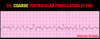
Ventricular Fibrillation
A chaotic rhythm that begins in the ventricles with no organized depolarization; the muscle quivers, like a bag of worms, with no effective contraction (no pulse)
Two types:
a) Coarse ventricular fibrillation
b) Fine ventricular fibrillation
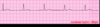
Coarse Ventricular Fibrillation (V-Fib)
Waveforms equal or greater than 3mm in amplitude
Fine Ventricular Fibrillation (V-Fib)
Waveforms less than 3mm in amplitude

Describe conduction pathway of an eletrical impulse in ventricular fibrillation.
1) Impulses originate from multiple automaticity foci in the ventricles
2) the impulses DO NOT follow any specific conduction pathway and DO NOT provide organized depolarization of both atria and ventricles
Ventricular Fibrillation
Rate
Rhythm
P wave
PR interval
QRS Complex
Rate: unable to determine rate (no discernable waves or complexes)
Rhythm: irregularly irregular
P wave: not discernable
PR Interval: n/a
QRS Complex: not discernable
Ventricular Fibrillation
Causes
Adverse Effects
Treatment
Causes: (same as V-Tach and PVC)
- 3 big causes:
- hypoxia - poor oxygenation can make automaticity focus become irritable
- heart disease - ACS, CHF, etc.
- hypokalemia - electrolyte imbalance
- Other causes:
- normal variant
- increased sympathetic tone - stress/anxiety, exercise, etc.
- Medications - Digitalis, sympathomimetics, antidepressants
- Drugs - caffeine, tobacco
- Acid-base imbalance
Adverse Effects:
- no cardiac output
- profound shock, unconsciousness and/or death if not treated
- Coarse v-fib changes progressively to fine v-fib the longer it lasts
Treatment: CPR (if pulseless); defibrillation
_*artifact can mimic v-fib and therefore always check the patient’s pulse before beginning treatment*_
Asystole (Flat line)
No impulse has originated as all pacemakers have failed
A total absence of cardiac electrical activity

Describe the conduction pathway of an electrical impulse in asystole.
No impulse is generated in any cardiac tissue, no impulse follows any conduction pathway
Asystole
Rate
Rhythm
P wave
PR Interval
QRS Complex
Rate: 0
Rhythm: n/a
P wave: none (if only th eP waves are present, rhythm is ventricular standstill)
PR Interval: n/a
QRS Complex: n/a
Asystole
Causes
Adverse Effects
Treatment
Causes: same as V-Tach and V-fib
- 3 big causes:
- hypoxia - poor oxygenation can make automaticity focus become irritable
- heart disease - ACS, CHF, etc.
- hypokalemia - electrolyte imbalance
- Other causes:
- normal variant
- increased sympathetic tone - stress/anxiety, exercise, etc.
- Medications - Digitalis, sympathomimetics, antidepressants
- Drugs - caffeine, tobacco
- Acid-base imbalance
Adverse Effects: Death if untreated
Treatment: CPR
Ventricular Standstill
Occurs as the SA node is still functional, even though there are no impulses passing through to the ventricles
Only atrial depolarization is present and there is no ventricular depolarization (if there is no ventricular depolarization, there is no ventricular contraction and therefore no pulse)

Defibrillation
- A delivery of an unsynchronized electrical current through the myocardium over a very brief period to terminate a cardiac dysrhythmia
- electrical current (shock) attempts to simultaneously depolarize ventricular cells causing momentary asystole, which provides an opportunity for the heart’s natural pacemakers to resume function
- this pacemaker with the highest degree of automaticity should assume repsonsibility for pacing the heart

Adult defib pad placement
pads are placed in specific locations in order to allow the path of current to pass through the heart
electrical current is measured in joules
What are the shockable rhythms?
pulseless ventricular tachycardia
and
ventricular fibrillation
