Nervous System - Brain and Spinal Cord Flashcards
(28 cards)
how is the nervous system structurally divided?
CNS - Brain and Spinal Cord
Peripheral NS - Cranial and spinal nerves
a) Somatic NS
b) Visceral NS

which is longer - spinal cord or spinal column?
spinal column
draw and label a neuron

- which part of neurons make up white matter?
- which part of neurons make up grey matter?
white matter: axons
draw and label a spinal cord cross section
- which bit houses sensory nerves?
dorsal root houses sensory nerves (i think)

what surrounds the CNS? (3)
dura mater, arachnoid mater and pia mater
(outer —> inner)

how many spinal nerves are there?
31
label the spinal cord and the fibres that enter and exit the spinal cord

(from left to right)
- spinal nerve
- dorsal root ganglion
- dorsal root (opposite: ventral root)
- dorsal / ventral rootlets
- (raised block): grey matter)
- white matter

- the spinal cord is made up of what?
grey and white matter
what is grey matter of the spinal cord made up of?
what is white matter of the spinal cord made up of?
what is the arrangement of white and grey matter within the spinal cord?
what is the arrangement of white and grey matter within the brain cord?
grey: neuronal cell bodies and unmyelinated axons
white: myelinated axons
spinal cord arrangement: white matter surrounds the centrally distributed grey matter
brain arrangement: white matter in deeper areas, with surrounding gret matter
what are the 5 sections of the spinal column? - which vertebrae are for which?
- cervical (neck): 7 vert C1-C7
- thoracic (mid back) : 12 vert: T1-T12
- Lumbar (lower back) : 5 vert: L1-L5
- Sacrum: 5 fused vert
- Coccyx: 4 fused vert
= 33 vertabrae

what and where is the cauda equina?
what and where is the cervical enlargement?
what and where is the lumbar enlargement?
cauda equina: typically levels L1-L5
cervical enlargement: C4-T1 level. It represents the origin of the brachial plexus.
lumbar enlargement: Between T11 and L1 representing the origin of the lumbar and sacral plexi.
what are dermatomes?
A dermatome is an area of skin that is supplied by a single spinal nerve and these nerves transmit sensations, such as pain, from the skin to the CNS. Dermatomes have a segmented distribution throughout the body. The exact dermatome pattern can vary from person to person and some overlap between neighbouring dermatomes may also occur.
what is the name of the foramen through which spinal cord passes?
foramen magnum
what is the name of the shock absobers between vertabrae?
intervertebral discs
what are the main anatomical components of a typical vertabrae?
- vertebral body
- transverse process
- spinous process
- superior articular processes
- vertabral foramen

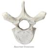
which type of vertabrae is this?

lumbar vert

which vertabrae is this?

sacrum

which vertabrae is this?
thoracic (heart shaped body)
which vertabrae is this?

cervical vert.

how mamy spinal nerves are there?
how are the split?
There are 31 pairs of spinal nerves and are named according to their origin:
8 pairs cervical (C1-C8)
12 pairs thoracic (T1-T12)
5 pairs lumbar (L1-L5)
5 pairs sacral (S1-S5)
1 pair coccygeal (C0)
where does the spinal cord extend from / to? / terminate at?
what happens to spinal cord after this?
- foramen magnum at base of the skull -> L1/L2 vertabrae.
- terminates at the: conus medullaris
- Below the L1 / L2 the spinal nerves are bunched as cauda equina (The collection of nerves at the end of the spinal cord is known as the cauda equina, due to its resemblance to a horse’s tail)


